. 2016 Mar 29;113(13):3621-6.
doi: 10.1073/pnas.1601911113. Epub 2016 Mar 14.

Human mesenchymal stromal cells reduce influenza A H5N1-associated acute lung injury in vitro and in vivo

Affiliations

Human mesenchymal stromal cells reduce influenza A H5N1-associated acute lung injury in vitro and in vivo

Michael C W Chan et al. Proc Natl Acad Sci U S A. .

Abstract

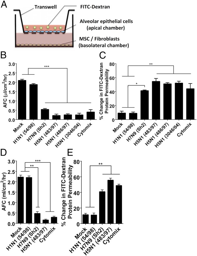
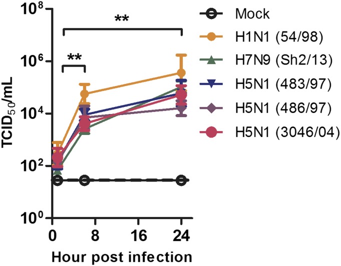
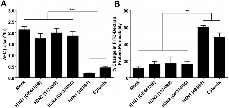
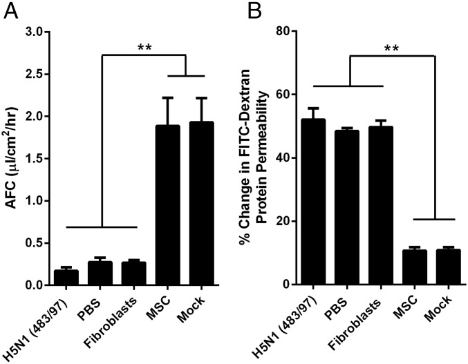
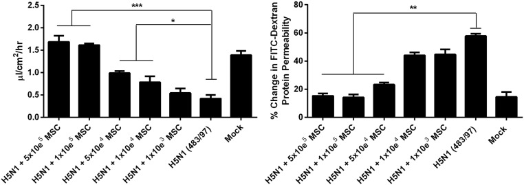
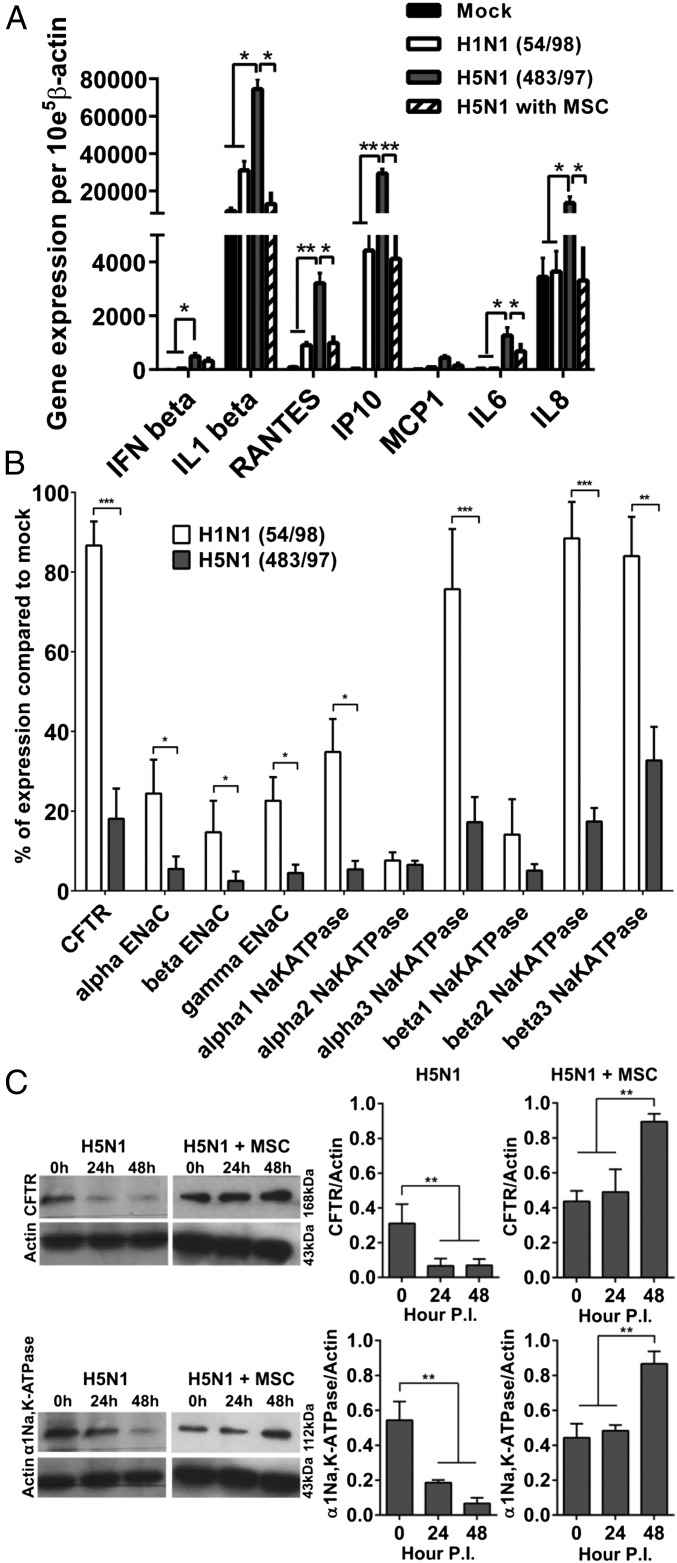
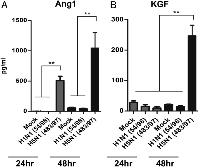
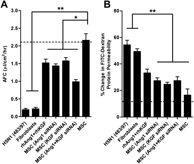
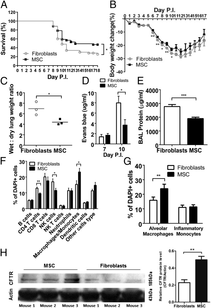
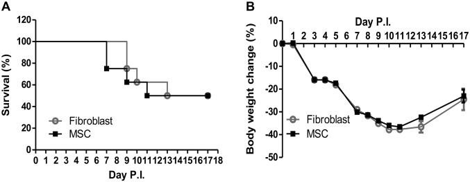
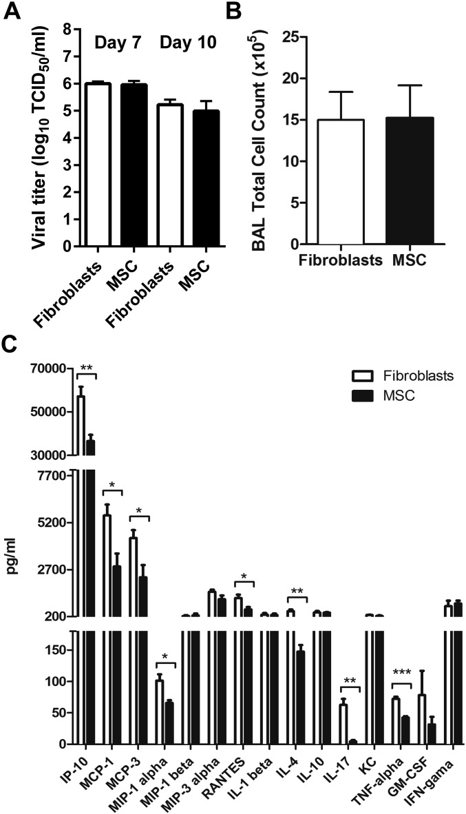
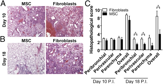
Influenza can cause acute lung injury. Because immune responses often play a role, antivirals may not ensure a successful outcome. To identify pathogenic mechanisms and potential adjunctive therapeutic options, we compared the extent to which avian influenza A/H5N1 virus and seasonal influenza A/H1N1 virus impair alveolar fluid clearance and protein permeability in an in vitro model of acute lung injury, defined the role of virus-induced soluble mediators in these injury effects, and demonstrated that the effects are prevented or reduced by bone marrow-derived multipotent mesenchymal stromal cells. We verified the in vivo relevance of these findings in mice experimentally infected with influenza A/H5N1. We found that, in vitro, the alveolar epithelium's protein permeability and fluid clearance were dysregulated by soluble immune mediators released upon infection with avian (A/Hong Kong/483/97, H5N1) but not seasonal (A/Hong Kong/54/98, H1N1) influenza virus. The reduced alveolar fluid transport associated with down-regulation of sodium and chloride transporters was prevented or reduced by coculture with mesenchymal stromal cells. In vivo, treatment of aged H5N1-infected mice with mesenchymal stromal cells increased their likelihood of survival. We conclude that mesenchymal stromal cells significantly reduce the impairment of alveolar fluid clearance induced by A/H5N1 infection in vitro and prevent or reduce A/H5N1-associated acute lung injury in vivo. This potential adjunctive therapy for severe influenza-induced lung disease warrants rapid clinical investigation.

Keywords: acute lung injury; alveolar fluid clearance; avian; influenza; mesenchymal stromal cells.


Conflict of interest statement

The authors declare no conflict of interest.

Figures

Fig. 1.
Fig. 1.
Highly pathogenic influenza viruses significantly reduce human alveolar fluid clearance (AFC) and significantly increase human alveolar protein permeability (APP). (A) Schematic diagram of the in vitro lung injury model. FITC-dextran, fluorescein isothiocyanate-dextran; MSC, mesenchymal stromal cells. (B and C) Infection with HPAI H5N1 (A/HongKong/483/97, A/HongKong/486/97, and A/HongKong/3046 /04) and H7N9 (A/Shanghai/2/2013) viruses (MOI = 0.1) significantly (B) reduced the AFC and (C) increased the APP of human alveolar epithelial cells compared with infection with LP influenza H1N1 virus (A/HongKong/54/98). Cytomix containing proinflammatory cytokines (IL-1β, TNF-α, and IFN-γ) was used as an injury-inducing positive control. Data are the mean ± SE from three experiments. *P < 0.05; **P < 0.01; ***P < 0.005. (D and E) The effect of conditioned medium (virus-free supernatant) from cells infected with H5N1 and H7N9 influenza viruses affected the AFC and APP of alveolar epithelial cells comparably with the live influenza viruses and (D) reduced the AFC and (E) increased the APP significantly more than supernatant from cells infected with H1N1 virus. Data are the mean ± SE from three experiments. **P < 0.01; ***P < 0.005.
Fig. S1.
Fig. S1.
In vitro replication kinetics of HPAI H5N1 (A/HongKong/483/97, A/Hong Kong/486/97, A/HongKong/3046/04) influenza viruses, HPAI H7N9 (A/Shanghai/2/2013) influenza virus, and an LP influenza virus (A/HongKong/54/98) in human alveolar epithelial cells. All viruses replicated productively, and a statistically significant increase in titers was detected at 8 h and 24 h p.i. Data are the mean ± SE from three experiments. **P < 0.01.
Fig. S2.
Fig. S2.
Highly pathogenic influenza H5N1 virus significantly reduces human alveolar fluid clearance (AFC) and significantly increases human alveolar protein permeability (APP) compared with low pathogenic seasonal influenza viruses. (A and B) HPAI H5N1 (A/HongKong/483/97) virus infection significantly (A) reduced the AFC and (B) increased the APP of human alveolar epithelial cells compared with infection with the LP influenza viruses A/Oklahoma/447/08 (H1N1), A/HongKong/1174/99 (H3N2), and A/Oklahoma/370/05 (H3N2).
Fig. 2.
Fig. 2.
MSCs prevent or reduce H5N1 virus impairment of alveolar epithelial AFC and APP. Alveolar epithelial cell monolayers on the apical chamber of transwell culture inserts were infected with H5N1 (A/HongKong/483/97) influenza virus (MOI = 0.1). Cells cocultured with MSCs in the basolateral chamber showed a significantly greater increase in net AFC (A) and decrease in APP (B) across the transwell inserts than did untreated infected cells, mock-cocultured cells, or cells cocultured with fibroblasts. Data represent the mean ± SE from three experiments. **P < 0.01.
Fig. S3.
Fig. S3.
The effect of MSCs on H5N1-impaired AFC (Left) and APP (Right) is dependent on the number of MSC used for coculture. Alveolar epithelial cell monolayers on the apical chamber of transwell culture inserts were infected with H5N1 (A/HongKong/483/97) influenza virus (MOI = 0.1). Influenza virus-infected cells cocultured with different cell numbers of MSCs in the basolateral chamber. Data represent the mean ± SE from three experiments, *P < 0.05; **P < 0.01; ***P < 0.005.
Fig. 3.
Fig. 3.
H5N1 influenza virus infection of alveolar epithelium induces greater cytokine and chemokine expression and lower sodium and chloride transporter expression than does H1N1 virus infection whereas coculture with MSCs prevents or reduces the H5N1 effects. See Fig. 2 for experimental details. (A) Alveolar epithelial cells infected with HPAI H5N1 virus showed greater gene expression of proinflammatory cytokines (IL1-β; IL-6; IL-8) and chemokines (RANTES; IP-10) than did cells infected with H1N1 virus whereas coculture with MSCs reduced expression of cytokines and chemokines. (B) H5N1 virus infection down-regulated expression of the major sodium and chloride transporters (with the exception of α2-Na,K-ATPase and β1-Na,K-ATPase) to a greater extent than infection with H1N1 virus. Data are percent expression compared with that in mock-infected cells. (C) Western blot analysis showed reduced expression of CFTR and α1-Na,K-ATPase protein in H5N1 virus-infected alveolar epithelial cells at 24 h p.i. and further reduction of α1-Na,K-ATPase at 48 h p.i. With MSC coculture, expression of CFTR and α1-Na,K-ATPase was increased at 24 h p.i. and increased more substantially at 48 h p.i. Shown is mean ± SE protein expression relative to β-actin expression in three experiments. *P < 0.05; **P < 0.01; ***P < 0.005.
Fig. 4.
Fig. 4.
Growth factor secretion by mesenchymal stromal cells is enhanced by coculture with H5N1 influenza virus-infected alveolar epithelial cells and contributes to reduction of AFC and APP impairment. Secretion of (A) Ang1 and (B) KGF by MSCs was increased significantly more by coculture with alveolar epithelium infected with H5N1 (A/HongKong/483/97) influenza virus vs. H1N1 (A/HongKong/54/98) virus, as measured by ELISA. Mock, mock infection. Data are the mean ± SE from three experiments. **P < 0.01.
Fig. S4.
Fig. S4.
Growth factor secretion by mesenchymal stromal cells contributes to reduction of AFC and APP impairment. (A and B) Ang1 and KGF mRNA was knocked down in MSCs by using 300 nM Ang1 and/or 100 nM KGF siRNA. H5N1-infected alveolar epithelium cocultured with MSCs with knock-down of Ang1, KGF, or both showed (A) less increase of AFC and (B) less decrease of APP than did cells cocultured with intact MSCs. Addition of 100 ng/mL recombinant human rhAng1 and rhKGF proteins to H5N1 virus–infected alveolar epithelium in the absence of MSCs reduced the H5N1 virus-mediated (A) AFC decrease and (B) APP increase. Dotted lines indicate AFC and APP in mock-infected cells. Data are the mean ± SE from three experiments. *P < 0.05; **P < 0.01.
Fig. 5.
Fig. 5.
Effects of treatment with MSC in an aged animal model of highly pathogenic H5N1 influenza virus infection. All experiments were carried out in aged (8–12 mo) mice inoculated with A/Hong Kong/486/1997(H5N1) virus and injected i.v. with 5 × 10⁵ MSC/100 μL or mouse fibroblasts (control) on day 5 p.i. (A) Survival of mice injected with MSC (n = 51) or mouse fibroblasts (n = 50). Data are the means from three experiments. (B) Percent body weight change of mice injected with MSCs (n = 79) or mouse fibroblasts (n = 83). Data are the mean from three experiments. (C) Wet-to-dry lung weight ratio of mice treated with MSCs (n = 3) or mouse fibroblasts (n = 3) on day 7 p.i. (D) Lung tissue concentration of Evans blue dye at days 7 and 10 p.i. in mice treated with MSCs (n = 4) or mouse fibroblasts (n = 4). (E) Total protein concentration (indicating lung injury) at day 7 p.i. in BAL fluid from mice treated with MSCs (n = 4) or mouse fibroblasts (n = 4). (F) Immune cell profile of BAL fluid on day 7 p.i. from mice treated with MSCs (n = 5) or mouse fibroblasts (n = 5). (G) Mean (± SD) percentage of alveolar macrophages and inflammatory monocytes at day 7 p.i. in BAL fluid from mice treated with MSCs (n = 5) or mouse fibroblasts (n = 5). Counts were repeated at least twice. (H) CFTR transporter protein at day 10 p.i. in lung lysates of virus-infected mice injected with MSCs (n = 3) or mouse fibroblasts (n = 3). In all comparisons, *P < 0.05; **P < 0.01; ***P < 0.005.
Fig. S5.
Fig. S5.
Effects of treatment with MSCs in a young animal model of highly pathogenic H5N1 influenza virus infection. All experiments were carried out in aged (6–8 wk) mice inoculated with A/Hong Kong/486/1997(H5N1) virus and injected i.v. with 5 × 10⁵ MSCs/100 μL or mouse fibroblasts (control) on day 5 p.i. MSC treatment of young mice 5 d after inoculation with H5N1 influenza virus showed no significant improvement in (A) survival or (B) body weight change. Young mice infected with A/HongKong/486/1997 (H5N1) virus were i.v. injected with 5 × 10⁵ MSCs/100 μL (n = 16) or with mouse fibroblasts (controls, n = 25).
Fig. S6.
Fig. S6.
Effects of treatment with MSCs in an aged animal model of highly pathogenic H5N1 influenza virus infection. All experiments were carried out in aged (8–12 mo) mice inoculated with A/Hong Kong/486/1997(H5N1) virus and injected i.v. with 5 × 10⁵ MSCs/100 μL or mouse fibroblasts (control) on day 5 p.i. (A) Comparable lung virus titers at days 7 and 10 p.i. in mice injected with MSCs (n = 3) or mouse fibroblasts (n = 3). (B) Mean (± SD) cell count at day 7 p.i. in BAL fluid of mice treated with MSCs (n = 5) or mouse fibroblasts (n = 5). Cell counts were repeated at least twice. (C) Concentration of major proinflammatory cytokines and chemokines in BAL fluid at day 7 p.i. in mice treated with MSCs (n = 4) or mouse fibroblasts (n = 4). In all comparisons, *P < 0.05; **P < 0.01; ***P < 0.005.
Fig. 6.
Fig. 6.
Improved lung pathology and histopathology scores of aged mice treated with MSCs after inoculation with highly pathogenic H5N1 virus. (A and B) Lung tissue at (A) day 10 p.i. and (B) day 18 p.i. from H5N1 virus-infected mice after treatment with MSCs (n = 4) or mouse fibroblasts (n = 4) on day 5 p.i. Hematoxylin/eosin stain. (Magnification: 100×.) (C) Histopathology scores of peribronchiolar, perivascular, and parenchymal lung tissues at days 10 and 18 p.i. from influenza virus-infected mice treated with MSCs or mouse fibroblasts. *P < 0.05.

References

    1. Chan MC, et al. Proinflammatory cytokine responses induced by influenza A (H5N1) viruses in primary human alveolar and bronchial epithelial cells. Respir Res. 2005;6:135. - PMC - PubMed
    1. Perrone LA, Plowden JK, García-Sastre A, Katz JM, Tumpey TM. H5N1 and 1918 pandemic influenza virus infection results in early and excessive infiltration of macrophages and neutrophils in the lungs of mice. PLoS Pathog. 2008;4(8):e1000115. - PMC - PubMed
    1. Kandun IN, et al. Factors associated with case fatality of human H5N1 virus infections in Indonesia: A case series. Lancet. 2008;372(9640):744–749. - PubMed
    1. Howard WA, Peiris M, Hayden FG. Report of the 'mechanisms of lung injury and immunomodulator interventions in influenza' workshop, 21 March 2010, Ventura, California, USA. Influenza Other Respir Viruses. 2011;5(6):453–454, e458–e475. - PMC - PubMed
    1. van Riel D, et al. H5N1 virus attachment to lower respiratory tract. Science. 2006;312(5772):399. - PubMed

Publication types

MeSH terms