

Toxic Epidermal Necrolysis in Recessive Dystrophic Epidermolysis Bullosa following Bone Marrow Transplantation
- PMID: 26976809
- PMCID: PMC5322426
- DOI: 10.1016/j.jpeds.2016.02.037
Toxic Epidermal Necrolysis in Recessive Dystrophic Epidermolysis Bullosa following Bone Marrow Transplantation
Abstract
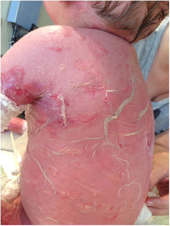
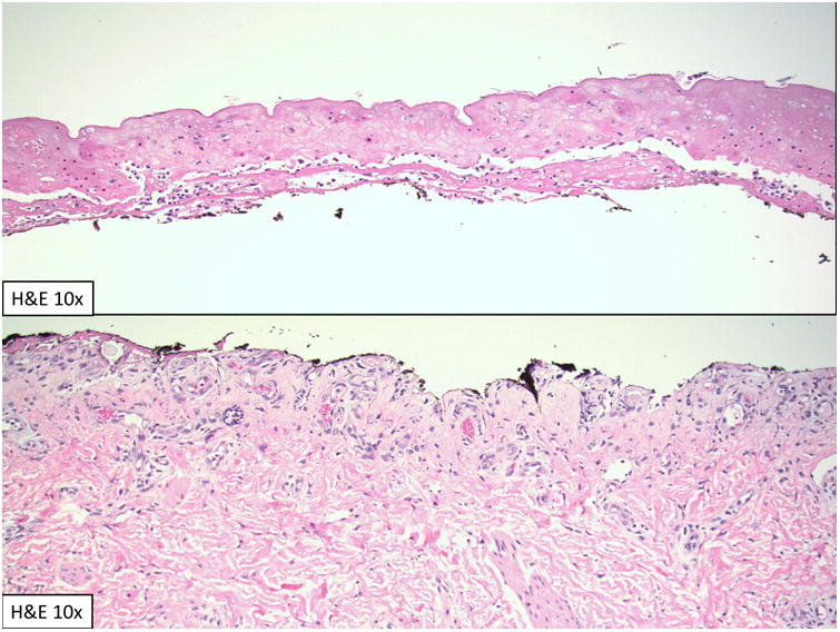
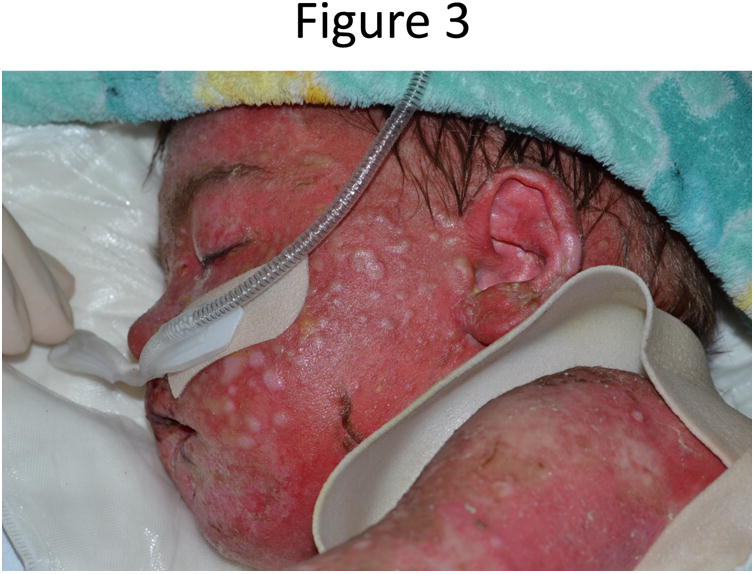
A 3-year-old child with recessive dystrophic epidermolysis bullosa treated with bone marrow transplantation subsequently developed body-wide epidermal detachment distinct from his epidermolysis bullosa. Toxic epidermal necrolysis was diagnosed by examination and skin biopsy. Although graft-vs-host disease was considered, he had no features of this diagnosis by laboratory studies or skin biopsy, and he improved without addition of further immune suppressants. Throughout the episode, the patient was maintained on cyclosporine A, a component of his transplant regimen, and also a reported therapy for toxic epidermal necrolysis. He had full recovery. Re-epithelialization occurred in a unique folliculocentric pattern, which we postulate was related to the patient's mesenchymal stem cell infusion, received as an adjunct to his marrow transplantation.
Keywords: bone marrow transplantation; epidermolysis bullosa; mesenchymal stem cells; toxic epidermal necrolysis.
Copyright © 2016 Elsevier Inc. All rights reserved.
Figures
References
-
- Hamilton GM, Fish J. Pediatric Toxic Epidermal Necrolysis: An Institutional Review of Patients Admitted to an Intensive Care Unit. J Burn Care Res. 2013;34(6):e351–8. - PubMed
-
- Ferrandiz-Pulido C, Garcia-Patos V. A review of causes of Stevens–Johnson syndrome and toxic epidermal necrolysis in children. Arch Dis Child. 2013;98(12):998–1003. - PubMed
-
- Takeda H, Mitsuhashi Y, Kondo S, et al. Toxic epidermal necrolysis possibly linked to hyperacute graft-versus- host disease after allogeneic bone marrow transplantation. Journal of Dermatology. 1997;24(10):635–641. - PubMed
-
- Villada G, Roujeau JC, Cordonnier C, et al. Toxic epidermal necrolysis after bone marrow transplantation: study of nine cases. J Am Acad Dermatol. 1990;23(5):870–875. - PubMed
-
- Macedo FI, Faris J, Lum LG, et al. Extensive Toxic Epidermal Necrolysis Versus Acute Graft Versus Host Disease After Allogenic Hematopoietic Stem-Cell Transplantation: Challenges in Diagnosis and Management. J Burn Care Res. 2014;35(6):e431–435. - PubMed
Publication types
MeSH terms
Substances
Grants and funding
LinkOut - more resources
Full Text Sources
Other Literature Sources
Medical
