. 2016 Apr;147(4):353-68.
doi: 10.1085/jgp.201511510. Epub 2016 Mar 14.

Stabilization of a prokaryotic LAT transporter by random mutagenesis

Affiliations

Stabilization of a prokaryotic LAT transporter by random mutagenesis

Arturo Rodríguez-Banqueri et al. J Gen Physiol. 2016 Apr.

Abstract

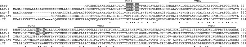
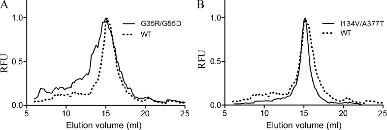
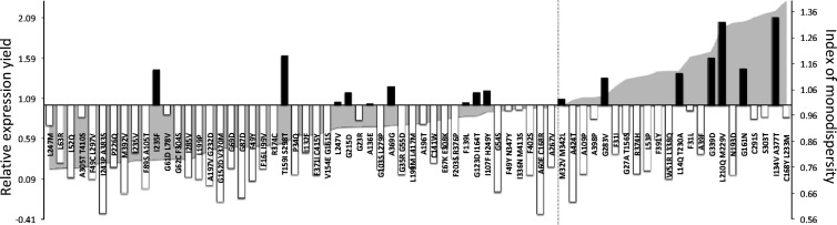
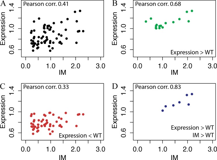
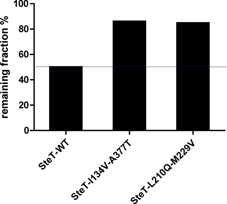
The knowledge of three-dimensional structures at atomic resolution of membrane transport proteins has improved considerably our understanding of their physiological roles and pathological implications. However, most structural biology techniques require an optimal candidate within a protein family for structural determination with (a) reasonable production in heterologous hosts and (b) good stability in detergent micelles. SteT, the Bacillus subtilis L-serine/L-threonine exchanger is the best-known prokaryotic paradigm of the mammalian L-amino acid transporter (LAT) family. Unfortunately, SteT's lousy stability after extracting from the membrane prevents its structural characterization. Here, we have used an approach based on random mutagenesis to engineer stability in SteT. Using a split GFP complementation assay as reporter of protein expression and membrane insertion, we created a library of 70 SteT mutants each containing random replacements of one or two residues situated in the transmembrane domains. Analysis of expression and monodispersity in detergent of this library permitted the identification of evolved versions of SteT with a significant increase in both expression yield and stability in detergent with respect to wild type. In addition, these experiments revealed a correlation between the yield of expression and the stability in detergent micelles. Finally, and based on protein delipidation and relipidation assays together with transport experiments, possible mechanisms of SteT stabilization are discussed. Besides optimizing a member of the LAT family for structural determination, our work proposes a new approach that can be used to optimize any membrane protein of interest.


Figures

Figure 1.
Figure 1.
Multiple alignments of SteT and human LATs. The figure only shows the alignment of the regions where SteT and human LATs are more conserved (TMDs 1, 2, 6, 7, and 8). The full sequence of these transporters have ∼30% amino acid identity (Reig et al., 2007). Lines above the sequences define the SteT TMDs. Residues totally conserved among these transporters are indicated by an asterisk. Residues of LAT-1 predicted to interact with the substrate (Geier et al., 2013) and conserved in SteT are highlighted with a gray background.
Figure 2.
Figure 2.
Examples of normalized FSEC profiles of SteT-GFP variants. To compare the monodispersity in DDM of each SteT mutant, FSEC chromatograms were normalized and overlapped. (A) A typical result of a double mutation (G35R/G55R) that causes a decrease of monodispersity in DDM. (B) The improving effect of the double mutation I134V/A377T on SteT monodispersity in DDM. RFU, relative fluorescent units.
Figure 3.
Figure 3.
Analysis of expression and monodispersity in DDM of SteT random mutants. The values of relative expression yield in E. coli of each mutant with respect to WT (left axis) are represented as shadow areas. These values were calculated using the split GFP assay and normalized with respect to the corresponding value of SteT WT. Mutants situated on the left of the dashed line show lower expression yield than WT, whereas mutants that show better expression yield are situated on the right side of this line. Vertical bars in the graph represent the values of the IM of a giving mutant (see Materials and methods for description). White bars correspond to mutants with worse monodispersity in DDM than WT (IM <1) and black bars correspond to mutants with better monodispersity in DDM than WT (IM >1).
Figure 4.
Figure 4.
Correlation between expression and stability in detergent of the SteT random mutants. (A) Values of expression and IM from Fig. 3 were plotted and analyzed using Pearson’s coefficients. (B–D) Mutants were also analyzed separately according the following criteria: expression levels higher than WT (B), expression levels lower than WT (C), and expression levels higher than WT and IM >1 (D). The numerical values of Pearson’s coefficients of each dataset are indicated in each panel.
Figure 5.
Figure 5.
Normalized FSEC profiles of SteT WT, I134V/A377T, and L210Q/M229V solubilized in DDM, DM, Cymal-6, and OG. (A–H) Normalized FSECs of SteT-I134V/A377T (A–D) and SteT-L210Q/M229V (E–H) were overlapped with the corresponding normalized FSEC of SteT WT (dashed lines). SteT variants containing the GFP in the C-terminal end were solubilized from the membrane with DDM (A and E), DM (B and F), Cymal-6 (C and G), or OG (D and H) and injected into the gel filtration column equilibrated with a buffer containing 2× CMC of DDM. RFU, relative fluorescent units.
Figure 6.
Figure 6.
Position of the amino acid replaced in SteT mutants L210Q/M229V and I134V/A377T. Amino acid substitutions were modeled in a SteT 3-D structural model (Bartoccioni et al., 2010) and represented as spheres. Transmembrane segments are represented as cylinders, and only the TMDs containing mutations are numbered. TMDs 6 and 7 containing, respectively, the double substitution L210Q and M229V are colored in red, and TMDs 4 and 10 containing, respectively, the substitutions I134V and A377T are drawn in green. Residue K295 (TMD8), implicated in substrate recognition in SteT (Bartoccioni et al., 2010), is also labeled and represented as blue spheres. (A) A lateral perspective of the molecule. (B) A periplasmic view after 60° rotation of A through the x axis. The gray shadow square in A represents the putative position of the surrounding membrane.
Figure 7.
Figure 7.
Transport activity of SteT WT and the double mutants I134V/A377T and L210Q/M229V. Transport activity of each SteT version was analyzed from the time-dependent uptake curves of 10 µM radiolabeled l-Ser into proteoliposomes formed after reconstituting each detergent-purified version of SteT, loaded with 4 mM l-Ser. The data points correspond to the net transport activity of each SteT version and were calculated after subtracting the data of nonloaded proteoliposomes from the data of proteoliposomes loaded with l-Ser. Each data dataset is the mean of three independent experiments, and the SD is also represented. The figure also shows the GFP fluorescence in SDS-PAGE gels of the three proteins after reconstituting in proteoliposomes. SteT monomer fused to the GFP is the main band between 42 and 72 kD.
Figure 8.
Figure 8.
SEC profiles of DDM-purified SteT WT, I134V/A377T, and L210Q/M229V. Purified proteins from His-tag affinity chromatography were concentrated up to 2 mg/ml and injected in a Superdex 200 50/1 50G column. (A and B) Monodispersity in these experimental conditions was analyzed by overlapping the normalized chromatograms of SteT WT with I134V/A377T (A) and L210Q/M229V (B).
Figure 9.
Figure 9.
SEC profiles of purified SteT I134V/A377T and L210Q/M229V in DM, NG, Cymal-6, and OG. Affinity purified proteins in DDM were concentrated up to 4 mg/ml and injected into a Superdex 200 50/1 50G column equilibrated with 2× CMC of the indicated detergent.
Figure 10.
Figure 10.
Stability of purified SteT WT, I134V/A377T, and L210Q/M229V after thermal denaturation. 2 mg/ml of purified proteins in DDM were incubated at 50°C for 30 min. The bars indicate the remaining percentage of each indicated SteT version present in the supernatant and measured after ultracentrifugation.
Figure 11.
Figure 11.
Effect of E. coli membrane lipids in the monodispersity of purified SteT I134V/A377T, L210Q/M229V, and WT. (A) SteT I134V/A377T was solubilized from the membrane with 1% (wt/vol) DDM and immobilized in Ni-NTA beads for affinity purification. Beads were divided and poured into four columns and washed with 5 column volumes of 0.02, 0.03, 0.04, and 0.05% (wt/vol) DDM, respectively, in each column. Each protein sample was eluted from the affinity column and subjected to gel filtration chromatography in a Superdex 200 10/30 column equilibrated with the same concentration of DDM used in the affinity purification. By gradually increasing the concentration of DDM during purification, SteT I134V/A377T became less monodisperse as a result of the delipidation effect of DDM. (B) Solubilized SteT I134V/A377T in 1% (wt/vol) DDM was immobilized in Ni-NTA beads for affinity purification. Beads were divided into three columns and washed with buffer containing no lipid or 0.05 and 0.2 mg/ml E. coli lipids, respectively, in each column, always in the presence of 0.05% (wt/vol) DDM (delipidating conditions). Each protein sample was eluted from the affinity column and subjected to gel filtration chromatography in a Superdex 200 10/30 column equilibrated with 0.05% (wt/vol) DDM and the same amount of E. coli lipids used in the affinity purification. As seen in the figure, even in the presence of delipidating conditions (washing the Ni-NTA beads with 0.05% [wt/vol] DDM), the presence of E. coli lipids kept SteT I134V/A377T monodisperse in a concentration-dependent manner. (C and D) Solubilized SteT WT (C) and L210Q/M229V (D) in 1% (wt/vol) DDM were immobilized in Ni-NTA beads for affinity purification. As in B, beads were divided into three columns and washed with buffer containing no lipid or 0.05 and 0.2 mg/ml E. coli lipids, respectively, in the presence of 0.02% and 0.05% (wt/vol) DDM for WT and L210Q/M229V, respectively. Each protein sample was eluted from the affinity column and subjected to gel filtration chromatography in a Superdex 200 10/30 column equilibrated with 0.05% (wt/vol) DDM (0.02% in the case of WT) and the same amount of E. coli lipids used in the affinity purification. Even in the presence of 0.2 mg/ml E. coli lipids (and lower concentration of DDM in the case of WT: 0.02% [wt/vol] instead of 0.05%), both SteT variants eluted in a polydisperse manner.
Figure 12.
Figure 12.
Position of mutants G161N and A339D in a SteT 3-D model. Amino acid substitutions G161N and A339D were modeled in a SteT 3-D structural model (Bartoccioni et al., 2010) and represented as spheres. TMDs are represented as cylinders, and for clarity, only some of them are numbered. The G161N replacement (TMD5) introduces a new asparagine residue suggested to interact with TMD8. Similarly, the introduced aspartate in the A339D mutation (TMD9) is suggested to interact with residues in TMDs 3 and/or 4. (A) A lateral perspective of the molecule. (B) A periplasmic view after 45° rotation of A through the x axis.

Similar articles

Cited by

References

    1. Abramson J., Smirnova I., Kasho V., Verner G., Kaback H.R., and Iwata S.. 2003. Structure and mechanism of the lactose permease of Escherichia coli. Science. 301:610–615. 10.1126/science.1088196 - DOI - PubMed
    1. Barák I., and Muchová K.. 2013. The role of lipid domains in bacterial cell processes. Int. J. Mol. Sci. 14:4050–4065. 10.3390/ijms14024050 - DOI - PMC - PubMed
    1. Bartoccioni P., Del Rio C., Ratera M., Kowalczyk L., Baldwin J.M., Zorzano A., Quick M., Baldwin S.A., Vázquez-Ibar J.L., and Palacín M.. 2010. 
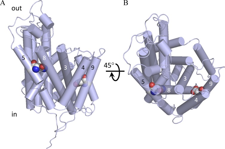
Role of transmembrane domain 8 in substrate selectivity and translocation of SteT, a member of the L-amino acid transporter (LAT) family. J. Biol. Chem. 285:28764–28776. 10.1074/jbc.M110.116632 - DOI - PMC - PubMed
    1. Bill R.M., Henderson P.J.F., Iwata S., Kunji E.R.S., Michel H., Neutze R., Newstead S., Poolman B., Tate C.G., and Vogel H.. 2011. Overcoming barriers to membrane protein structure determination. Nat. Biotechnol. 29:335–340. 10.1038/nbt.1833 - DOI - PubMed
    1. Bröer S., and Palacín M.. 2011. The role of amino acid transporters in inherited and acquired diseases. Biochem. J. 436:193–211. 10.1042/BJ20101912 - DOI - PubMed

Publication types

MeSH terms

Substances

LinkOut - more resources