. 2016 May 15;473(10):1343-53.
doi: 10.1042/BCJ20160046. Epub 2016 Mar 14.

Human DC-SIGN binds specific human milk glycans

Affiliations

Human DC-SIGN binds specific human milk glycans

Alexander J Noll et al. Biochem J. .

Abstract

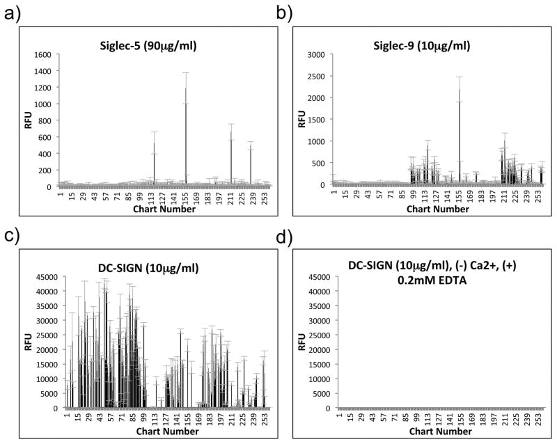
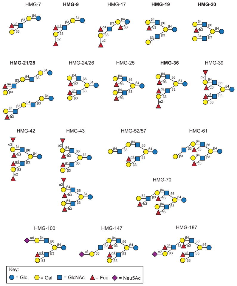
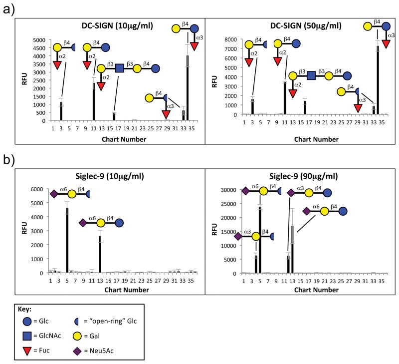
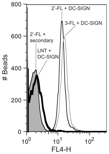
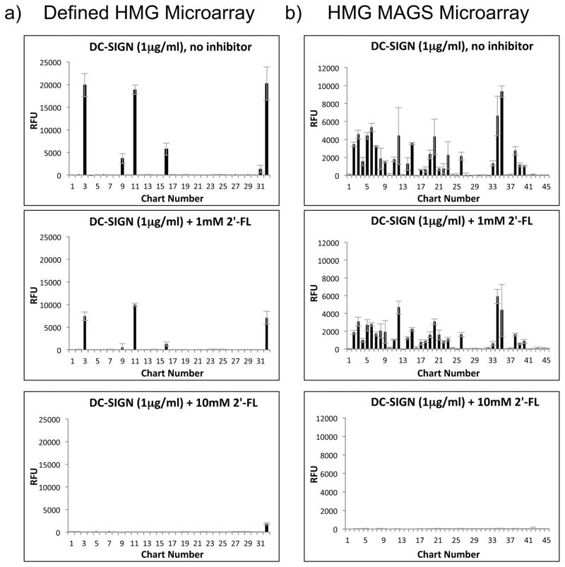
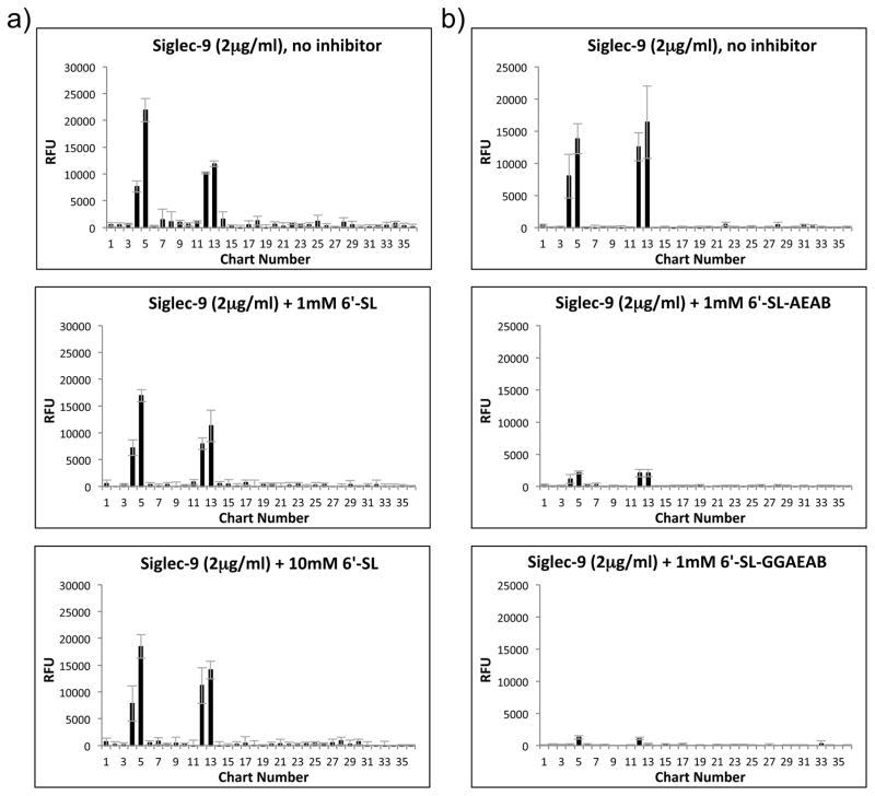
Human milk glycans (HMGs) are prebiotics, pathogen receptor decoys and regulators of host physiology and immune responses. Mechanistically, human lectins (glycan-binding proteins, hGBP) expressed by dendritic cells (DCs) are of major interest, as these cells directly contact HMGs. To explore such interactions, we screened many C-type lectins and sialic acid-binding immunoglobulin-like lectins (Siglecs) expressed by DCs for glycan binding on microarrays presenting over 200 HMGs. Unexpectedly, DC-specific intercellular adhesion molecule-3-grabbing non-integrin (DC-SIGN) showed robust binding to many HMGs, whereas other C-type lectins failed to bind, and Siglec-5 and Siglec-9 showed weak binding to a few glycans. By contrast, most hGBP bound to multiple glycans on other microarrays lacking HMGs. An α-linked fucose residue was characteristic of HMGs bound by DC-SIGN. Binding of DC-SIGN to the simple HMGs 2'-fucosyl-lactose (2'-FL) and 3-fucosyl-lactose (3-FL) was confirmed by flow cytometry to beads conjugated with 2'-FL or 3-FL, as well as the ability of the free glycans to inhibit DC-SIGN binding. 2'-FL had an IC50 of ∼1 mM for DC-SIGN, which is within the physiological concentration of 2'-FL in human milk. These results demonstrate that DC-SIGN among the many hGBP expressed by DCs binds to α-fucosylated HMGs, and suggest that such interactions may be important in influencing immune responses in the developing infant.

Keywords: dendritic cell-specific intercellular adhesion molecule-3-grabbing non-integrin (DC-SIGN); glycan microarrays; glycan recognition; glycan-binding proteins; human milk glycans; lectins.


Conflict of interest statement

Declarations of Interest

R.D.C. and D.F.S. are consultants for Abbott Nutrition. G.D.M. and R.H.B. are employees of Abbott. The other authors declare that they have no conflict of interest in the work reported.

Figures

Fig. 1
Fig. 1. HM-SGM-v2 Data for Siglec-5, Siglec-9, and DC-SIGN
Siglec-5, Siglec-9, and DC-SIGN were screened on the HM-SGM-v2 at multiple concentrations. The results for 90μg/ml Siglec-5 (a), 10μg/ml Siglec-9 (b), and 10μg/ml DC-SIGN with (c) or without (d) Ca2+ are shown; Alexa Fluor 488-labeled anti-human IgG was used for detection. Refer to Supplementary File 1 for the results at all concentrations screened. For DC-SIGN without Ca2+ (d), Ca2+ was omitted from the binding buffer and 0.2mM EDTA was added.
Fig. 2
Fig. 2. Proposed Structures of HMGs Bound by DC-SIGN on the HM-SGM-v2
A portion of the HM-SGM-v2 structures bound by DC-SIGN were printed on a separate microarray and interrogated by metadata-assisted glycan sequencing (MAGS), where multiple lectins and antibodies specific for particular glycan determinants were screened. Proposed structures for these HMGs samples are shown. HMG samples in bold-face font were further analyzed by multi-dimensional mass spectrometry (MSn) to more accurately determine the structures(s) within these samples; HMG-20, -21, and -28 were previously sequenced by MSn [21, 28] and HMGs-9, -19, and -36 were also by sequenced by MSn in a more recent manuscript [33]. Refer to Supplementary File 2 for the lectin and antibody screening data.
Fig. 3
Fig. 3. Defined HMGs Microarray Screening Data for DC-SIGN and Siglec-9
10μg/ml and 50μg/ml DC-SIGN (a) and 10μg/ml and 90μg/ml Siglec-9 (b) were screened on the defined HMGs microarray, and Alexa Fluor 488-labeled anti-human IgG was used for detection. Graphs on the left show the lower concentrations and the right graphs show the higher concentrations. Refer to Supplementary File 4 for the raw data for these screenings.
Fig. 4
Fig. 4. DC-SIGN Binding to HMGs-Derivatized Microspheres
DC-SIGN was incubated with microspheres (beads) derivatized with 2′-FL, 3-FL, or LNT. Alexa Fluor 633-labeled anti-human IgG was used for detection of recombinant DC-SIGN. 2′-FL microspheres incubated with the Alexa Fluor 633-labeled anti-human IgG alone was used as the negative control. All samples were analyzed by Flow Cytometry with a 633nm laser and FL-4 filter for detection. Histograms of DC-SIGN binding to LNT beads (thick line), 2′-FL beads (thin black line), and 3-FL beads (thin grey line), as well as secondary antibody alone binding to 2′-FL beads (filled line), are shown.
Fig. 5
Fig. 5. Inhibition of DC-SIGN Binding to HMGs Microarrays with Free, Underivatized HMGs
1μg/ml DC-SIGN was preincubated with or without 0.1, 1, or 10mM of free, underivatized 2′-FL and then screened on the defined HMGs microarray (a) or the HMGs MAGS microarray (b) described in Supplementary File 2. The results for 0mM 2′FL (no inhibitor), 1mM 2′-FL, and 10mM 2′-FL on both microarrays are shown; refer to Supplementary File 5 for the results of all other screenings, including 0.1mM 2′-FL.
Fig. 6
Fig. 6. Inhibition of Siglec-9 Binding to Defined HMGs Microarray with Free, Underivatized HMGs and Free, Derivatized 6′-Sialyllactose
2μg/ml Siglec-9 was preincubated with 1mM or 10mM free, underivatized 6′-SL or no inhibitor and screened on the defined HMGs microarray (a). 2μg/ml Siglec-9 was preincubated with free 1mM 6′-SL-AEAB (Neu5Acα2-6Galβ1-4Glcitol-AEAB) or 6′-SL-GGAEAB (Neu5Acα2-6Galβ1-4Glc-GGAEAB) [25] or no inhibitor and screened on the defined HMGs microarray (b). Parts a and b of this figure were performed on separate slides but on the same day and at the same time. Refer to Supplementary File 6 for all other free inhibition of Siglec-9 binding to the defined HMGs microarray.

References

    1. Kunz C, Rudloff S, Baier W, Klein N, Strobel S. Oligosaccharides in human milk: structural, functional, and metabolic aspects. Annu Rev Nutr. 2000;20:699–722. - PubMed
    1. Urashima T, Asakuma S, Leo F, Fukuda K, Messer M, Oftedal OT. The predominance of type I oligosaccharides is a feature specific to human breast milk. Adv Nutr. 2012;3:473S–482S. - PMC - PubMed
    1. Newburg DS. Glycobiology of human milk. Biochemistry (Mosc) 2013;78:771–785. - PubMed
    1. Newburg DS, Ruiz-Palacios GM, Morrow AL. Human milk glycans protect infants against enteric pathogens. Annu Rev Nutr. 2005;25:37–58. - PubMed
    1. Zivkovic AM, German JB, Lebrilla CB, Mills DA. Human milk glycobiome and its impact on the infant gastrointestinal microbiota. Proc Natl Acad Sci U S A. 2011;108(Suppl 1):4653–4658. - PMC - PubMed

Publication types

MeSH terms