. 2016 Mar;28(3):786-803.
doi: 10.1105/tpc.15.00608. Epub 2016 Mar 14.

Functionally Similar WRKY Proteins Regulate Vacuolar Acidification in Petunia and Hair Development in Arabidopsis

Affiliations

Functionally Similar WRKY Proteins Regulate Vacuolar Acidification in Petunia and Hair Development in Arabidopsis

Walter Verweij et al. Plant Cell. 2016 Mar.

Abstract

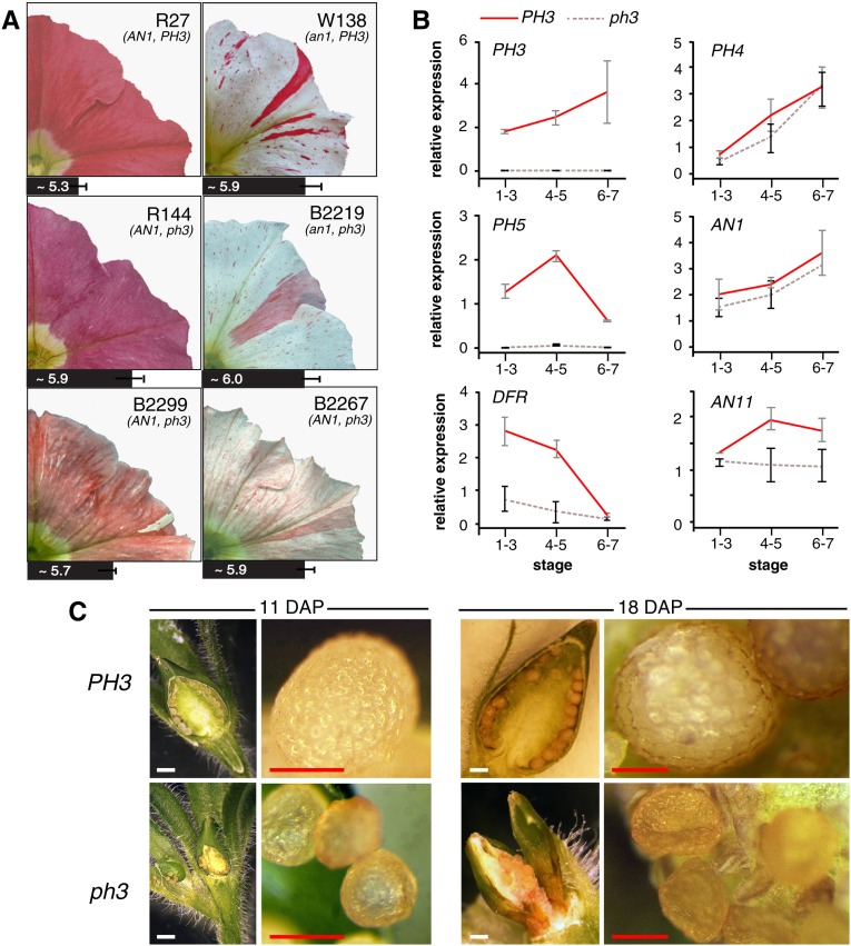
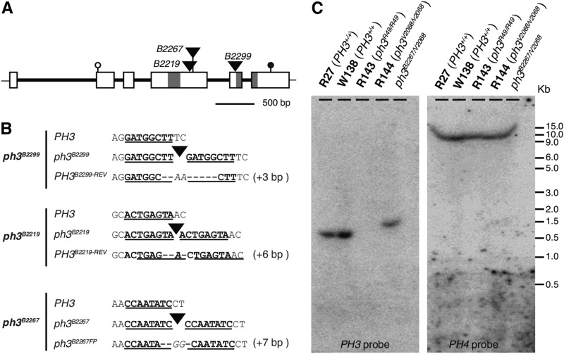
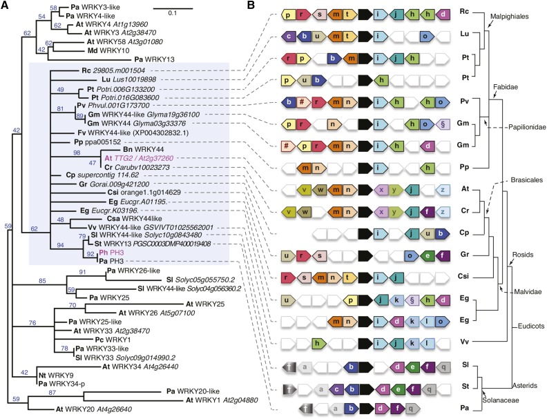
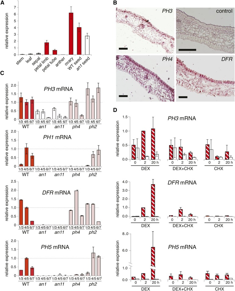
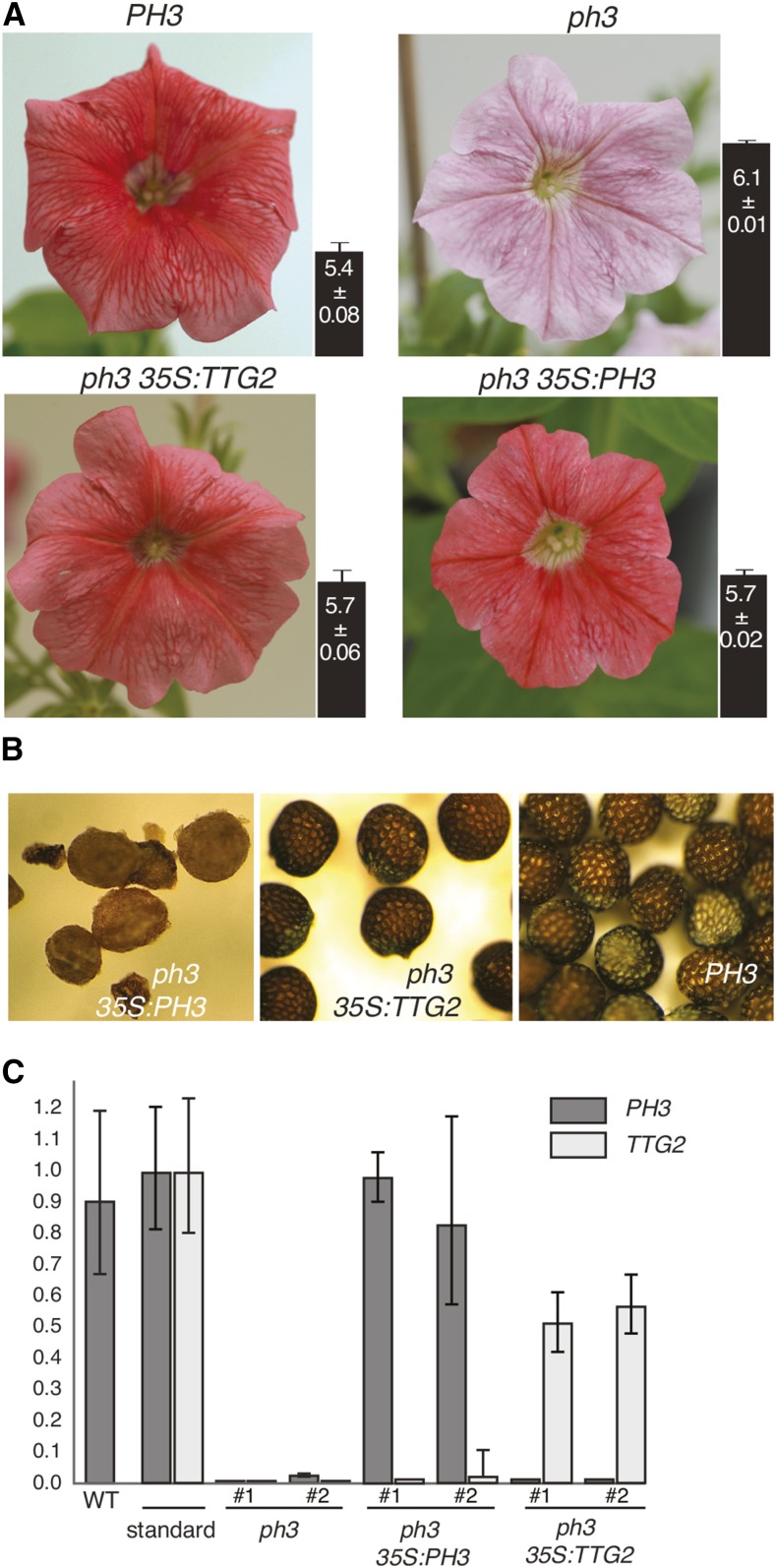
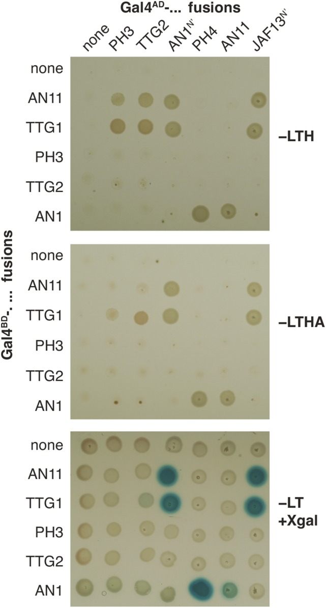
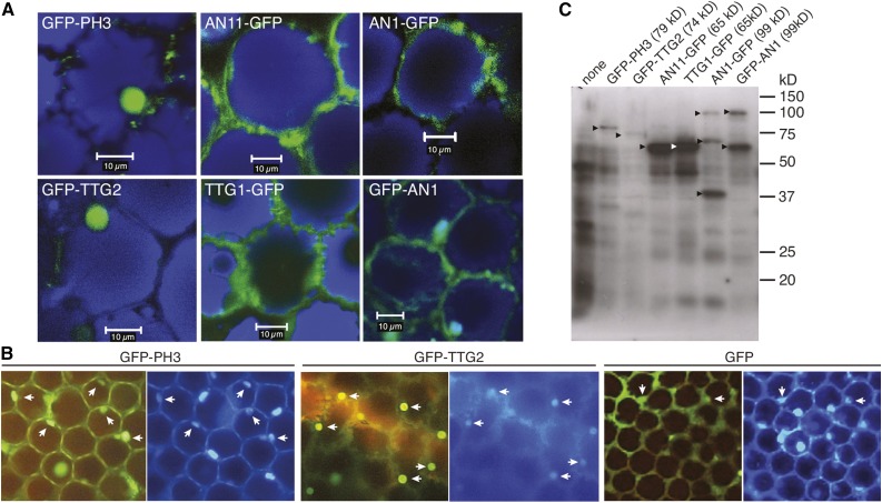
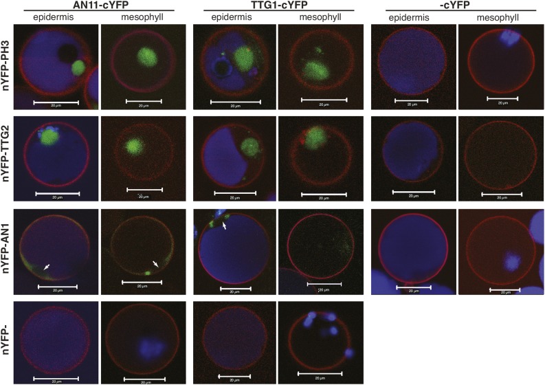
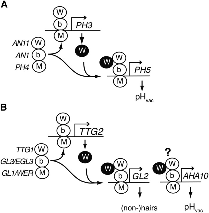
The WD40 proteins ANTHOCYANIN11 (AN11) from petunia (Petunia hybrida) and TRANSPARENT TESTA GLABRA1 (TTG1) from Arabidopsis thaliana and associated basic helix-loop-helix (bHLH) and MYB transcription factors activate a variety of differentiation processes. In petunia petals, AN11 and the bHLH protein AN1 activate, together with the MYB protein AN2, anthocyanin biosynthesis and, together with the MYB protein PH4, distinct genes, such as PH1 and PH5, that acidify the vacuole. To understand how AN1 and AN11 activate anthocyanin biosynthetic and PH genes independently, we isolated PH3. We found that PH3 is a target gene of the AN11-AN1-PH4 complex and encodes a WRKY protein that can bind to AN11 and is required, in a feed-forward loop, together with AN11-AN1-PH4 for transcription of PH5. PH3 is highly similar to TTG2, which regulates hair development, tannin accumulation, and mucilage production in Arabidopsis. Like PH3, TTG2 can bind to petunia AN11 and the Arabidopsis homolog TTG1, complement ph3 in petunia, and reactivate the PH3 target gene PH5. Our findings show that the specificity of WD40-bHLH-MYB complexes is in part determined by interacting proteins, such as PH3 and TTG2, and reveal an unanticipated similarity in the regulatory circuitry that controls petunia vacuolar acidification and Arabidopsis hair development.


Figures

Figure 1.
Figure 1.
Phenotype of ph3 Mutants. (A) Petal limbs of the parental lines R27 (AN1+/+ PH3+/+) and W138 (an1m/m, PH3+/+), the derived stable recessive ph3 mutant R144 (AN1+/+ ph3V2068/V2068), and three transposon-tagged ph3 mutants B2267-1 (AN1+/m ph3V2068/B2267), B2219-1 (an1m/m ph3V2068/B2219), and B2299-1 (AN1m/Rev ph3V2068/B2299). The bars under the images represent the pH of the crude petal extract (n = 3, mean ± sd). (B) Real-time RT-PCR analysis of mRNA from PH3, the regulatory genes, PH4, AN1, and AN11, and the structural genes PH5 and DFR in petals of lines R27 (PH3+) and R144 (ph3) from flowers of developmental stages 1 to 3 (small bud and uncolored petals), 4 to 5 (fully expanded bud and colored petals), and 6 to 7 (fully opened flower with still closed anthers and flower with dehiscent anthers). Relative expression is indicated as the mean ± sd of three biological replicates. (C) Seedpods of a PH3 and a ph3 mutant at 11 and 18 d after pollination (DAP). White bars = 1 mm, and red bars = 0.2 mm.
Figure 2.
Figure 2.
Analysis of PH3 and Mutant Alleles. (A) Structure of PH3 and mutant alleles. Exons and introns are indicated by rectangles and thick lines, start and stop codons by open and close circles, and dTPH1 insertions in three distinct ph3 alleles by triangles. Regions encoding two WRKY domains are indicated by gray shading (B) Sequence alterations in three distinct transposon-tagged ph3 alleles and derived excision alleles. For each insertion allele, the sequence of the progenitor wild-type allele is shown above the sequence of the excision alleles. Target site duplication is indicated by boldface font and underlining. Nucleotides that were deleted during excisions are indicated by dashes and extra nucleotides that were inserted in italics. (C) DNA gel blot analysis of the PH3 and PH4 loci in different genotypes. The blot was first hybridized with a cDNA PH3 probe spanning nucleotide 404 to 1407 and subsequently stripped and rehybridized with the full-length PH4 cDNA as a control for DNA loading.
Figure 3.
Figure 3.
PH3 Is a WRKY Protein and Homologous to Arabidopsis TTG2. (A) Phylogenetic tree of a selection of type I WRKY proteins. Blue numbers on tree branches indicate percentage of bootstrap support (1500 replicates). The blue shaded rectangle indicates the clade containing TTG2/PH3 homologs. Prefixes denote the species of origin: Arabidopsis thaliana (At), Brassica napus (Bn), Capsella rubella (Cr), Carica papaya (Cp), Citrus sinensis (Csi), Cucumis sativum (Csa), Eucalyptus grandis (Eg), Fragaria vesca (Fv), Glycine max (Gc), Gossypium raimondii (Gr), Hordeum vulgare (Hv), Ipomea batatas (Ib), Linum usitatissimum (Lu), Malus domestica (Md), Nicotiana tabacum (Nt), Petroselinum crispum (Pc), Petunia axillaris (Pa), Petunia hybrida (Ph), Phaseolus vulgaris (Pv), Populus trichocarpa (Pt), Prunus persica (Pp), Ricinus communis (Rc), Solanum lycopersicum (Sl), Solanum tuberosum (St), and Vitis vinifera (Vv). (B) Diagram showing relationships between genes immediately surrounding putative TTG2/PH3 homologs from distinct species inferred by Phytozome (Goodstein et al., 2012). Analysis of the P. axillaris region was done manually using unpublished sequence data from the petunia platform. The arrows denote genes and direction of transcription. Black arrows indicate PH3/TTG2 homologs. Similarity among flanking genes is indicated by similar colors and symbols/letters. Relationships of the different (groups of) species of origin are indicated on the right.
Figure 4.
Figure 4.
Expression Pattern and Genetic Regulation of PH3. (A) Real-time RT-PCR analysis of PH3 mRNA in organs from stage 5-7 flowers (stage 5, fully elongated bud still closed; stage 6, almost fully open flower; stage 7, fully open flower with dehiscent anthers) of the wild-type line R27 and seeds from R27 and the isogenic an1 line W225. GAPDH mRNA served as a constitutive control. (B) In situ hybridization of PH3, DFR, and AN1 in petals. The negative control was the DFR sense probe. (C) Real-time RT-PCR of DFR, PH3, and PH5 mRNAs at different flower developmental stages in petals of the wild-type line R27 (red bar) and isogenic lines with an1, an11 (white bars), ph4, or ph2 mutations (purplish bars). (D) Real-time RT-PCR analysis of PH3 mRNA in the petal limbs of detached stage 4 flowers of an an1 mutant (white bars) and an1 p35S:AN1-GR transgenic line (hatched bars) that were treated for 0, 2, or 24 h with DEX and/or cycloheximide (CHX).
Figure 5.
Figure 5.
Expression of p35S:PH3 and p35S:TTG2 in Petunia. (A) Flowers of PH3 wild type, ph3 mutant, and ph3 mutants complemented by p35S:PH3 or p35S:TTG2. The pH values of the crude petal extract are indicated by the bars next to the pictures (mean ± sd, n = 4). (B) Seeds of ph3 p35S:PH3 and ph3 p35S:TTG2 transformants with red petals and a PH3+ line. (C) Real-time RT-PCR analysis of the expression of the PH3 endogene, p35S:PH3, and p35S:TTG2 in four transgenic lines with red flowers (shown in [A]). The standards for PH3 and TTG2 were obtained by spiking leaf cDNA with 1 fg (10−15 g) of a PH3 or TTG2 cDNA fragment, respectively.
Figure 6.
Figure 6.
Interaction of PH3 and TTG2 with WD40 Proteins. Yeast strains expressing the indicated GAL4AD and GAL4BD fusions were grown on dropout media lacking leucine (−L), tryptophan (−T), histidine (−H), and/or adenine (−A). Activation of the GAL4-regulated HIS and ADE reporter genes is seen as growth on –LTH and –LTHA media and of the lacZ gene as a blue staining after overlaying the cells with X-Gal.
Figure 7.
Figure 7.
Cellular Localization of PH3 and TTG2 Compared with AN1 and AN11. (A) Confocal laser scanning micrographs of agroinfected wild-type petunia petals expressing the indicated GFP fusions. GFP fluorescence is shown in green, and fluorescence of anthocyanins in blue. Bars = 10 μm. (B) Fluorescence micrographs of agroinfected petunia petals expressing the distinct GFP fusions, after staining with DAPI. White arrowheads show that the DAPI fluorescence corresponds to the dots of GFP fluorescence from the PH3 and TTG2 constructs and confirms that these WRKY proteins localize in the nucleus. (C) Immunoblot analysis of GFP fusion proteins expressed in agroinfected petunia petals detected with anti-GFP. Arrowheads indicate the expected molecular weight for the different fusion products.
Figure 8.
Figure 8.
BiFC Showing Interaction of WD Proteins with bHLH and WRKY Partners. Images are confocal laser scanning micrographs of petunia petal protoplasts derived from the epidermis, which contains anthocyanins (blue signal), or from the mesophyll with different combinations of the cYFP and nYFP fusions. Transformed cells were marked with a third construct, expressing the plasma membrane marker RFP-AtSYP122 (red signal). Bars = 20 μm.
Figure 9.
Figure 9.
Position of PH3 and TTG2 in the Regulatory Circuitries That Control Vacuolar Acidification and Hair Development. (A) Regulatory circuit containing PH3 in petunia. (B) Regulatory circuit containing TTG2 in Arabidopsis. Complexes of MYB, bHLH, and proteins are shown as white ovals, marked with M, b, and W, respectively; the encoding genes are indicated on the left in italics. WRKY proteins are indicated as a black oval, marked W. The question mark indicates that regulation of AHA10 is inferred but not experimentally proven.

References

    1. Albert N.W., Lewis D.H., Zhang H., Schwinn K.E., Jameson P.E., Davies K.M. (2011). Members of an R2R3-MYB transcription factor family in petunia are developmentally and environmentally regulated to control complex floral and vegetative pigmentation patterning. Plant J. 65: 771–784. - PubMed
    1. Appelhagen I., Nordholt N., Seidel T., Spelt K., Koes R., Quattrochio F., Sagasser M., Weisshaar B. (2015). TRANSPARENT TESTA 13 is a tonoplast P3A -ATPase required for vacuolar deposition of proanthocyanidins in Arabidopsis thaliana seeds. Plant J. 82: 840–849. - PubMed
    1. Assaad F.F., Qiu J.L., Youngs H., Ehrhardt D., Zimmerli L., Kalde M., Wanner G., Peck S.C., Edwards H., Ramonell K., Somerville C.R., Thordal-Christensen H. (2004). The PEN1 syntaxin defines a novel cellular compartment upon fungal attack and is required for the timely assembly of papillae. Mol. Biol. Cell 15: 5118–5129. - PMC - PubMed
    1. Balkunde R., Bouyer D., Hülskamp M. (2011). Nuclear trapping by GL3 controls intercellular transport and redistribution of TTG1 protein in Arabidopsis. Development 138: 5039–5048. - PubMed
    1. Baudry A., Heim M.A., Dubreucq B., Caboche M., Weisshaar B., Lepiniec L. (2004). TT2, TT8, and TTG1 synergistically specify the expression of BANYULS and proanthocyanidin biosynthesis in Arabidopsis thaliana. Plant J. 39: 366–380. - PubMed

Publication types

MeSH terms

LinkOut - more resources