. 2016 Feb 26;3(2):e207.
doi: 10.1212/NXI.0000000000000207. eCollection 2016 Apr.

Immunoadsorption therapy in autoimmune encephalitides

Affiliations

Immunoadsorption therapy in autoimmune encephalitides

Müjgan Dogan Onugoren et al. Neurol Neuroimmunol Neuroinflamm. .

Abstract

Objective: It was hypothesized that in encephalitides with autoantibodies directed to CNS surface antigens an antibody-removing intervention might speed up recovery.

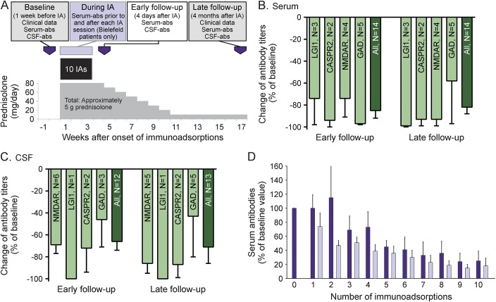
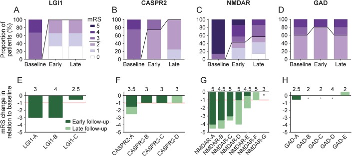
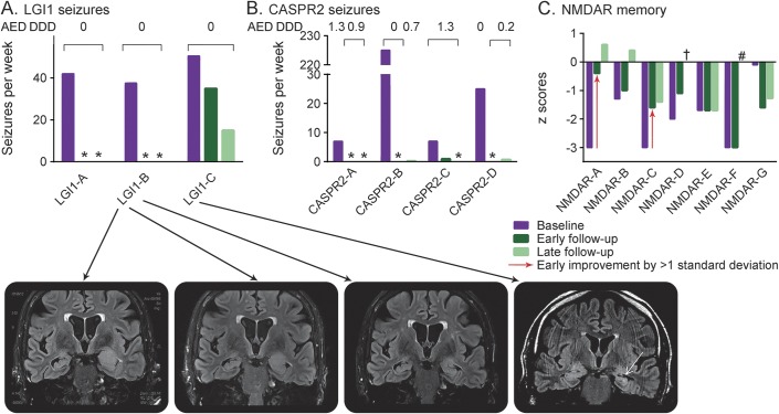
Methods: The outcome of autoimmune encephalitis in 19 patients with antibodies against surface antigens (leucine-rich, glioma inactivated 1 [LGI1], n = 3; contactin-associated protein-2 [CASPR2], n = 4; NMDA receptor [NMDAR], n = 7) and intracellular antigens (glutamic acid decarboxylase [GAD], n = 5) after immunoadsorption in addition to corticosteroid therapy was evaluated retrospectively. Modified Rankin scale (mRS) scores and data on seizures, memory, and antibody titers directly after immunoadsorption (early follow-up) and after a median of 4 months (late follow-up) were compiled.

Results: Immediately after immunoadsorption, 9 of 14 patients with antibodies against LGI1, CASPR2, or NMDAR (64%), but none with GAD antibodies, had improved by at least one mRS point. Five of the 7 patients with LGI1 or CASRP2 antibodies had become seizure-free, and 2 patients with NMDAR antibodies had a memory improvement of more than 1 SD of a normal control population. At late follow-up, 12 of 14 patients with surface antibodies had improved (86%), and none of the patients with GAD antibodies.

Conclusions: It is suggested that addition of immunoadsorption to immunosuppression therapy in patients with surface antibodies may accelerate recovery. This supports the pathogenic role of surface antibodies.

Classification of evidence: This study provides Class IV evidence that immunoadsorption combined with immunosuppression therapy is effective in patients with autoimmune encephalitis with surface antibodies.


Figures

Figure 1
Figure 1. Standard treatment scheme and antibody titer changes
(A) Schematic depiction of the standard treatment schedule. (B–D) Changes of antibody titers in serum (B) and CSF (C) at early and late follow-up in relation to the antibody targets (means with standard errors of the means). (D) Antibody titers of all patients (pooled) from Bielefeld-Bethel (n = 15, means and standard errors of the means) on the immunoadsorption days directly prior to (dark purple bars) and after the immunoadsorptions (light purple bars). This shows the known sawtooth configuration that reflects the antibody redistribution between the vascular and extravascular compartments. Even after the fifth session, there is ongoing reduction of antibodies. CASPR2 = contactin-associated protein-2; GAD = glutamic acid decarboxylase; IA = immunoadsorption; LGI1 = leucine-rich glioma inactivated protein 1; NMDAR = NMDA receptor.
Figure 2
Figure 2. Change in patients' clinical performance at early and late follow-up in relation to baseline
(A–D) Outcome is depicted as in the largest existing anti–NMDA receptor (NMDAR) encephalitis series 3. Modified Rankin Scale (mRS) values of the present series that lie in between whole numbers (because of the averaging procedure in case of divergent ratings) are rounded up to be as conservative as possible. The mRS values are given as shades of purple. The line in between mRS 2 and 3 separates patients who can look after themselves (mRS ≤ 2) from those who cannot live independently (mRS > 2). (E–H) Individual mRS changes are given (reduction indicates clinical improvement). Responder patients are those with mRS change of at least −1 (red lines). The dark green parts of the columns are changes from baseline to early follow-up; the light green parts are those that evolved in addition to that until late follow-up. Above each bar, baseline mRS values are indicated. Patient NMDAR-F improved between early and late follow-up while she underwent radio-chemotherapy of her small cell lung cancer. *No change in mRS. CASPR2 = contactin-associated protein-2; GAD = glutamic acid decarboxylase; LGI1 = leucine-rich glioma inactivated protein 1.
Figure 3
Figure 3. Impressively rapid changes in seizure frequency and memory performance
Seizures in patients with (A) leucine-rich glioma inactivated protein 1 (LGI1) antibodies and (B) contactin-associated protein-2 (CASPR2) antibodies. The antiepileptic drug defined daily dose (AED DDD) figures show that this was not due to anticonvulsive therapy. (C) Memory performance of anti–NMDA receptor (NMDAR) patients with 2 patients improving by >1 SD already at early follow-up (red arrows). Note the preservation of hippocampal volume in patient LGI1-B in contrast to the hippocampal sclerosis that was present from the beginning of IA in patient LGI1-C. This is a possible explanation for his incomplete response. *Seizure-free. †Value 0. #No data. AED DDD according to WHO (http://www.whocc.no/atc_ddd_index/; accessed September 2, 2015).

References

    1. Vincent A, Bien CG, Irani SR, Waters P. Autoantibodies associated with diseases of the CNS: new developments and future challenges. Lancet Neurol 2011;10:759–772. - PubMed
    1. Lancaster E, Dalmau J. Neuronal autoantigens: pathogenesis, associated disorders and antibody testing. Nat Rev Neurol 2012;8:380–390. - PMC - PubMed
    1. Titulaer MJ, McCracken L, Gabilondo I, et al. Treatment and prognostic factors for long-term outcome in patients with anti-NMDA receptor encephalitis: an observational cohort study. Lancet Neurol 2013;12:157–165. - PMC - PubMed
    1. Lancaster E, Lai M, Peng X, et al. Antibodies to the GABA(B) receptor in limbic encephalitis with seizures: case series and characterisation of the antigen. Lancet Neurol 2010;9:67–76. - PMC - PubMed
    1. Irani SR, Alexander S, Waters P, et al. Antibodies to Kv1 potassium channel-complex proteins leucine-rich, glioma inactivated 1 protein and contactin-associated protein-2 in limbic encephalitis, Morvan's syndrome and acquired neuromyotonia. Brain 2010;133:2734–2748. - PMC - PubMed