

A test-retest dataset for assessing long-term reliability of brain morphology and resting-state brain activity
- PMID: 26978040
- PMCID: PMC4792176
- DOI: 10.1038/sdata.2016.16
A test-retest dataset for assessing long-term reliability of brain morphology and resting-state brain activity
Abstract
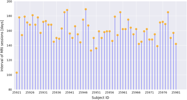
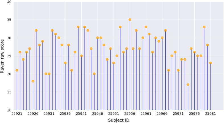
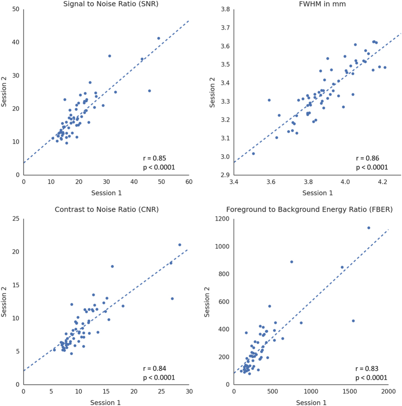
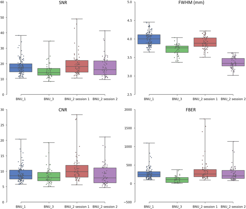
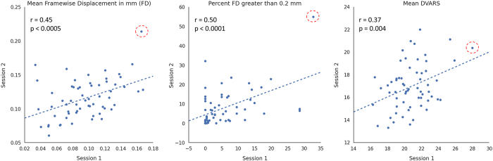
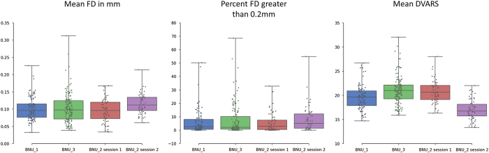
We present a test-retest dataset for evaluation of long-term reliability of measures from structural and resting-state functional magnetic resonance imaging (sMRI and rfMRI) scans. The repeated scan dataset was collected from 61 healthy adults in two sessions using highly similar imaging parameters at an interval of 103-189 days. However, as the imaging parameters were not completely identical, the reliability estimated from this dataset shall reflect the lower bounds of the true reliability of sMRI/rfMRI measures. Furthermore, in conjunction with other test-retest datasets, our dataset may help explore the impact of different imaging parameters on reliability of sMRI/rfMRI measures, which is especially critical for assessing datasets collected from multiple centers. In addition, intelligence quotient (IQ) was measured for each participant using Raven's Advanced Progressive Matrices. The data can thus be used for purposes other than assessing reliability of sMRI/rfMRI alone. For example, data from each single session could be used to associate structural and functional measures of the brain with the IQ metrics to explore brain-IQ association.
Conflict of interest statement
The authors declare no competing financial interests.
Figures
Comment in
-
Commentary: A test-retest dataset for assessing long-term reliability of brain morphology and resting-state brain activity.Front Neurosci. 2017 Feb 22;11:85. doi: 10.3389/fnins.2017.00085. eCollection 2017. Front Neurosci. 2017. PMID: 28275335 Free PMC article. No abstract available.
References
Data Citations
-
- Liu J., Zhen Z., Huang L. 2014. Functional Connectomes Project International Neuroimaging Data-Sharing Initiative. http://dx.doi.org/10.15387/fcp_indi.corr.bnu2 - DOI
References
-
- Aue T., Lavelle L. A. & Cacioppo J. T. Great expectations: What can fMRI research tell us about psychological phenomena? Int. J. Psychophysiol. 73, 10–16 (2009). - PubMed
-
- Smith K. Brain imaging: fMRI 2.0. Nature 484, 24–26 (2012). - PubMed
-
- Biswal B., Zerrin Yetkin F., Haughton V. M. & Hyde J. S. Functional connectivity in the motor cortex of resting human brain using echo-planar mri. Magn. Reson. Med. 34, 537–541 (1995). - PubMed
-
- Lenroot R. K. & Giedd J. N. Brain development in children and adolescents: Insights from anatomical magnetic resonance imaging. Neurosci. Biobehav. Rev. 30, 718–729 (2006). - PubMed
-
- Fox M. D. & Raichle M. E. Spontaneous fluctuations in brain activity observed with functional magnetic resonance imaging. Nat. Rev. Neurosci. 8, 700–711 (2007). - PubMed
Publication types
MeSH terms
LinkOut - more resources
Full Text Sources
Other Literature Sources
Medical
