. 2016 Mar 15;11(3):e0149986.
doi: 10.1371/journal.pone.0149986. eCollection 2016.

Time-Resolved Visualisation of Nearly-Native Influenza A Virus Progeny Ribonucleoproteins and Their Individual Components in Live Infected Cells

Affiliations

Time-Resolved Visualisation of Nearly-Native Influenza A Virus Progeny Ribonucleoproteins and Their Individual Components in Live Infected Cells

Sergiy Avilov et al. PLoS One. .

Abstract

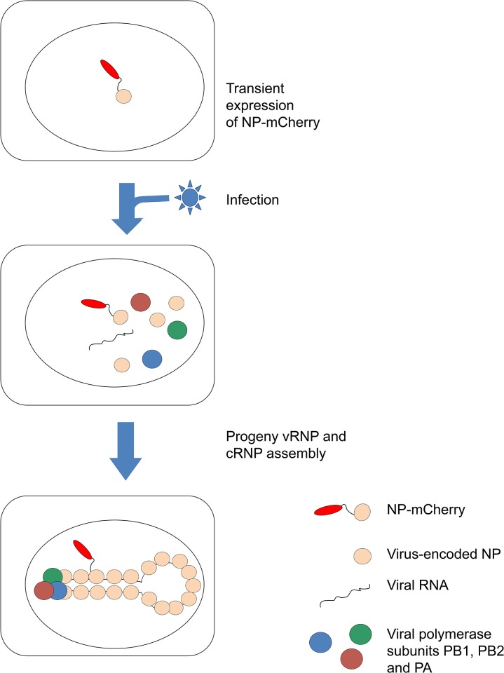
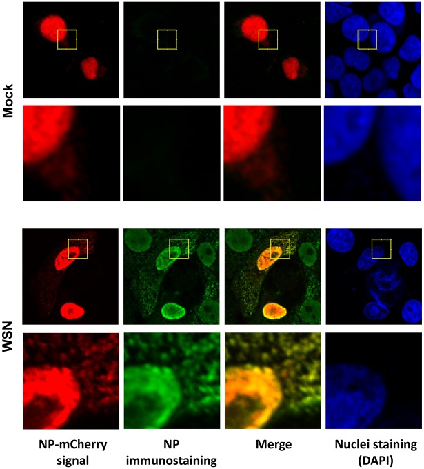
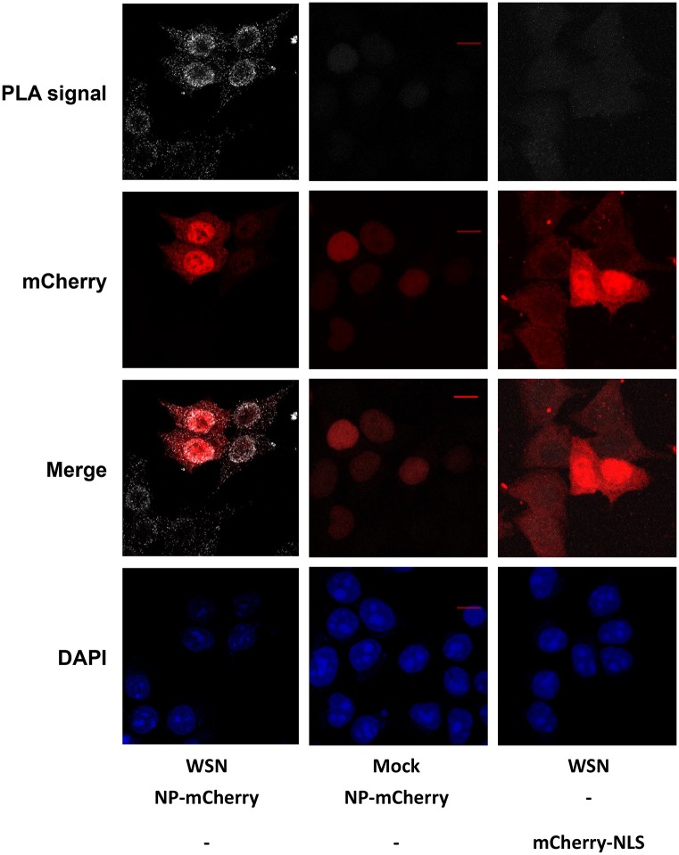
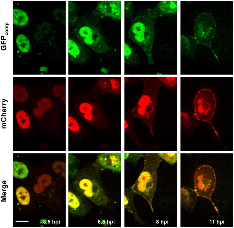
Influenza viruses are a global health concern because of the permanent threat of novel emerging strains potentially capable of causing pandemics. Viral ribonucleoproteins (vRNPs) containing genomic RNA segments, nucleoprotein oligomers, and the viral polymerase, play a central role in the viral replication cycle. Our knowledge about critical events such as vRNP assembly and interactions with other viral and cellular proteins is poor and could be substantially improved by time lapse imaging of the infected cells. However, such studies are limited by the difficulty to achieve live-cell compatible labeling of active vRNPs. Previously we designed the first unimpaired recombinant influenza WSN-PB2-GFP11 virus allowing fluorescent labeling of the PB2 subunit of the viral polymerase (Avilov et al., J.Virol. 2012). Here, we simultaneously labeled the viral PB2 protein using the above-mentioned strategy, and virus-encoded progeny RNPs through spontaneous incorporation of transiently expressed NP-mCherry fusion proteins during RNP assembly in live infected cells. This dual labeling enabled us to visualize progeny vRNPs throughout the infection cycle and to characterize independently the mobility, oligomerization status and interactions of vRNP components in the nuclei of live infected cells.


Conflict of interest statement

Competing Interests: The authors have declared that no competing interests exist.

Figures

Fig 1
Fig 1. Principle of the labeling of influenza vRNPs with transiently expressed NP-mCherry proteins.
Image is not to scale. Neither the real stoechiometry nor the spatial organization of a vRNP is represented. For clarity, only a single NP-mCherry incorporated into an RNP complex is shown, although incorporation of multiple NP-mCherry molecules per RNP might also occur.
Fig 2
Fig 2. Colocalization of transiently expressed NP-mCherry with vRNPs in infected A549 cells.
Mock-infected cells (top panels) or cells infected with the WSN-wt influenza virus (bottom panels) are shown. Cells were fixed at 6 hpi and stained with the anti-NP monoclonal antibody clone 3/1. Scale bar: 10 μm. Pseudocolors: red, mCherry; green, NP; blue, nuclei staining (DAPI).
Fig 3
Fig 3. Detection of proximity between NP-mCherry and vRNPs in infected cells by proximity ligation assay.
The anti-NP monoclonal antibody 3/1 and a rabbit antibody recognizing mCherry were used. Maximal intensity projections of the z-stacks acquired with a laser scanning confocal microscope are shown. Scale bar: 10 μm. Pseudocolors: white, PLA signal; red, mCherry; blue, nuclei staining (DAPI).
Fig 4
Fig 4. Live Vero cells transfected with GFP1-10 and NP-mCherry and infected with the WSN-PB2-GFP11 virus.
Selected frames from S1 Movie at the indicated times post-infection are shown. Pseudocolors: green, PB2-GFPcomp; red, NP-mCherry. Scale bar, 10 μm. Time-lapse series of single optical slices were acquired with a Nipkow spinning disk microscope.
Fig 5
Fig 5. Fluorescence correlation spectroscopy data for GFPcomp-labeled species (green) and mCherry-labeled species (red).
The experimental autocorrelation data, fitted curves (in black), and residuals, are shown for an individual representative HEK-293T cell. A. Control cell transiently expressing MBD-GFPcomp and mCherry and infected with the WSN-wt virus; single-component translational diffusion model. Arrow points to the inflection point of the autocorrelation curve. B. Cell transfected with GFP1-10 and NP-mCherry and infected with the WSN-PB2-GFP11 virus. Two-component translational diffusion model.
Fig 6
Fig 6. FLIM-FRET microscopy in live HEK-293T cells.
A. The principle of FLIM-FRET assay with GFPcomp and mCherry as the FRET donor and acceptor, respectively, is schematically drawn; a representative fluorescence decay dataset fitted curve (in red) and residuals for a single pixel within the nucleus of a cell infected with the WSN-PB2-GFP11 virus are shown. B. Fluorescence intensity (left and middle panels) and mean GFPcomp fluorescence lifetime (right panels) images of the infected and/or transfected cells. Graphs to the right of the micrographs show the distributions of mean GFPcomp fluorescence lifetime values (occurrence of pixels with a given mean lifetime) in the nuclei pointed by yellow arrowheads in GFPcomp intensity images (middle right panels). Sketches on the far right show the “observable”, fluorescently labeled species for each sample; virus ideographs indicate viral infection, either with WSN-wt (in positive and negative controls) or with WSN-PB2-GFP11 (in other infected samples).

Similar articles

Cited by

References

    1. Yoon SW, Webby RJ, Webster RG. Evolution and ecology of influenza A viruses. Current topics in microbiology and immunology. 2014;385:359–75. Epub 2014/07/06. 10.1007/82_2014_396 . - DOI - PubMed
    1. Schrauwen EJ, Fouchier RA. Host adaptation and transmission of influenza A viruses in mammals. Emerging microbes & infections. 2014;3(2):e9 Epub 2014/02/01. 10.1038/emi.2014.9 - DOI - PMC - PubMed
    1. Arranz R, Coloma R, Chichon FJ, Conesa JJ, Carrascosa JL, Valpuesta JM, et al. The Structure of Native Influenza Virion Ribonucleoproteins. Science. 2012;338(6114):1634–7. 10.1126/science.1228172 . - DOI - PubMed
    1. Moeller A, Kirchdoerfer RN, Potter CS, Carragher B, Wilson IA. Organization of the Influenza Virus Replication Machinery. Science. 2012;338(6114):1631–4. 10.1126/science.1227270 . - DOI - PMC - PubMed
    1. Ortin J, Martin-Benito J. The RNA synthesis machinery of negative-stranded RNA viruses. Virology. 2015;479–480:532–44. Epub 2015/04/01. 10.1016/j.virol.2015.03.018 . - DOI - PubMed

Publication types

Substances

LinkOut - more resources