

A Preliminary Study on Sinus Fungus Ball with MicroCT and X-Ray Fluorescence Technique
- PMID: 26978273
- PMCID: PMC4792473
- DOI: 10.1371/journal.pone.0148515
A Preliminary Study on Sinus Fungus Ball with MicroCT and X-Ray Fluorescence Technique
Abstract
Background: Sinus fungus ball, an accumulation of fungal dense concretions, is a common disease in practice, and might cause fatal complications or lead to death once converted into invasive type. Early preoperative diagnosis of this disease can lead to appropriate treatment for patients and prevent multiple surgical procedures. Up to now, the diagnostic criteria of sinus fungus ball have been defined and computed tomography (CT) scan was considered as a valuable preoperative diagnostic tool. However, the sensitivity of clinical CT is only about 62%. Thus, investigating the factors which influence sensitivity is necessary for clinical CT to be a more valuable preoperative diagnosis tool. Furthermore, CT scan usually presents micro-calcifications or spots with metallic density in sinus fungus ball. Previous literatures show that there are some metallic elements such as calcium and zinc in fungus ball, and they concluded that endodontic treatment has a strong correlation with the development of maxillary sinus fungus ball and zinc ion was an exogenous risk factor. But the pathogenesis of sinus fungus ball still remains unclear because fungus ball can also develop in other non-maxillary sinuses or the maxillary sinus without root canal treatment. Is zinc ion the endogenous factor? Study on this point might be also helpful for investigating the pathogenesis of sinus fungus ball. In this paper, we tried to investigate the factors which influence the sensitivity of clinical CT by imaging sinus fungus ball with microCT. The origin of zinc ion was also studied through elements test for different fungal ball samples using x-ray fluorescence technique.
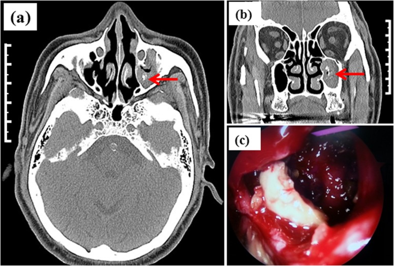
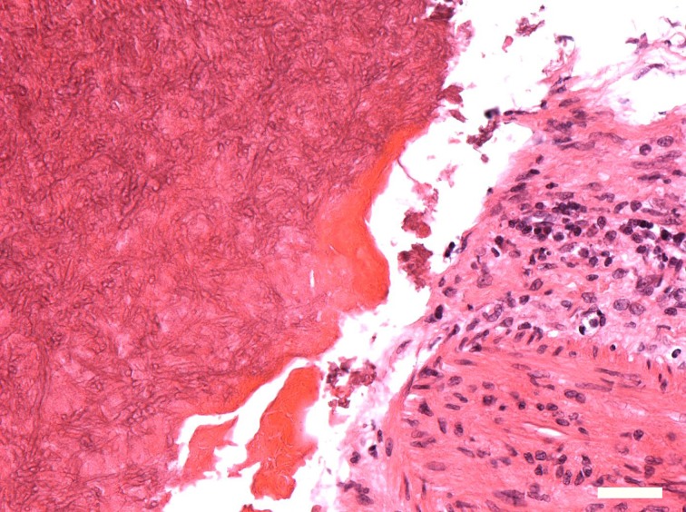
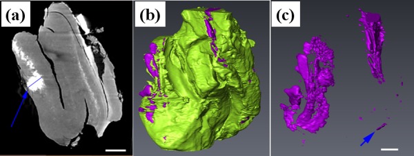
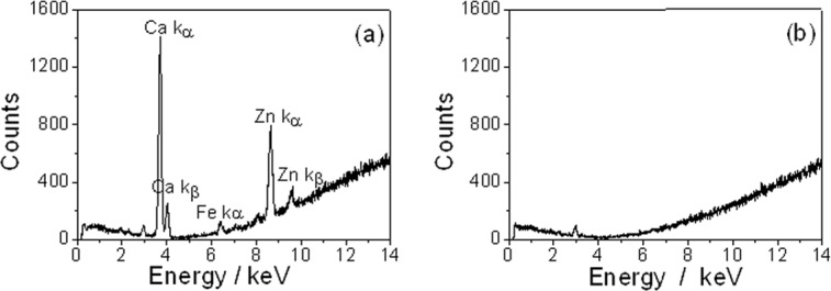
Methods: Specimens including fungal ball material and sinus mucosa from patients confirmed by pathological findings were extracted after surgery. All fungal ball specimens came from sphenoid sinus, ethmoidal sinus and maxillary sinus with or without previous endodontic treatment respectively. All of them were imaged by microCT with spatial resolution up to 5μm to acquire three-dimensional structure, and then the heavy metal elements were detected with x-ray fluorescence spectrometer analysis.
Result: High concentration of zinc and calcium were detected in all fungal ball specimens compared to sinus mucosa membrane. Particles with different size varied from disperse to density, which have similar shape to the result of clinical CT but with different size, were found in three-dimensional reconstruction results of microCT.
Conclusions: Spatial resolution is an influent factor of clinical CT sensitivity for sinus fungus ball. Improving the resolution of clinical CT will help to improve its sensitivity. Besides iatrogenic endodontic materials, endogenous metal elements of zinc and calcium might associate with the growth of fungal ball and the micro-calcifications or spots with metallic density of CT imaging.
Conflict of interest statement
Figures
Similar articles
-
Maxillary fungus ball: zinc-oxide endodontic materials as a risk factor.Acta Otorhinolaryngol Ital. 2015 Apr;35(2):93-6. Acta Otorhinolaryngol Ital. 2015. PMID: 26019392 Free PMC article.
-
Fungus ball of the paranasal sinuses: Analysis of our serie of patients.Acta Otorrinolaringol Esp. 2016 Jul-Aug;67(4):220-5. doi: 10.1016/j.otorri.2015.09.005. Epub 2015 Dec 19. Acta Otorrinolaringol Esp. 2016. PMID: 26708329 English, Spanish.
-
Radiologic characteristics of sinonasal fungus ball: an analysis of 119 cases.Acta Radiol. 2011 Sep 1;52(7):790-5. doi: 10.1258/ar.2011.110021. Epub 2011 Apr 27. Acta Radiol. 2011. PMID: 21525111
-
Fungal Sinusitis.Neuroimaging Clin N Am. 2015 Nov;25(4):569-76. doi: 10.1016/j.nic.2015.07.004. Epub 2015 Aug 21. Neuroimaging Clin N Am. 2015. PMID: 26476380 Review.
-
Mucormycosis (Mucor fungus ball) of the maxillary sinus.Ear Nose Throat J. 2014 Oct-Nov;93(10-11):E18-22. Ear Nose Throat J. 2014. PMID: 25397383 Review.
Cited by
-
Maxillary Sinusitis Following Orthognathic Surgery: Should It Be Considered Odontogenic Sinusitis?Clin Case Rep. 2024 Nov 29;12(12):e9654. doi: 10.1002/ccr3.9654. eCollection 2024 Dec. Clin Case Rep. 2024. PMID: 39619300 Free PMC article.
-
Differences in clinical and imaging presentation of maxillary sinus fungus ball with and without intralesional hyperdensity.Sci Rep. 2021 Dec 14;11(1):23945. doi: 10.1038/s41598-021-03507-1. Sci Rep. 2021. PMID: 34907314 Free PMC article.
-
Characteristic features of fungus ball in the maxillary sinus and the location of intralesional calcifications on computed tomographic images: A report of 2 cases.Imaging Sci Dent. 2020 Dec;50(4):377-384. doi: 10.5624/isd.2020.50.4.377. Epub 2020 Dec 15. Imaging Sci Dent. 2020. PMID: 33409149 Free PMC article.
-
High CT Attenuation Values Relative to the Brainstem Predict Fungal Hyphae Within the Sinus.Front Surg. 2022 Jun 16;9:876340. doi: 10.3389/fsurg.2022.876340. eCollection 2022. Front Surg. 2022. PMID: 35784936 Free PMC article.
-
Aspergillus fumigatus biofilm formation on different bone substitutes used in maxillary sinus augmentation: an in vitro analysis.Int J Implant Dent. 2019 Jun 20;5(1):22. doi: 10.1186/s40729-019-0175-5. Int J Implant Dent. 2019. PMID: 31218468 Free PMC article.
References
-
- Dhong HJ, Jung JY, Park JH. Diagnostic accuracy in sinus fungus balls: CT scan and operative findings. Am J Rhinol. 2000; 14:227–231. - PubMed
-
- deShazo RD, O'Brien M, Chapin K, Soto-Aguilar M, Swain R, Lyons M, et al. Criteria for the diagnosis of sinus mycetoma. J Allergy ClinImmunol. 1997; 99:475–485. - PubMed
Publication types
MeSH terms
LinkOut - more resources
Full Text Sources
Other Literature Sources
Medical
