. 2016 Mar 11;17(3):359.
doi: 10.3390/ijms17030359.

Co-Expression and Co-Localization of Cartilage Glycoproteins CHI3L1 and Lubricin in Osteoarthritic Cartilage: Morphological, Immunohistochemical and Gene Expression Profiles

Affiliations

Co-Expression and Co-Localization of Cartilage Glycoproteins CHI3L1 and Lubricin in Osteoarthritic Cartilage: Morphological, Immunohistochemical and Gene Expression Profiles

Marta Anna Szychlinska et al. Int J Mol Sci. .

Abstract

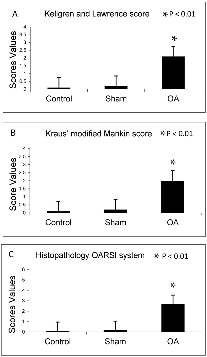
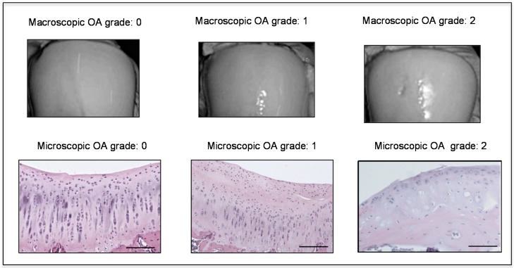
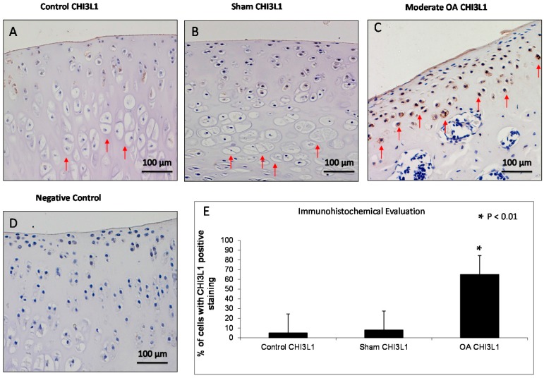
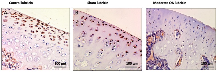
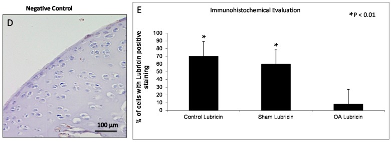
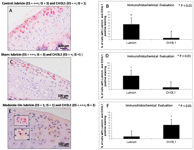
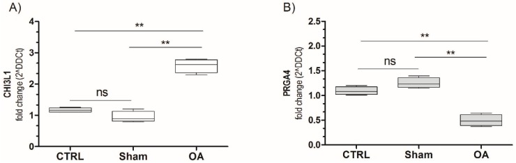
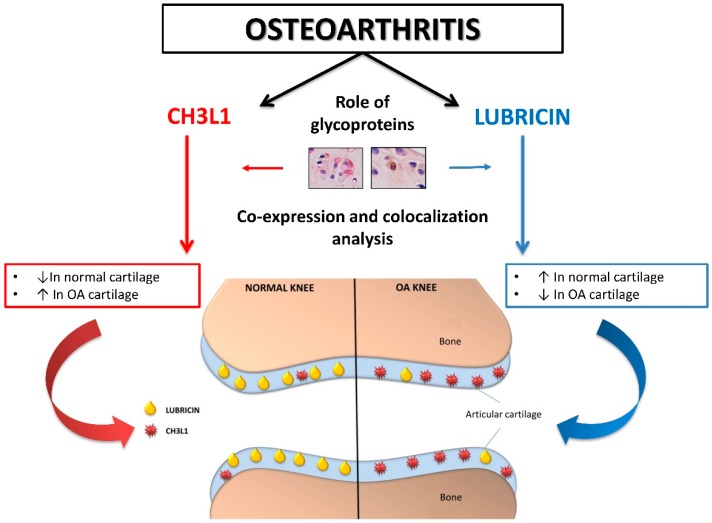
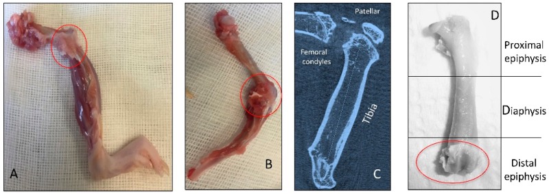
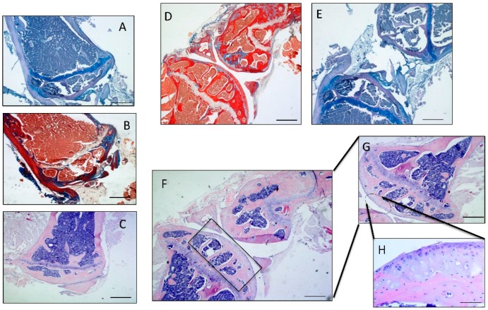
Osteoarthritis is the most common human arthritis characterized by degeneration of articular cartilage. Several studies reported that levels of human cartilage glycoprotein chitinase 3-like-1 (CHI3L1) are known as a potential marker for the activation of chondrocytes and the progression of Osteoarthritis (OA), whereas lubricin appears to be chondroprotective. The aim of this study was to investigate the co-expression and co-localization of CHI3L1 and lubricin in normal and osteoarthritic rat articular cartilage to correlate their modified expression to a specific grade of OA. Samples of normal and osteoarthritic rat articular cartilage were analyzed by the Kellgren-Lawrence OA severity scores, the Kraus' modified Mankin score and the Histopathology Osteoarthritis Research Society International (OARSI) system for histomorphometric evaluations, and through CHI3L1 and lubricin gene expression, immunohistochemistry and double immuno-staining analysis. The immunoexpression and the mRNA levels of lubricin increased in normal cartilage and decreased in OA cartilage (normal vs. OA, p < 0.01). By contrast, the immunoexpression and the mRNA levels of CHI3L1 increased in OA cartilage and decreased in normal cartilage (normal vs. OA, p < 0.01). Our findings are consistent with reports suggesting that these two glycoproteins are functionally associated with the development of OA and in particular with grade 2/3 of OA, suggesting that in the future they could be helpful to stage the severity and progression of the disease.

Keywords: CHI3L1; Immunohistochemistry; Lubricin; Osteoarthritis; anterior cruciate ligament transection (ACLT); mRNA.


Figures

Figure 1
Figure 1
(A) Kellgren and Lawrence score among groups; (B) Kraus’ modified Mankin score among groups; and (C) Histopathology Osteoarthritis Research Society International (OARSI) system among groups. Results are presented as the mean ± SEM. Analysis Of Variance (ANOVA), was used to evaluate the significance of the results. * p < 0.01, when compared to the control groups. OA: Osteoarthritis.
Figure 2
Figure 2
Macroscopic and microscopic articular cartilage degeneration between OA grade 0 to OA grade 2 according to macroscopic Kraus’ modified Mankin score and microscopic histopathology OARSI system, Magnifications ×20, scale bars 100 µm.
Figure 3
Figure 3
Evaluation of chitinase 3-like-1 (CHI3L1) immunostaining. (A,B) CHI3L1 immunohistochemistry specimen from control (A) and sham (B) rat femoral articular cartilage (without anterior cruciate ligament transection (ACLT)) showed a weak/absent (Extent Score (ES) = +; Intensity immunostaining (IS) = 1) immunostaining in cartilage superficial, middle and deep zone in which hypertrophic chondrocytes are evident (red arrows); (C) CHI3L1 immunohistochemistry specimen from moderate OA rat femoral articular cartilage (with ACLT) exhibited a strong (ES = +++; IS = 3) immunostaining in middle and deep cartilage zone (red arrows) and a reduction of cartilage thickness of the superficial and the middle zones is evident and in the deep zone the chondrocytes are not hypertrophic and are not arranged in columns; (D) The negative control treated with PBS without the primary antibody (CHI3L1) did not show immunostaining (ES = 0; IS = 0). (AD) Magnifications ×20; Scale bars: 100 µm; (E) Immunohistochemical evaluation graph: percentage of CHI3L1 positive cells out of the total number of cells counted in control groups and in OA group. Results are presented as the mean ± SEM. ANOVA was used to evaluate the significance of the results. * p < 0.01, when compared to the control groups.
Figure 4
Figure 4
Evaluation of lubricin immunostaining. (A,B) Lubricin immunohistochemistry specimen from control (A) and sham (B) cartilage (without ACLT) showed a very strong (ES = +++; IS = 4) immunostaining in chondrocytes from superficial and middle zone of rat femoral articular cartilage; (C) Lubricin immunohistochemistry specimen from moderate OA cartilage (with ACLT) showed a weak/absent (ES = +; IS = 1) immunostaining in chondrocytes from rat femoral articular cartilage (superficial, middle and deep zone); (D) The negative control treated with PBS without the primary antibody (lubricin) did not show immunostaining (ES = 0; IS = 0). (AD) Magnifications ×20; Scale bars: 100 µm; (E) Immunohistochemical evaluation graph: percentage of lubricin positive cells out of the total number of cells counted in control groups and in the OA group. Results are presented as the mean ± SEM. ANOVA was used to evaluate the significance of the results. * p < 0.01, when compared to the control groups.
Figure 4
Figure 4
Evaluation of lubricin immunostaining. (A,B) Lubricin immunohistochemistry specimen from control (A) and sham (B) cartilage (without ACLT) showed a very strong (ES = +++; IS = 4) immunostaining in chondrocytes from superficial and middle zone of rat femoral articular cartilage; (C) Lubricin immunohistochemistry specimen from moderate OA cartilage (with ACLT) showed a weak/absent (ES = +; IS = 1) immunostaining in chondrocytes from rat femoral articular cartilage (superficial, middle and deep zone); (D) The negative control treated with PBS without the primary antibody (lubricin) did not show immunostaining (ES = 0; IS = 0). (AD) Magnifications ×20; Scale bars: 100 µm; (E) Immunohistochemical evaluation graph: percentage of lubricin positive cells out of the total number of cells counted in control groups and in the OA group. Results are presented as the mean ± SEM. ANOVA was used to evaluate the significance of the results. * p < 0.01, when compared to the control groups.
Figure 5
Figure 5
Evaluation of lubricin and CHI3L1 double staining in control and in moderate OA cartilage. (A,C) In control and in sham groups lubricin immunolabeling was strong (ES = +++; IS = 3, red staining, red arrows), instead the expression of CHI3L1 was weak/absent (ES = +; IS = 1, brown staining, brown arrow); (E) In moderate OA, lubricin immunolabeling was weak/absent (ES = +; IS = 1, red staining, red arrows), instead the expression of CHI3L1 was strong (ES = +++; IS = 3, brown staining, brown arrows); (A,C,E) Magnifications ×20; Scale bars: 100 µm; inserts: magnifications ×40; Scale bars: 50 µm; (B) Immunohistochemical evaluation graph: percentage of lubricin and CHI3L1-positive cells out of the total number of cells counted in the control group; (D) Immunohistochemical evaluation graph: percentage of lubricin and CHI3L1-positive cells out of the total number of cells counted in the sham group; (F) Immunohistochemical evaluation graph: percentage of lubricin and CHI3L1-positive cells out of the total number of cells counted in the moderate OA group. Results are presented as the mean ± SEM. Student’s t test, was used to evaluate the significance of the results. * p < 0.01, when compared lubricin vs. CHI3L1.
Figure 6
Figure 6
CHI3L1 (A) and PRG4 (B) mRNA expression in osteoarthritic rat cartilage model. Expression levels of CHI3L1 and PRG4 (lubricin) in cartilage of osteoarthritis rats. Total RNA was extracted as indicated in Materials and Method and CHI3L1/PRG4 expression was measured by real-time PCR. Data are expressed as mean ± SD of at least three independent experiments. ** p < 0.001, compared to sham and control. CTRL: control; ns: not significant.
Figure 7
Figure 7
Graphical representation of the involvement of two glycoproteins (CHI3L1 and lubricin) in normal and osteoarthritic articular cartilage. Red arrows (CHI3L1 pathway); Blue arrows (lubricin pathway).
Figure 8
Figure 8
Representation of in vivo explanted knee join bones (red circles). (A,B) Knee OA bones joint after explantation, not cleaned of soft tissues; (C) X-ray microtomography imaging of the OA knee bones joint; (D) OA femur cleaned of soft tissues.
Figure 9
Figure 9
(A) Micrograph of the OA distal epiphysis of the femur stained with Toluidine Blue, Magnification ×10; (B) Micrograph of the OA distal epiphysis of the femur stained with Masson’s Trichrome, Magnification ×10; (C) Micrograph of the OA distal epiphysis of the femur stained with Hematoxylin and Eosin, Magnification ×10; (D) Micrograph of the OA knee joint stained with Masson's Trichrome, Magnification ×5; (E) Micrograph of the OA knee joint stained with Toluidine Blue, Magnification ×5; (F) Micrograph of the OA knee joint stained with Hematoxylin and Eosin, Magnification ×5; (G) Magnification of J ×10; (H) Magnification ×20, Scale bars: 100 µm.

References

    1. Giunta S., Castorina A., Marzagalli R., Szychlinska M.A., Pichler K., Mobasheri A., Musumeci G. Ameliorative effects of PACAP against cartilage degeneration. Morphological, immunohistochemical and biochemical evidence from in vivo and in vitro models of rat osteoarthritis. Int. J. Mol. Sci. 2015;16:5922–5944. doi: 10.3390/ijms16035922. - DOI - PMC - PubMed
    1. Mobasheri A., Matta C., Zákány R., Musumeci G. Chondrosenescence: Definition, hallmarks and potential role in the pathogenesis of osteoarthritis. Maturitas. 2015;80:237–244. doi: 10.1016/j.maturitas.2014.12.003. - DOI - PubMed
    1. Musumeci G., Aiello F.C., Szychlinska M.A., di Rosa M., Castrogiovanni P., Mobasheri A. Osteoarthritis in the XXIst century: Risk factors and behaviours that influence disease onset and progression. Int. J. Mol. Sci. 2015;16:6093–6112. doi: 10.3390/ijms16036093. - DOI - PMC - PubMed
    1. Musumeci G., Szychlinska M.A., Mobasheri A. Age-related degeneration of articular cartilage in the pathogenesis of osteoarthritis: Molecular markers of senescent chondrocytes. Histol. Histopathol. 2015;30:1–12. doi: 10.7243/2055-091X-2-1. - DOI - PubMed
    1. Di Rosa M., Malaguarnera G., de Gregorio C., D’Amico F., Mazzarino M.C., Malaguarnera L. Modulation of chitotriosidase during macrophage differentiation. Cell. Biochem. Biophys. 2013;66:239–247. doi: 10.1007/s12013-012-9471-x. - DOI - PubMed

Publication types

LinkOut - more resources