Review
. 2015 Jul-Sep;59(3):137-40.

ANOMALOUS HEAD POSTURES IN STRABISMUS AND NYSTAGMUS DIAGNOSIS AND MANAGEMENT

Review

ANOMALOUS HEAD POSTURES IN STRABISMUS AND NYSTAGMUS DIAGNOSIS AND MANAGEMENT

Luminita Teodorescu. Rom J Ophthalmol. 2015 Jul-Sep.

Abstract

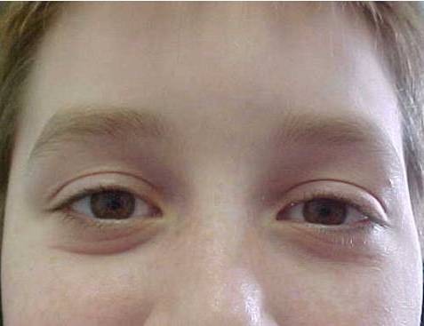
Abnormal head positions are adopted in order to improve visual acuity, to avoid diplopia or to obtain a more comfortable binocular vision. The head can be turned or tilted toward right or left, with the chin rotated up or downwards or combination of these positions. The ophthalmologic examination including the assessment of versions leads to the diagnosis. When versions are free, the cause may be congenital nystagmus or strabismus with large angle. When versions are limited we suspect paralytic or restrictive strabismus. The head tilted to one shoulder suggests cyclotropia (IV Nerve Palsy) or congenital nystagmus. We present few of the above cases. An adequate surgical treatment can improve or correct the ocular deviation, diplopia and the abnormal head posture.

Conclusions: The abnormal head posture must be assessed and treated early in order to correct the ocular position and head posture. All patient presenting abnormal head position HAD TO BE investigated by an ophthalmologist.


Figures

Fig. 1
Fig. 1
Congenital esotropia with manifest nystagmus, left eye fixing in adduction, head turned towards left, the direction of the left fixing eye – pre-op.
Fig. 2
Fig. 2
Post-operative Fig. after bi-lateral medial rectus recession
Fig. 3
Fig. 3
Infantile nystagmus with left face turn
Fig. 4
Fig. 4
Post-operative after left Medial Rectus recession 7 mm, right lateral rectus recession 10 mm
Fig. 5
Fig. 5
Congenital nystagmus with combined vertical and horizontal null zone, chin-up and left face turn position
Fig. 6
Fig. 6
Post-operative after left medial rectus recession 8.5 mm, right lateral rectus recession 12 mm, bi-inferior rectus recession 7 mm, bi-superior rectus resection 6 mm.
Fig. 7
Fig. 7
(up or in the middle), 8 (left), 9 (right): Right VI nerve palsy after trauma, no abduction in the right eye
Fig. 10
Fig. 10
(up), 11(left), 12(right): After right medial rectus recession 6,5 mm and half-tendon transfer of the superior and inferior to the lateral rectus insertion.
Fig. 13
Fig. 13
(up), 14 (left), 15(right): Right III Nerve Palsy, superior branch, exotropia and hypotropia, left head turn, no adduction
Fig. 16
Fig. 16
(up), 17 (left), 18 (right) Post-op, Right lateral rectus recession 14 mm, right medial rectus resection 9 mm with up-ward insertion
Fig. 19
Fig. 19
Left Congenital IV nerve Palsy; Right head tilted, facial asymmetry
Fig. 20
Fig. 20
Left hypertropia in primary position
Fig. 21
Fig. 21
Bielschowsky head tilted test positive: hypertropia increases in tilting the head towards to side of the palsy
Fig. 22
Fig. 22
Post-op, head straight, ortophoria, after left inferior oblique myectomy and left superior rectus recession
Fig. 23
Fig. 23
Normal head tilted test

References

    1. Noorden G. K, Helveston E. M. Strabismus. A decision making approach. St. Louis: Mosby; 1996. pp. 52–60.
    1. Helveston E. M. "Strabismus minute" Orbis Cybersight, Anomalous Head Posture and Strabismus. New York: 1999. Lecture 8 of 48 .
    1. Helveston E. M. "Strabismus minute" Orbis Cybersight, Surgery Options for Nystagmus. New York: 1999. Lecture 21 of 48.

LinkOut - more resources