. 2016 Jun 2:324:469-484.
doi: 10.1016/j.neuroscience.2016.03.014. Epub 2016 Mar 12.

Altered metabolic activity in the developing brain of rats predisposed to high versus low depression-like behavior

Affiliations

Altered metabolic activity in the developing brain of rats predisposed to high versus low depression-like behavior

Chelsea R McCoy et al. Neuroscience. .

Abstract

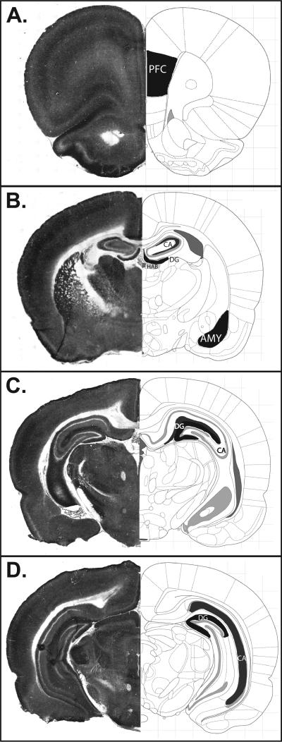
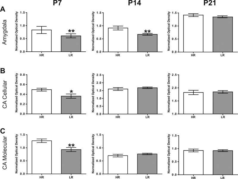
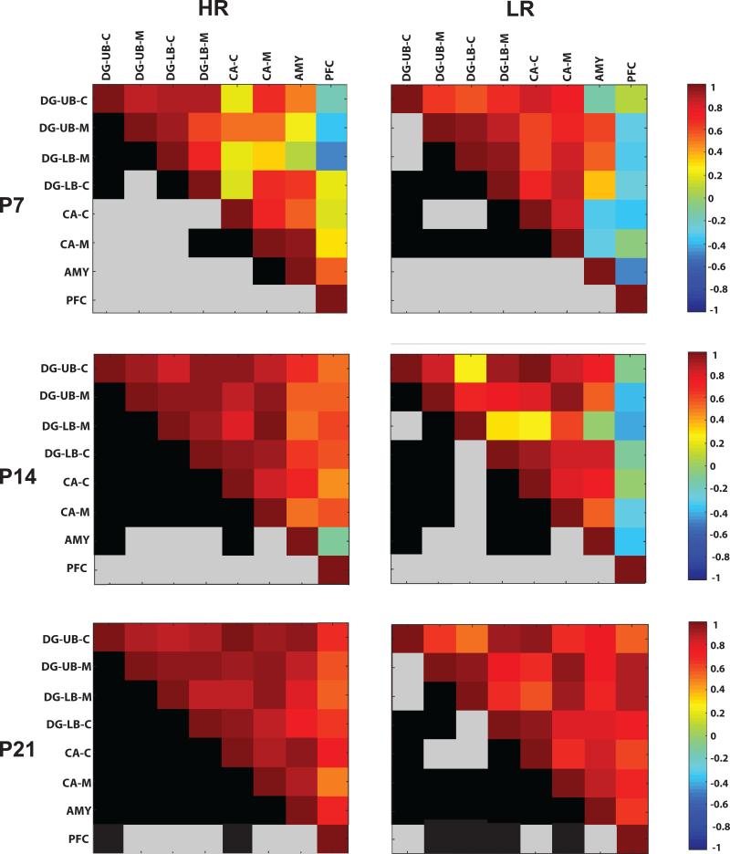
Individual differences in human temperament can increase the risk of psychiatric disorders like depression and anxiety. Our laboratory utilized a rat model of temperamental differences to assess neurodevelopmental factors underlying emotional behavior differences. Rats selectively bred for low novelty exploration (Low Responders, LR) display high levels of anxiety- and depression-like behavior compared to High Novelty Responder (HR) rats. Using transcriptome profiling, the present study uncovered vast gene expression differences in the early postnatal HR versus LR limbic brain, including changes in genes involved in cellular metabolism. These data led us to hypothesize that rats prone to high (versus low) anxiety/depression-like behavior exhibit distinct patterns of brain metabolism during the first weeks of life, which may reflect disparate patterns of synaptogenesis and brain circuit development. Thus, in a second experiment we examined activity of cytochrome C oxidase (COX), an enzyme responsible for ATP production and a correlate of metabolic activity, to explore functional energetic differences in the HR/LR early postnatal brain. We found that HR rats display higher COX activity in the amygdala and specific hippocampal subregions compared to LRs during the first 2 weeks of life. Correlational analysis examining COX levels across several brain regions and multiple early postnatal time points suggested desynchronization in the developmental timeline of the limbic HR versus LR brain during the first two postnatal weeks. These early divergent COX activity levels may reflect altered circuitry or synaptic activity in the early postnatal HR/LR brain, which could contribute to the emergence of their distinct behavioral phenotypes.

Keywords: amygdala; anxiety; cytochrome C oxidase; metabolism; neurodevelopment; transcriptome.


Figures

Figure 1
Figure 1. HR/LR rats exhibit widespread gene expression differences in the early postnatal brain
(A) Number of differentially expressed genes in the HR/LR hippocampus, amygdala, and prefrontal cortex at postnatal day (P) 7, P14, and P21. (B) Number of genes up-regulated or down-regulated in the early postnatal HR/LR amygdala (left) and hippocampus (right). Filtering for these gene lists was based on p-value < 0.05 fold change > 1.5 with multiple test corrections. (C) PCA of transcriptome in developing HR and LR amygdala and hippocampus. PCA was used to assess gene expression profiles from each amygdala or hippocampal sample dissected from HR and LR pup brains collected at P7, P14, and P21. The major component of variance in both brain regions separated animals by developmental age. Top panels depict samples colored based on developmental ages P7 (pink), P14 (blue), and P21 (orange). The second major component of variance was attributable to HR/LR phenotype. Thus, bottom panels depict samples colored based on HR (green) and LR (red) phenotypes. Green and red ellipses highlight the variablity of samples from each phenotype at each developmental age.
Figure 2
Figure 2. Pathways related to mitochondria and cellular metabolism are altered in the early postnatal HR/LR amygdala and hippocampus
(A-B) Top gene ontology cellular component enrichment analysis identified top terms that were altered in the early postnatal HR versus LR amygdala (A) and hippocampus (B). (C) Percentage of genes within several metabolic pathways that were differentially expressed in HR versus LR amygdala at P7, P14, and P21. (D) Percentage of genes within each metabolic pathway that were differentially expressed in HR/LR hippocampus at P7, P14, and P21. Enrichment analysis and analysis of metabolic pathways were performed on altered genes across development ages that were filtered at p < 0.05 and FC > 1.2.
Figure 3
Figure 3. Representative Cytochrome c Oxidase (COX) staining and corresponding atlas images depicting brain regions analyzed
Representative tissue sections processed for COX histochemistry at the level of the (A) prefrontal cortex (PFC; bregma +2.76 mm); (B) rostral hippocampus (HPC) and amygdala (AMY; bregma −2.04 mm); (C) mid-hippocampus (bregma −4.44 mm); and (D) caudal hippocampus (bregma −5.40 mm). Regions of interests are shown in black and dark grey on the atlas images, which were adapted from “The Rat Brain” (6th edition) by Paxinos and Watson (2006).
Figure 4
Figure 4. Cytochrome c oxidase (COX) activity in the early postnatal HR versus LR limbic brain
COX activity from tissue sections normalized to a dot blot of known COX protein amount was measured from amygdala (A), cellular and molecular layers of Cornu ammonis (CA; B-C) at three early postnatal time points: postnatal day (P) 7, 14 and 21. (A) At P7 and P14, HR pups exhibited greater COX activity in the amygdala compared to LR pups. (B-C) In the cellular and molecular layers of CA, HR showed increased COX activity at P7. ** indicates p-value < 0.01, * indicates p-value < 0.05.
Figure 5
Figure 5. Inter-correlations of COX activity within limbic brain regions of HR (left) and LR (right) at postnatal day (P) 7, 14 and 21 (shown top to bottom)
The top half of each square depicts a colormap showing how COX activity within select brain regions (i.e., sublayers of the dentate gyrus, CA region of the hippocampus, amygdala, or prefrontal cortex) correlate with the other measures. The mirrored bottom half of each square is color-coded in black and gray. A black square indicates that the correlation for that measure was statistically significant (p<0.05) whereas a gray square indicates that the correlation was not significant. Abbreviations: DG-UB-C, dentate gyrus upper blade cellular layer; DG-UB-M. dentate gyrus upper blade molecular layer; DG-LB-M, dentate gyrus lower blade molecular layer; DG-LB-C, dentate gyrus lower blade cellular layer; CA-C, cornu ammonis cellular layer; CA-M, cornu ammonis molecular layer; AMY, amygdala; PFC, prefrontal cortex.
Figure 6
Figure 6. Multidimensional scaling analysis of HR/LR COX activity differences in the first weeks of life
Following calculation of region specific pair-wise differences between correlation matrices of HR/LR brain regions at P7/14/ 21, multidimensional scaling expresses the data on a 2-dimensional plot where the distance between two given brain regions represents their association strength. Abbreviations: DG-UB-C, dentate gyrus upper blade cellular layer; DG-UB-M. dentate gyrus upper blade molecular layer; DG-LB-M, dentate gyrus lower blade molecular layer; DG-LB-C, dentate gyrus lower blade cellular layer; CA-C, cornu ammonis cellular layer; CA-M, cornu ammonis molecular layer; AMY, amygdala; PFC, prefrontal cortex.

References

    1. Adzic M, Lukic I, Mitic M, Djordjevic J, Elakovic I, Djordjevic A, Krstic-Demonacos M, Matic G, Radojcic M. Brain region- and sex-specific modulation of mitochondrial glucocorticoid receptor phosphorylation in fluoxetine treated stressed rats: effects on energy metabolism. Psychoneuroendocrinology. 2013;38:2914–2924. - PubMed
    1. Anand A, Li Y, Wang Y, Lowe MJ, Dzemidzic M. Resting state corticolimbic connectivity abnormalities in unmedicated bipolar disorder and unipolar depression. Psychiatry Res. 2009;171:189–198. - PMC - PubMed
    1. Babcock DF, Hille B. Mitochondrial oversight of cellular Ca2+ signaling. Curr Opin Neurobiol. 1998;8:398–404. - PubMed
    1. Bannerman DM, Deacon RM, Offen S, Friswell J, Grubb M, Rawlins JN. Double dissociation of function within the hippocampus: spatial memory and hyponeophagia. Behav Neurosci. 2002;116:884–901. - PubMed
    1. Bayer SA. Development of the hippocampal region in the rat. I. Neurogenesis examined with 3H-thymidine autoradiography. J Comp Neurol. 1980a;190:87–114. - PubMed

Publication types

Substances