In vivo acoustic and photoacoustic focusing of circulating cells
- PMID: 26979811
- PMCID: PMC4793240
- DOI: 10.1038/srep21531
In vivo acoustic and photoacoustic focusing of circulating cells
Abstract
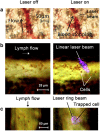
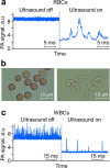
In vivo flow cytometry using vessels as natural tubes with native cell flows has revolutionized the study of rare circulating tumor cells in a complex blood background. However, the presence of many blood cells in the detection volume makes it difficult to count each cell in this volume. We introduce method for manipulation of circulating cells in vivo with the use of gradient acoustic forces induced by ultrasound and photoacoustic waves. In a murine model, we demonstrated cell trapping, redirecting and focusing in blood and lymph flow into a tight stream, noninvasive wall-free transportation of blood, and the potential for photoacoustic detection of sickle cells without labeling and of leukocytes targeted by functionalized nanoparticles. Integration of cell focusing with intravital imaging methods may provide a versatile biological tool for single-cell analysis in circulation, with a focus on in vivo needleless blood tests, and preclinical studies of human diseases in animal models.
Figures


References
-
- Shapiro H. M. Practical Flow Cytometry. (Wiley-Liss. New York, 2003).
-
- Fulwyler M. J. Electronic separation of biological cells by volume. Science 150, 910–911 (1965). - PubMed
-
- Davies K. E. et al.. Cloning of a representative genomic library of the human X chromosome after sorting by flow cytometry. Nature 293, 374–376 (1981). - PubMed
-
- Krutzik P. O. & Nolan G. P. Fluorescent cell barcoding in flow cytometry allows high-throughput drug screening and signaling profiling. Nat. Methods 3, 361–368 (2006). - PubMed
-
- Rufer N. et al.. Telomere length dynamics in human lymphocyte subpopulations measured by flow cytometry. Nature Biotechnol. 16, 743–747 (1998). - PubMed
Publication types
MeSH terms
Grants and funding
LinkOut - more resources
Full Text Sources
Other Literature Sources

