Comparative Study
. 2016 Jun 1;110(3):319-30.
doi: 10.1093/cvr/cvw054. Epub 2016 Mar 14.

Exosomes induce and reverse monocrotaline-induced pulmonary hypertension in mice

Affiliations
Comparative Study

Exosomes induce and reverse monocrotaline-induced pulmonary hypertension in mice

Jason M Aliotta et al. Cardiovasc Res. .

Abstract

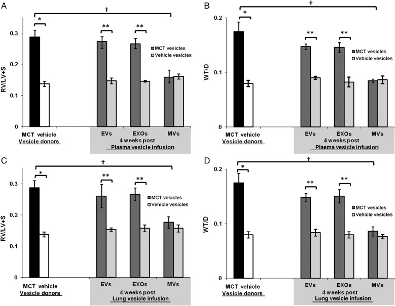
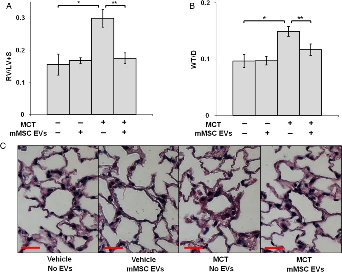
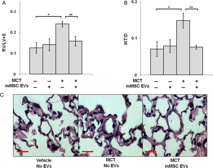
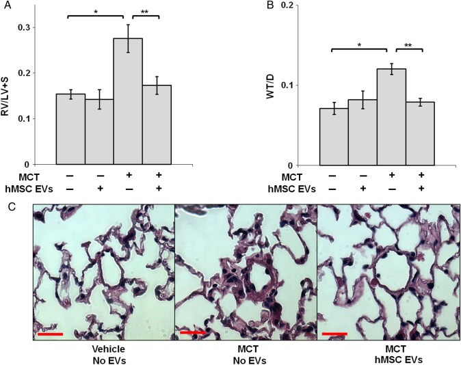
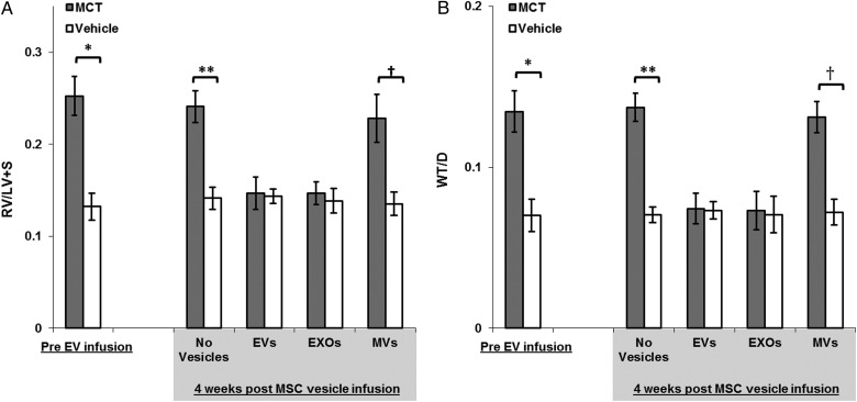
Aims: Extracellular vesicles (EVs) from mice with monocrotaline (MCT)-induced pulmonary hypertension (PH) induce PH in healthy mice, and the exosomes (EXO) fraction of EVs from mesenchymal stem cells (MSCs) can blunt the development of hypoxic PH. We sought to determine whether the EXO fraction of EVs is responsible for modulating pulmonary vascular responses and whether differences in EXO-miR content explains the differential effects of EXOs from MSCs and mice with MCT-PH.

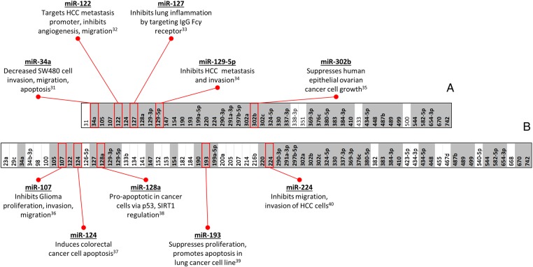
Methods and results: Plasma, lung EVs from MCT-PH, and control mice were divided into EXO (exosome), microvesicle (MV) fractions and injected into healthy mice. EVs from MSCs were divided into EXO, MV fractions and injected into MCT-treated mice. PH was assessed by right ventricle-to-left ventricle + septum (RV/LV + S) ratio and pulmonary arterial wall thickness-to-diameter (WT/D) ratio. miR microarray analyses were also performed on all EXO populations. EXOs but not MVs from MCT-injured mice increased RV/LV + S, WT/D ratios in healthy mice. MSC-EXOs prevented any increase in RV/LV + S, WT/D ratios when given at the time of MCT injection and reversed the increase in these ratios when given after MCT administration. EXOs from MCT-injured mice and patients with idiopathic pulmonary arterial hypertension (IPAH) contained increased levels of miRs-19b,-20a,-20b, and -145, whereas miRs isolated from MSC-EXOs had increased levels of anti-inflammatory, anti-proliferative miRs including miRs-34a,-122,-124, and -127.

Conclusion: These findings suggest that circulating or MSC-EXOs may modulate pulmonary hypertensive effects based on their miR cargo. The ability of MSC-EXOs to reverse MCT-PH offers a promising potential target for new PAH therapies.

Keywords: Exosomes; Extracellular vesicles; Mesenchymal stem cells; MicroRNA; Pulmonary hypertension.


Figures

Figure 1
Figure 1
EXOs from MCT-injured mice induce RV hypertrophy, pulmonary vascular remodeling. (AD) RV/LV + S, WT/D ratios of MCT-injured (black bars) and vehicle-injected mice (white bars). Plasma and lung vesicles harvested from these mice were infused into healthy mice. RV/LV + S, WT/D ratios 4 weeks after healthy mice were infused with (A and B) plasma- or (C and D) lung EVs, -EXOs, or -MVs isolated from MCT-injured (dark grey bars), vehicle-injected (light grey bars) mice. n = 5 mice per group; *vs. vehicle-injected mice (P < 0.0001 for all comparisons), **vs. mice injected with vesicles from vehicle-injected mice (P < 0.0001 for all comparisons), vs. MCT-injured mice (P < 0.0001 for all comparisons).
Figure 2
Figure 2
Prevention of MCT-induced RV hypertrophy, pulmonary vascular remodelling with mMSC-EVs. (A) RV/LV + S, (B) WT/D ratios of MCT-injured or vehicle-injected mice that received concurrent mMSC-EV or vehicle infusions. n = 8 mice per group; *vs. vehicle-injected mice treated with vehicle (P < 0.0001 for all comparisons); **vs. MCT-injured mice treated with vehicle (P < 0.0001 RV/LV + S ratio, P = 0.0094 WT/D ratio). (C) Haematoxylin and eosin staining of lung sections from each group. Red bar = 20 µm.
Figure 3
Figure 3
Reversal of MCT-induced RV hypertrophy, pulmonary vascular remodelling with mMSC-EVs. (A) RV/LV + S, (B) WT/D ratios 4 weeks after MCT-injured or vehicle-injected mice received mMSC-EV or vehicle infusions. n = 10 mice per group; *vs. vehicle-injected mice treated with vehicle (P < 0.0001 for all comparisons); **vs. MCT-injured mice treated with vehicle (P < 0.0001 for all comparisons). (C) Haematoxylin and eosin staining of lung sections from each group. Red bar = 20 µm.
Figure 4
Figure 4
Reversal of MCT-induced RV hypertrophy, pulmonary vascular remodelling with hMSC-EVs. (A) RV/LV + S, (B) WT/D ratios 4 weeks after MCT-injured or vehicle-injected mice received hMSC-EV or vehicle infusions. n = 5 mice per group; *vs. vehicle-injected mice treated with vehicle (P < 0.0001 for all comparisons); **vs. MCT-injured mice treated with vehicle (P < 0.0001 for all comparisons). (C) Haematoxylin and eosin staining of lung sections from each group. Red bar = 20 µm.
Figure 5
Figure 5
EV populations that do not reverse MCT-induced RV hypertrophy, pulmonary vascular remodelling. (A and B) RV/LV + S, WT/D ratios of MCT-injured (grey bars) and vehicle-injected mice (white bars) pre-EV infusion. Mice were then infused with murine bone marrow-derived lineage-depleted cell EVs (Lin-EVs), murine lung EVs, or vehicle (no EVs). RV/LV + S, WT/D ratios 4 weeks after MCT-injured (grey bars) or vehicle-injected (white bars) mice received EV or vehicle infusions. n = 5 mice per group; *vs. vehicle-injected mice (P < 0.001 for all comparisons); **vs. vehicle-treated mice treated with vehicle (P < 0.001 for all comparisons).
Figure 6
Figure 6
Reversal of MCT-induced RV hypertrophy, pulmonary vascular remodelling with mMSC-EXOs. (A and B) RV/LV + S, WT/D ratios of MCT-injured (grey bars) and vehicle-injected mice (white bars) pre-EV infusion. Mice were then infused with mMSC-EVs, mMSC-EXOs, mMSC-MVs, or vehicle (no vesicles). RV/LV + S, WT/D ratios 4 weeks after MCT-injured (grey bars) or vehicle-injected (white bars) mice received EVs, EXOs, MVs, or vehicle. n = 5 mice per group; *vs. vehicle-injected mice (P < 0.001 for all comparisons); **vs. vehicle-injected mice treated with vehicle (P < 0.001 for all comparisons); vs. vehicle-injected mice treated with mMSC-MVs (P < 0.001 for all comparisons).
Figure 7
Figure 7
mMSC-derived EXO-based miRs. miRs unique to or up-regulated (greater than two-fold) in mMSC-EXOs (PH-reversing EXOs) compared with (A) EXO-based miRs from the plasma of MCT-injured mice (PH-inducing EXOs), (B) EXO-based miRs from Lin− cells (EXOs that neither reverse nor induce PH). miRs in bold font/highlighted in grey are common to both comparisons.

Comment in

References

    1. Rabinovitch M, Guignabert C, Humbert M, Nicolls MR. Inflammation and immunity in the pathogenesis of pulmonary arterial hypertension. Circ Res 2014;115:165–175. - PMC - PubMed
    1. Mathew R. Pathogenesis of pulmonary hypertension: a case for caveolin-1 and cell membrane integrity. Am J Physiol Heart Circ Physiol 2014;306:H15–H25. - PubMed
    1. Bienertova-Vasku J, Novak J, Vasku A. MicroRNAs in pulmonary arterial hypertension: pathogenesis, diagnosis and treatment. J Am Soc Hypertens 2015;9:221–234. - PubMed
    1. Guignabert C, Tu L, Girerd B, Ricard N, Huertas A, Montani D, Humbert M. New molecular targets of pulmonary vascular remodeling in pulmonary arterial hypertension: importance of endothelial communication. Chest 2015;147:529–537. - PubMed
    1. Grant JS, White K, MacLean MR, Baker AH. MicroRNAs in pulmonary arterial remodeling. Cell Mol Life Sci 2013;70:4479–4494. - PMC - PubMed

Publication types

MeSH terms