Comparative Study
. 2016 Mar 15:14:73.
doi: 10.1186/s12967-016-0825-9.

Advantages of pure platelet-rich plasma compared with leukocyte- and platelet-rich plasma in promoting repair of bone defects

Affiliations
Comparative Study

Advantages of pure platelet-rich plasma compared with leukocyte- and platelet-rich plasma in promoting repair of bone defects

Wenjing Yin et al. J Transl Med. .

Abstract

Background: High levels of pro-inflammatory cytokines in leukocyte- and platelet-rich plasma (L-PRP) may activate the nuclear factor κB (NF-κB) pathway to counter the beneficial effect of the growth factors on bone regeneration. However, to date, no relevant studies have substantiated this.

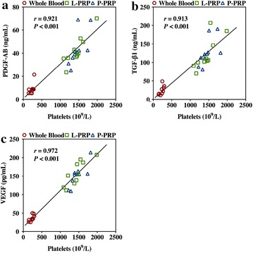
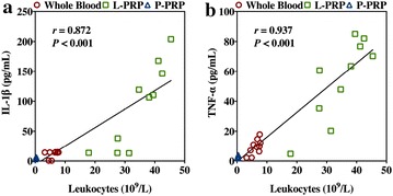
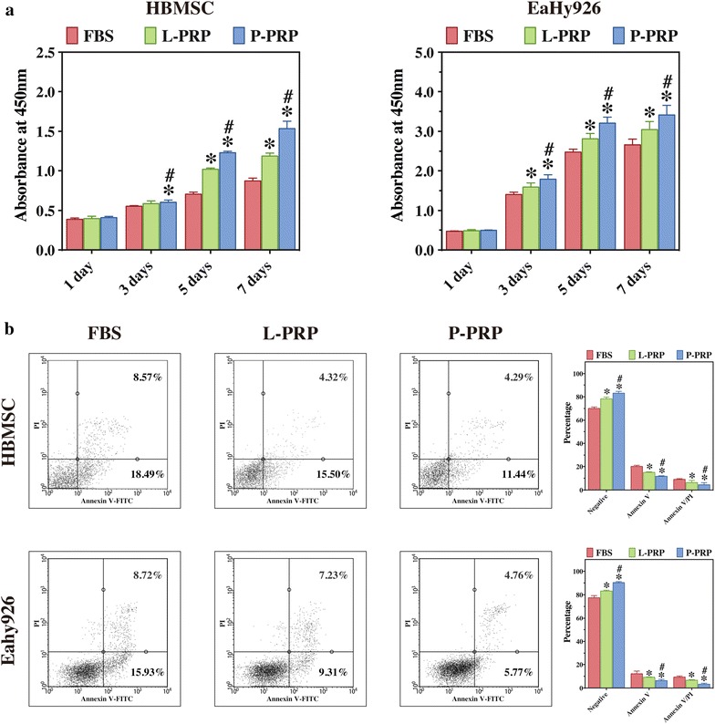
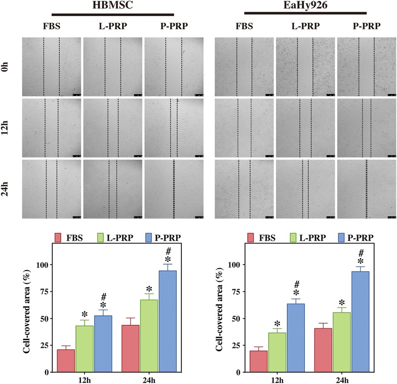
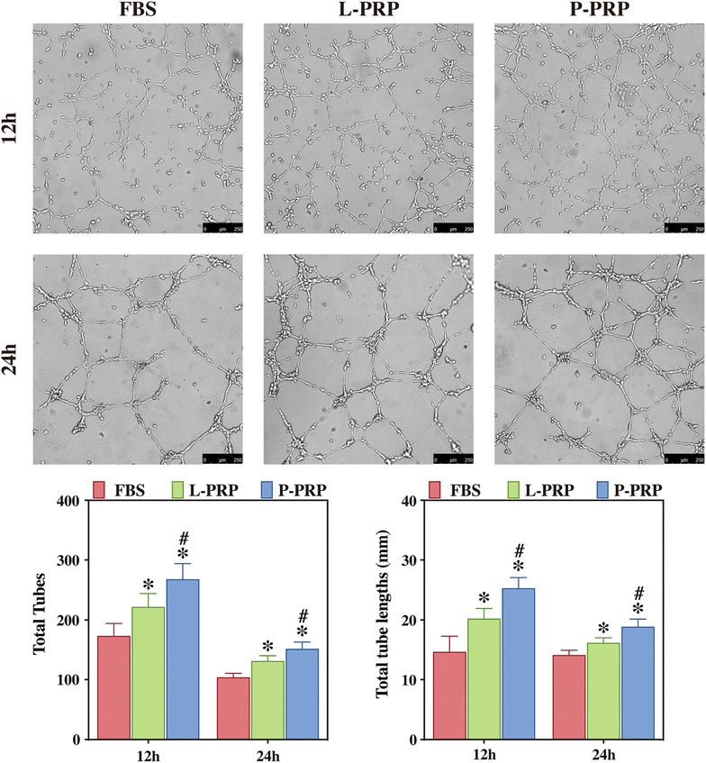
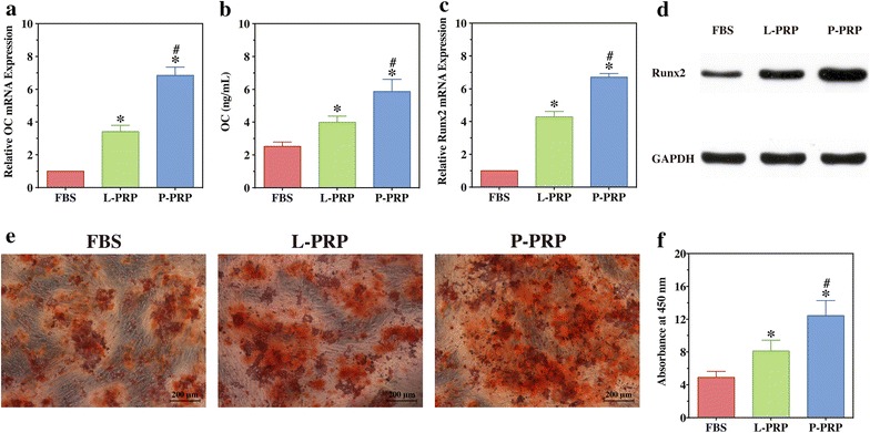
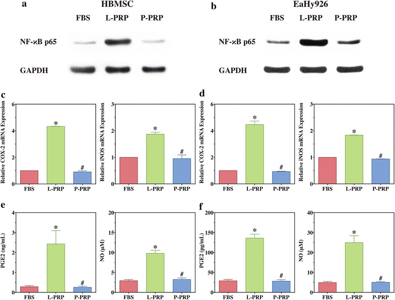
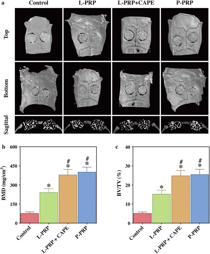
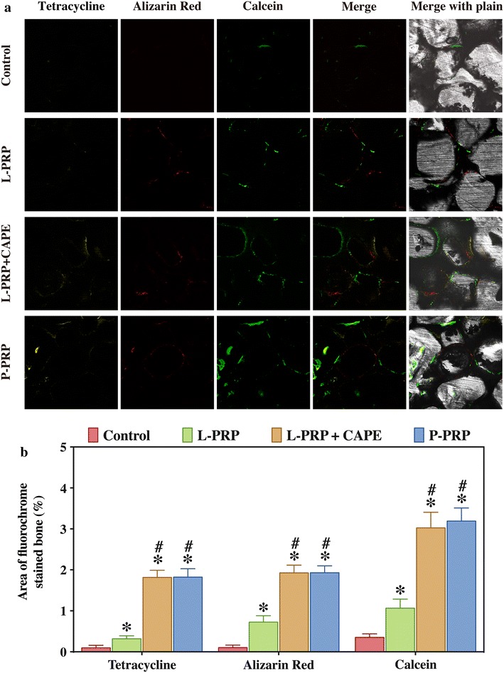
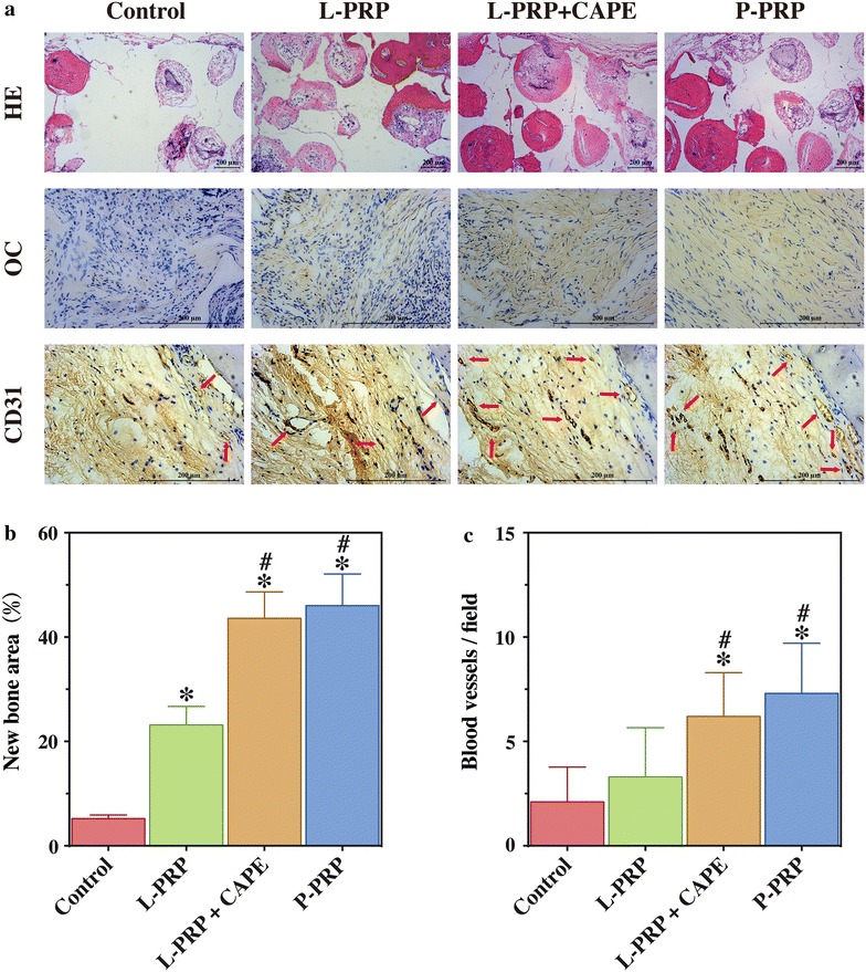
Methods: L-PRP and pure platelet-rich plasma (P-PRP) were isolated. The in vitro effects of L-PRP and P-PRP on the proliferation, viability and migration of human bone marrow-derived mesenchymal stem cells (HBMSCs) and EaHy926, tube formation of EaHy926, and osteogenic differentiation of HBMSCs were assessed by cell counting, flow cytometry, scratch assay, tube formation assay, and real-time quantitative polymerase chain reaction (RT-PCR), western blotting and Alizarin red staining, respectively. The in vitro effects of L-PRP and P-PRP on the nuclear translocation of NF-κB p65, mRNA expression of inducible nitric oxide synthase and cyclooxygenase-2, and production of prostaglandin E2 and nitric oxid were assessed by western blotting, RT-PCR, enzyme-linked immunosorbent assay and Griess reaction, respectively. The in vivo effects of L-PRP or P-PRP preprocessed β-tricalcium phosphate (β-TCP) on the calvarial defects in rats were assessed by histological and immunofluorescence examinations.

Results: P-PRP, which had similar platelet and growth factors concentrations but significantly lower concentrations of leukocytes and pro-inflammatory cytokines compared with L-PRP, promoted the proliferation, viability and migration of HBMSCs and EaHy926, tube formation of EaHy926 and osteogenic differentiation of HBMSCs in vitro, compared with L-PRP. The implantation of P-PRP preprocessed β-TCP also yielded better histological results than the implantation of L-PRP preprocessed β-TCP in vivo. Moreover, L-PRP treatment resulted in the activation of the NF-κB pathway in HBMSCs and EaHy926 in vitro while the postoperative delivery of caffeic acid phenethyl ester, an inhibitor of NF-κB activation, enhanced the histological results of the implantation of L-PRP preprocessed β-TCP in vivo.

Conclusions: Leukocytes in L-PRP may activate the NF-κB pathway via the increased pro-inflammatory cytokines to induce the inferior effects on bone regeneration of L-PRP compared with P-PRP. Hence, P-PRP may be more suitable for bone regeneration compared with L-PRP, and the combined use of P-PRP and β-TCP represents a safe, simple, and effective alternative option for autogenous bone graft in the treatment of bone defects.

Keywords: Animal model; Bone regeneration; Leukocyte- and platelet-rich plasma; Nuclear factor κB; Platelet-rich plasma; Pure platelet-rich plasma.


Figures

Fig. 1
Fig. 1
Correlations between platelet concentration and PDGF-AB concentration (a), TGF-β1 concentration (b), and VEGF concentration (c)
Fig. 2
Fig. 2
Correlations between leukocyte concentration and IL-1β concentration (a), and TNF-α concentration (b)
Fig. 3
Fig. 3
Effects of PRPs on the proliferation and viability of HBMSCs and EaHy926. a cell proliferation was analyzed with CCK-8 assay; b cell viability was analyzed with Annexin V/PI staining and flow cytometry quantification. Compared with FBS, both L-PRP and P-PRP promoted the proliferation and viability of HBMSCs and EaHy926, with P-PRP showing greater effects. Bars represent the means and standard deviation (n = 5); * indicates the statistically significant difference between PRPs and FBS (P < 0.05); # indicates the statistically significant difference between P-PRP and L-PRP (P < 0.05)
Fig. 4
Fig. 4
Effects of PRPs on the migration of HBMSCs and EaHy926. Cell migration was analyzed using Culture-Inserts and quantified using WimScratch software. Compared with FBS, both L-PRP and P-PRP promoted the migration of HBMSCs and EaHy926, with P-PRP showing greater effects. Bars represent the means and standard deviation (n = 5) and scales represent 250 μm; * indicates the statistically significant difference between PRPs and FBS (P < 0.05); # indicates the statistically significant difference between P-PRP and L-PRP (P < 0.05)
Fig. 5
Fig. 5
Effects of PRPs on EaHy926 tube formation. EaHy926 tube formation was analyzed using μ-slide angiogenesis plates and WimTube software quantification. Compared with FBS, both L-PRP and P-PRP promoted the tube formation of EaHy926, with P-PRP showing greater effects. Bars represent the means and standard deviation (n = 5) and scales represent 250 μm; * indicates the statistically significant difference between PRPs and FBS (P < 0.05); # indicates the statistically significant difference between P-PRP and L-PRP (P < 0.05)
Fig. 6
Fig. 6
Effects of PRPs on the osteogenic differentiation of HBMSCs. a mRNA expression of OC of HBMSCs was detected by RT-PCR after osteogenic differentiation induction for 14 days; b OC concentration in conditioned medium was detected by ELISA after osteogenic differentiation induction for 14 days; c mRNA expression of Runx2 of HBMSCs was detected by RT-PCR after osteogenic differentiation induction for 14 days; d Runx2 expression was detected by western blotting after osteogenic differentiation induction for 14 days; e alizarin red staining of HBMSCs after osteogenic differentiation induction for 21 days; f, alizarin red staining was quantified by a colorimetric assay and the absorbance value was measured at 450 nm. Compared with FBS, both L-PRP and P-PRP improved the osteogenic differentiation of HBMSCs, with P-PRP showing greater effects. Bars represent the means and standard deviation (n = 3), and scales represent 200 μm; * indicates the statistically significant difference between PRPs and FBS (P < 0.05); # indicates the statistically significant difference between P-PRP and L-PRP (P < 0.05)
Fig. 7
Fig. 7
L-PRP induced activation of NF-κB in cells in vitro. Western blotting was performed to analyze expression of NF-κB p65 in the nucleus of HBMSCs (a) and EaHy926 (b); RT-PCR was conducted to detect mRNA expression of COX-2 and iNOS of HBMSCs (c) and EaHy926 (d); ELISA and Griess reaction were performed to determine PGE2 and NO production, respectively, of HBMSCs (e) and EaHy926 (f). Bars represent the means and standard deviation (n = 5); * indicates the statistically significant difference between PRPs and FBS (P < 0.05); # indicates the statistically significant difference between P-PRP and L-PRP (P < 0.05)
Fig. 8
Fig. 8
Micro-CT evaluation of bone regeneration in rat calvarial defects after 8 weeks postoperatively. a Top, bottom and cross-sectional views of the reconstructed images; b, c BMD (b) and BV/TV (c) of the regenerated bone in the defects. Bars represent the means and standard deviation (n = 10), and scales represent 200 μm; * indicates the statistically significant difference compared with the control group (P < 0.05); # indicates the statistically significant difference compared with the L-PRP group (P < 0.05)
Fig. 9
Fig. 9
Fluorochrome-labeling of new bone formation and mineralization in the defects. a Column 1 (yellow) shows the deposition of tetracycline at week 2, column 2 (red) shows the deposition of alizarin red at week 4, column 3 (green) shows the deposition of calcein at week 6, column 4 represents the merged images of the three fluorochromes for the same group, and column 5 represents the merged images of the three fluorochromes with a plain CLSM image for the same group; b the percentages of three fluorochrome areas in the defects. Bars represent the means and standard deviation (n = 10); * indicates the statistically significant difference compared with the control group (P < 0.05); # indicates the statistically significant difference compared with the L-PRP group (P < 0.05)
Fig. 10
Fig. 10
HE staining and immunohistochemical staining in the defects after 8 weeks postoperatively. a line 1, HE staining of new bone formation (red area) in the defects; line 2, the immunohistochemical staining of OC showed that there was almost no positive staining for OC in the control group, a limited amount in the L-PRP group, and a greater amount in the L-PRP + CAPE and P-PRP group; line 3, the immunohistochemical staining of CD31 showed that there were more new vessels, which were defined by positive CD31 staining and the typical round or oval structure (red arrows), in P-PRP and L-PRP + CAPE group than in the L-PRP and the control group (line 3); b quantitative analysis of the HE staining; c quantitative analysis of the CD31 staining. Bars represent the means and standard deviation (n = 10), and scales represent 200 μm; * indicates the statistically significant difference compared with the control group (P < 0.05); # indicates the statistically significant difference compared with the L-PRP group (P < 0.05)

References

    1. Dimitriou R, Jones E, McGonagle D, Giannoudis PV. Bone regeneration: current concepts and future directions. BMC Med. 2011;9:66. doi: 10.1186/1741-7015-9-66. - DOI - PMC - PubMed
    1. Calori GM, Colombo M, Mazza EL, Mazzola S, Malagoli E, Mineo GV. Incidence of donor site morbidity following harvesting from iliac crest or RIA graft. Injury. 2014;45(Suppl 6):116–120. doi: 10.1016/j.injury.2014.10.034. - DOI - PubMed
    1. Oryan A, Alidadi S, Moshiri A, Maffulli N. Bone regenerative medicine: classic options, novel strategies, and future directions. J Orthop Surg Res. 2014;9:18. doi: 10.1186/1749-799X-9-18. - DOI - PMC - PubMed
    1. Yu H, VandeVord PJ, Mao L, Matthew HW, Wooley PH, Yang SY. Improved tissue-engineered bone regeneration by endothelial cell mediated vascularization. Biomaterials. 2009;30:508–517. doi: 10.1016/j.biomaterials.2008.09.047. - DOI - PubMed
    1. Hirota M, Matsui Y, Mizuki N, Kishi T, Watanuki K, Ozawa T, et al. Combination with allogenic bone reduces early absorption of beta-tricalcium phosphate (beta-TCP) and enhances the role as a bone regeneration scaffold. Experimental animal study in rat mandibular bone defects. Dent Mater J. 2009;28:153–161. doi: 10.4012/dmj.28.153. - DOI - PubMed

Publication types

MeSH terms

Substances

LinkOut - more resources