. 2016 Mar 16:6:23286.
doi: 10.1038/srep23286.

P2X7 Receptor Suppression Preserves Blood-Brain Barrier through Inhibiting RhoA Activation after Experimental Intracerebral Hemorrhage in Rats

Affiliations

P2X7 Receptor Suppression Preserves Blood-Brain Barrier through Inhibiting RhoA Activation after Experimental Intracerebral Hemorrhage in Rats

Hengli Zhao et al. Sci Rep. .

Abstract

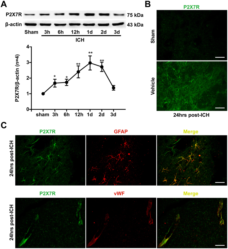
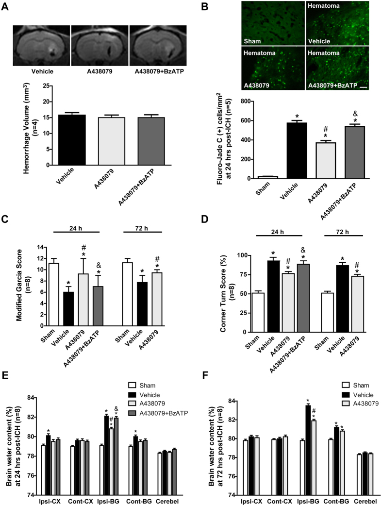
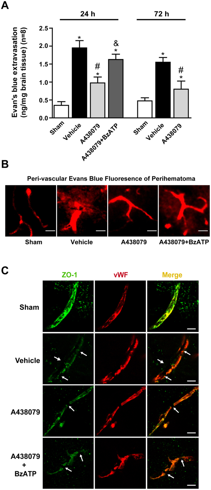
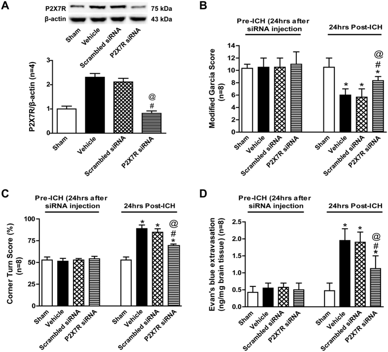
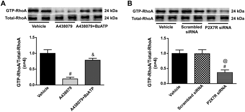
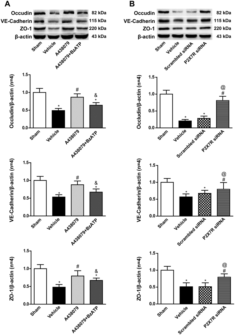
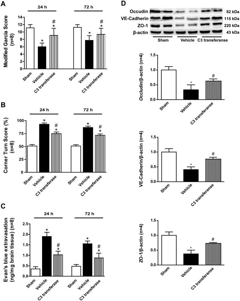
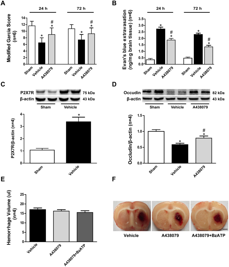
Blockading P2X7 receptor(P2X7R) provides neuroprotection toward various neurological disorders, including stroke, traumatic brain injury, and subarachnoid hemorrhage. However, whether and how P2X7 receptor suppression protects blood-brain barrier(BBB) after intracerebral hemorrhage(ICH) remains unexplored. In present study, intrastriatal autologous-blood injection was used to mimic ICH in rats. Selective P2X7R inhibitor A438079, P2X7R agonist BzATP, and P2X7R siRNA were administrated to evaluate the effects of P2X7R suppression. Selective RhoA inhibitor C3 transferase was administered to clarify the involvement of RhoA. Post-assessments, including neurological deficits, Fluoro-Jade C staining, brain edema, Evans blue extravasation and fluorescence, western blot, RhoA activity assay and immunohistochemistry were performed. Then the key results were verified in collagenase induced ICH model. We found that endogenous P2X7R increased at 3 hrs after ICH with peak at 24 hrs, then returned to normal at 72 hrs after ICH. Enhanced immunoreactivity was observed on the neurovascular structure around hematoma at 24 hrs after ICH, along with perivascular astrocytes and endothelial cells. Both A438079 and P2X7R siRNA alleviated neurological deficits, brain edema, and BBB disruption after ICH, in association with RhoA activation and down-regulated endothelial junction proteins. However, BzATP abolished those effects. In addition, C3 transferase reduced brain injury and increased endothelial junction proteins' expression after ICH. These data indicated P2X7R suppression could preserve BBB integrity after ICH through inhibiting RhoA activation.


Figures

Figure 1
Figure 1. Time course and spatial expression of P2X7 receptor after ICH.
(A) Representative bands and quantitative analyses of P2X7 receptor expression in the ipsilateral striatum. Relative densities of each protein have been normalized against the sham group. The cropped bands had been run under the same experimental conditions. (B) Representative immunofluorescence staining slices of P2X7 receptor (Green) in pericontusional striatum at 24 hours after ICH. Scalebar: 50 μm. (C) Representative immunofluorescence staining slices of P2X7 receptorwithglial fibrillary acidic protein (GFAP, red) and von Willebrand factor (vWF, red) in the perihematoma area at 24 hours following ICH. Triangle indicates hematoma area. Scale bar: 20 μm. *p < 0.05 vs. Sham; **p < 0.01 vs. Sham. n = 4 per group.
Figure 2
Figure 2. Effects of A438079 administration with/without BzATP pretreatment on outcomes at 24 and 72 hours after ICH.
(A) Representative T2-weighted images and quantitative analyses of hematoma size at 24 hours after ICH. (B) Representative Fluoro-Jade C staining images and quantitative analyses of Fluoro-Jade C positive neuron around hematoma at 24 hours after ICH. (C) Modified Garcia scores and (D) Corner turn score at 24 and 72 hours after ICH in each group. (E)Brain water content assessment at 24 hours after ICH (F) Brain water content assessment at 72 hours after ICH. Scale bar: 50μm. *p < 0.05 vs. Sham; #p < 0.05 vs. Vehicle; &p < 0.05 vs. A438079; n = 4 for T2-weighted images, n = 5 for Fluoro-Jade C staining, n = 8 for others. Ipsi-CX: ipsilateral cortex; Cont-CX: contralateral cortex; Ipsi-BG: ipsilateral basal ganglia; Cont-BG: contralateral basal ganglia; Cerebel: cerebellum.
Figure 3
Figure 3. Effects of A438079 administration with/without BzATP pretreatment on blood brain barrier integrity at 24 and 72 hours after ICH.
(A) Evans blue extravasation evaluation at 24 and 72 hours after ICH in each group. (B) Representative Evans blue fluorescence in ipsilateral striatum at 24 hours after ICH in each group. (C) Representative immunohistochemistry staining slices of zonula occluden-1 (ZO-1) and von Willebrand factor (vWF) at 24 hours after ICH. Arrow indicates the breakdown of continuous endothelia cell layer. *p < 0.05 vs. Sham; #p < 0.05 vs. Vehicle; &p < 0.05 vs. A438079. n = 8 per group.
Figure 4
Figure 4. Effects of P2X7 receptor siRNA on outcomes at 24 hours after ICH.
(A) Representative bands and quantitative analysis of the P2X7 receptor siRNA inhibiting effect. Relative densities of each protein have been normalized against the sham group. The cropped bands had been run under the same experimental conditions. (B)Modified Garcia scores and (C) Corner turn score at 24 hours after ICH in each group. (D) Evans blue extravasation evaluation at 24 hours after ICH in each group. *p < 0.05 vs Sham; #p < 0.05 vs Vehicle; @p < 0.05 vs Scramble siRNA. n = 8 per group. ICV = intracerebroventricular infusion.
Figure 5
Figure 5. Effects of P2X7 receptor modulation on RhoA activity at 24 hours after ICH.
(A) Representative bands and quantitative analysis of GTP-RhoA/Total-RhoA ratio after A438079 treatment with/without BzATP pretreatment at 24 hours after ICH in each group. (B) Representative bands and quantitative analysis of GTP-RhoA/Total-RhoA ratio P2X7 siRNA pretreatment at 24 hours after ICH in each group. Relative densities of each protein have been normalized against the vehicle group. The cropped bands had been run under the same experimental conditions. *p < 0.05 vs Sham; #p < 0.05 vs Vehicle; &p < 0.05 vs A438079; @p < 0.05 vs Scramble siRNA. n = 4 per group.
Figure 6
Figure 6. Effects of P2X7 receptor modulation on the expressions of endothelial junction proteins at 24 hours after ICH.
(A) Representative bands and quantitative analysis of the expressions of Occudin, VE-Cadherin and zonula occluden-1 (ZO-1) after A438079 treatment with/without BzATP pretreatment at 24 hours post-ICH in each group. (B) Representative bands and quantitative analysis of the expressions of Occudin, VE-Cadherin and zonula occluden-1 (ZO-1) with P2X7 siRNA pretreatment at 24 hours after ICH in each group. Relative densities of each protein have been normalized against the sham group. The cropped bands had been run under the same experimental conditions. *p < 0.05 vs Sham; #p < 0.05 vs Vehicle; &p < 0.05 vs A438079; @p < 0.05 vs Scramble siRNA. n = 4 per group.
Figure 7
Figure 7. Effects of RhoA inhibitor C3 transferase on the outcomes and the expressions of endothelial junction proteins after ICH.
(A) Modified Garcia scores and (B) Corner turn score at 24 and 72 hours after ICH in each group. (C) Evans blue extravasation evaluation at 24 and 72 hours after ICH in each group. (D) Representative bands and quantitative analysis of the expressions of Occudin, VE-Cadherin and ZO-1 after C3 transferase treatment at 24 hours post-ICH in each group. The cropped bands had been run under the same experimental conditions. *p < 0.05 vs Sham; #p < 0.05 vs Vehicle; n = 4 per group.
Figure 8
Figure 8. Effects of P2X7 receptor modulating exhibited the similar effects in collagenase induced ICH model.
(A) Modified Garcia scores and (B) Corner turn score at 24 and 72 hours after ICH in each group. (C) Representative bands and quantitative analyses of P2X7 receptor expression in the ipsilateral striatum. Relative densities of each protein have been normalized against the sham group. (D) Representative bands and quantitative analysis of the expressions of Occudin at 24 hours post-ICH in each group. The cropped bands had been run under the same experimental conditions. (E) Quantitative analyses of hematoma size among each group and (F) representative coronal autopsy images at 24 hours after ICH. *p < 0.05 vs Sham; #p < 0.05 vs Vehicle; n = 6 for modified Garcia score and Evan’s blue extravasation, n = 4 for others.

References

    1. Xi G., Strahle J., Hua Y. & Keep R. F. Progress in translational research on intracerebral hemorrhage: is there an end in sight? Progress in neurobiology 115, 45–63, 10.1016/j.pneurobio.2013.09.007 (2014). - DOI - PMC - PubMed
    1. Zhou Y., Wang Y., Wang J., Anne Stetler R. & Yang Q. W. Inflammation in intracerebral hemorrhage: from mechanisms to clinical translation. Progress in neurobiology 115, 25–44, 10.1016/j.pneurobio.2013.11.003 (2014). - DOI - PubMed
    1. Wang L. et al. Recombinant ADAMTS13 reduces tissue plasminogen activator-induced hemorrhage after stroke in mice. Annals of neurology 73, 189–198, 10.1002/ana.23762 (2013). - DOI - PubMed
    1. Vose L. R. et al. Treatment with thyroxine restores myelination and clinical recovery after intraventricular hemorrhage. The Journal of neuroscience: the official journal of the Society for Neuroscience 33, 17232–17246, 10.1523/JNEUROSCI.2713-13.2013 (2013). - DOI - PMC - PubMed
    1. Abbott N. J., Patabendige A. A., Dolman D. E., Yusof S. R. & Begley D. J. Structure and function of the blood-brain barrier. Neurobiology of disease 37, 13–25, 10.1016/j.nbd.2009.07.030 (2010). - DOI - PubMed

Publication types

MeSH terms

LinkOut - more resources