. 2016 Mar 16:6:23228.
doi: 10.1038/srep23228.

Distinct Biochemical Activities of Eyes absent During Drosophila Eye Development

Affiliations

Distinct Biochemical Activities of Eyes absent During Drosophila Eye Development

Meng Jin et al. Sci Rep. .

Abstract

Eyes absent (Eya) is a highly conserved transcriptional coactivator and protein phosphatase that plays vital roles in multiple developmental processes from Drosophila to humans. Eya proteins contain a PST (Proline-Serine-Threonine)-rich transactivation domain, a threonine phosphatase motif (TPM), and a tyrosine protein phosphatase domain. Using a genomic rescue system, we find that the PST domain is essential for Eya activity and Dac expression, and the TPM is required for full Eya function. We also find that the threonine phosphatase activity plays only a minor role during Drosophila eye development and the primary function of the PST and TPM domains is transactivation that can be largely substituted by the heterologous activation domain VP16. Along with our previous results that the tyrosine phosphatase activity of Eya is dispensable for normal Eya function in eye formation, we demonstrate that a primary function of Eya during Drosophila eye development is as a transcriptional coactivator. Moreover, the PST/TPM and the threonine phosphatase activity are not required for in vitro interaction between retinal determination factors. Finally, this work is the first report of an Eya-Ey physical interaction. These findings are particularly important because they highlight the need for an in vivo approach that accurately dissects protein function.


Figures

Figure 1
Figure 1. eya genomic rescue constructs rigorously define Eya functional domains.
(a) Schematic view of the eya genomic rescue (GR) constructs assayed in this study. The percent rescue indicated is for animals with one copy of each GR construct in an eya2 homozygous mutant background. At least 100 adult eyes were scored for each GR and the penetrance of the transgene-induced phenotypes for all GRs are 100%, with minor variation in expressivity. Each construct is inserted at the same genomic docking site such that all constructs are directly comparable. (b–k) Representative images of adult eyes for each genotype tested are shown. PST: grey box, TPM: purple box, Y4: red vertical lines, VP16: green box, ED1: orange box.
Figure 2
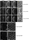
Figure 2. Threonine phosphatase activity is required for normal anterior expression of eya and cone cell development.
Expression of Ey (a–c), Eya (a’–c’), and So (a”–c”) proteins are shown in eya+GR, eyaY4GR, and eyaΔTPMGR rescued animals. (d–f) Adult plastic sections in flies rescued with one copy of eya+GR, eyaY4GR and eyaΔTPMGR, respectively. (g,h) Third instar eye discs by Cut staining. (I,j) Eye discs prepared from 48 hrs after puparium formation and stained with Dlg.
Figure 3
Figure 3. Loss of threonine phosphatase activity does not strongly affect Dac, Ato, or Cyclin B expression.
Dac (a,b), Ato (c,d), and the cell cycle marker Cyclin B (e,f) expression in animals rescued with a single copy of eya+GR or eyaY4GR are shown. Expression patterns and levels of Eya, Ato and Cyclin B are similar in both genotypes although minor disruption of Ato is observed.
Figure 4
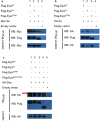
Figure 4. Threonine phosphatase activity is not required for Eya interaction with Ey, So, or Dac.
(a,b) Co-immunoprecipitation (co-IP) studies between wild-type and threonine phosphatase-dead Eya (EyaY4 and EyaΔTPM) and Ey or So are shown. Flag-tagged Eya was co-expressed with HA-Ey or Myc-So in S2 cells and co-IP with anti-Flag beads followed by western blotting (WB) was performed. Ey, So, and Eya (wild-type and mutants) were detected by anti-HA, anti-Myc, and anti-Flag antibodies, respectively. Lanes 1, 2, and 3 show that EyaWT, EyaY4 and EyaΔTPM can pull down So and Ey, respectively. Empty vector is the negative control (Lane 4). (c) co-IP analysis of Eya and Dac. Lane 1, Flag-EyaWT/HA-Dac; lane 2, Flag-EyaY4/HA-Dac; lane 3, Flag-EyaΔTPM/HA-Dac; lane 4 Flag-EyaΔPST/TPM/HA-Dac; lane 5, empty vector. Anti-FLAG is used for IP. The red arrow indicates Dac protein. All proteins are expressed at similar levels in crude cell lysates (the bottom panel of each set and data not shown). Western blots presented in a-c were cropped to improve clarity and full-length blots are presented in Supplementary Figs S7–9. All gels were run under the same experimental conditions.
Figure 5
Figure 5. eyaΔPST/TPMGR fails to rescue eya mutant defects even though mutant transcript and protein are detected.
(a) Lanes 1–3 show RT-PCR on RNA prepared from eyes discs 68 hrs after egg laying (AEL). Lane 1: wild-type; Lane 2: eya2; eyaΔPST/TPMGR; Lane 3: eya2; Lane 4: water. A truncated ΔPST/TPM transcript is readily detected (Lane 2). (b) An anti-Eya Western blot on extracts prepared from 68 hrs after egg laying (AEL) eye discs (n = 40/lane) from either eyacliIID/CyO; eya+GR/+ or eyacliIID/CyO; eyaΔPST/TPMGR/+ animals shows a readily detectable, truncated ΔPST/TPM protein (*). Heterozygous eyacliIID animals were used to obtain enough tissue for the experiment. Western blot presented in b is cropped to improve clarity and full-length blot is presented in Supplementary Fig. S10. (c–f) Eye discs prepared from larvae 68 hrs AEL are stained for Eya expression. (g–j) Eya staining of 56 hrs AEL eye discs.
Figure 6
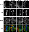
Figure 6. PST/TPM is required for normal Dac expression.
(a–d) Dac expression in Canton-S (a), eya2 (b), eya2; eya+GR (c) and eya2; eyaΔPST/TPMGR (d) eye imaginal discs from 68 hrs AEL. (e–h) Immunostaining of Dac on 56 hrs AEL eye discs. (i–k) Dac, Eya and GFP expression in eyaΔPST/TPM rescued eyacliIID null clones. Yellow arrow indicates one of the larger, more posterior clones in which Dac expression is reduced. (l) Merge of channels.
Figure 7
Figure 7. Overexpression of So in eya2; eyaΔPST/TPMGR animals causes increased Dac expression.
(a–d) Dac staining in third instar larvae after inducing so expression with ey-Gal4/UAS-so. Yellow arrows (d) indicate region of the disc in which Dac expression is increased. (e–h) Eya expression after ey-Gal4/UAS-so induction. eya2; eya+GR (a,e) and eya2 (b,f) are used as positive and negative controls.
Figure 8
Figure 8. The PST/TPM of Eya is necessary for ey repression posterior to the morphogenetic furrow.
(a–a’”) Wild-type clones. (b–b’”) eyacliIID null clones. (c–c’”) eya+GR rescued eyacliIID null clones. (d–d’”) eyaΔPST/TPMGR rescued eyacliIID null clones. Grayscale images of Elav, Ey, and GFP expression are shown in grayscale (a–d”) and as red, blue, and green, respectively, in a’”–d’”; Elav marks differentiating photoreceptors and complete loss of GFP expression marks homozygous mutant clones.
Figure 9
Figure 9. The PST/TPM domain is not required for interaction between Eya and Ey, So, or Dac.
(a,b) S2 cells were transfected as described in Materials and Methods, lysates were immunoprecipitated (IP) with anti-Flag beads, and then immunoblotted (WB) with anti-Myc, anti-HA, and anti-Flag antibodies to detect Myc-So, HA-Ey, and Flag-Eya, respectively. Co-IP for EyaΔPST/TPM and Dac is shown in Fig. 4c. Western blots presented in a-b were cropped to improve clarity and full-length blots are presented in Supplementary Fig. S11–12. All gels were run under the same experimental conditions.

References

    1. Pappu K. S. & Mardon G. Genetic control of retinal specification and determination in Drosophila. Int J Dev Biol 48, 913–924 (2004). - PubMed
    1. Czerny T. et al.. Twin of eyeless, a second Pax-6 gene of Drosophila, acts upstream of eyeless in the control of eye development. Mol Cell 3, 297–307 (1999). - PubMed
    1. Quiring R., Walldorf U., Kloter U. & Gehring W. J. Homology of the eyeless gene of Drosophila to the Small eye gene in mice and Aniridia in humans. Science 265, 785–789 (1994). - PubMed
    1. Bonini N. M., Leiserson W. M. & Benzer S. The eyes absent gene: genetic control of cell survival and differentiation in the developing Drosophila eye. Cell 72, 379–395 (1993). - PubMed
    1. Cheyette B. N. et al.. The Drosophila sine oculis locus encodes a homeodomain-containing protein required for the development of the entire visual system. Neuron 12, 977–996 (1994). - PubMed

Publication types

MeSH terms

LinkOut - more resources