. 2016 Mar;8(Suppl 2):S210-22.
doi: 10.3978/j.issn.2072-1439.2016.01.76.

Uniportal video-assisted thoracoscopic bronchoplastic and carinal sleeve procedures

Affiliations

Uniportal video-assisted thoracoscopic bronchoplastic and carinal sleeve procedures

Diego Gonzalez-Rivas et al. J Thorac Dis. 2016 Mar.

Abstract

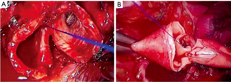
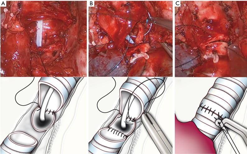
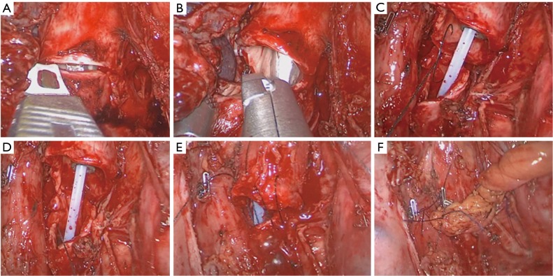
Despite of the recent advanced with the video-assisted thoracoscopic surgery (VATS), the most common approach for bronchial and carinal resection is still the open surgery. The technical difficulties, the steep learning curve and the concerns about performing an oncologic and safe reconstruction in advanced cases, are the main reasons for the low adoption of VATS for sleeve resections. Most of the authors use 3-4 incisions for thoracoscopic sleeve procedures. However these surgical techniques can be performed by a single incision approach by skilled uniportal VATS surgeons. The improvements of the surgical instruments, high definition cameras and recent 3D systems have greatly contributed to facilitate the adoption of uniportal VATS techniques for sleeve procedures. In this article we describe the technique of thoracoscopic bronchial sleeve, bronchovascular and carinal resections through a single incision approach.

Keywords: Sleeve lobectomy; bronchoplasty; carinal resection; double sleeve; uniportal video-assisted thoracoscopic surgery (VATS); vascular reconstruction.


Conflict of interest statement

Conflicts of Interest: The authors have no conflicts of interest to declare.

Figures

Figure 1
Figure 1
CT scan showing a tumor on the main RUL bronchus (A) and a tumor involving the left main pulmonary (B). CT, computed tomography; RUL, right upper lobe.
Figure 2
Figure 2
Surgical technique for thoracoscopic suturing by uniportal VATS (tying the knots) (12). VATS, video-assisted thoracoscopic surgery. Available online: http://www.asvide.com/articles/814
Figure 3
Figure 3
Surgical photo showing instrumentation for bronchial sleeve anastomosis.
Figure 4
Figure 4
Simple bronchoplasty for a right upper lobe endobronchial tumor (15). Available online: http://www.asvide.com/articles/815
Figure 5
Figure 5
Right upper sleeve resection (17). Available online: http://www.asvide.com/articles/816
Figure 6
Figure 6
Left upper lobe sleeve resection (20). Available online: http://www.asvide.com/articles/817
Figure 7
Figure 7
Drawing showing the sequence of lower lobe sleeve resection and anastomosis. (A) Exposure of left upper, lower and main bronchus and transection of LUL bronchus; (B) transection of the main bronchus using scissors; (C) a suture is placed at the angle of anterior portion of both ends of bronchi; (D) final result. LUL, left upper lobe.
Figure 8
Figure 8
Bronchus intermedius resection (sparing lung) (21). Available online: http://www.asvide.com/articles/818
Figure 9
Figure 9
Drawing showing bronchus intermedius bronchial resection and anastomosis (carcinoid tumor). (A) Bronchial distal resection by using scissors (artery is retracted); (B) anterior suture used for apposition of distal and proximal bronchial end; (C) final result.
Figure 10
Figure 10
Left upper sleeve resection and vascular reconstruction (25). Available online: http://www.asvide.com/articles/819
Figure 11
Figure 11
LUL double sleeve lobectomy. (A) Bronchial sleeve; (B) vascular sleeve. LUL, left upper lobe.
Figure 12
Figure 12
Combined drawings and surgical photos showing carinal reconstruction after right upper sleeve carinal resection. (A) Completed continuous suture of left side wall of the anastomosis between trachea and left main bronchus; (B) after creation of neocarina (left main to intermedius bronchus) the right side wall of the trachea, left main and bronchus intermedius is completed; (C) final result.
Figure 13
Figure 13
Surgical images of right carinal pneumonectomy. (A) The distal trachea is incised; (B) the circunferencial resection is completed by using scissors. (C) the running suture was started on the left wall of the trachea and left main bronchus; (D) the membranous portion was anastomosed after the left wall was completed; (E) the right side of the trachea and left main is performed; (F) a pericardial flap covered the anatomosis.

References

    1. Villamizar NR, Darrabie MD, Burfeind WR, et al. Thoracoscopic lobectomy is associated with lower morbidity compared with thoracotomy. J Thorac Cardiovasc Surg 2009;138:419-25. - PubMed
    1. Swanson SJ, Herndon JE, 2nd, D'Amico TA, et al. Video-assisted thoracic surgery lobectomy: report of CALGB 39802da prospective, multi-institution feasibility study. J Clin Oncol 2007;25:4993-7. - PubMed
    1. McKenna RJ, Jr, Houck W, Fuller CB. Video-assisted thoracic surgery lobectomy: experience with 1,100 cases. Ann Thorac Surg 2006;81:421-5; discussion 425-6. - PubMed
    1. Kaseda S, Aoki T, Hangai N, et al. Better pulmonary function and prognosis with video-assisted thoracic surgery than with thoracotomy. Ann Thorac Surg 2000;70:1644-6. - PubMed
    1. Yang X, Wang S, Qu J. Video-assisted thoracic surgery (VATS) compares favorably with thoracotomy for the treatment of lung cancer: a five-year outcome comparison. World J Surg 2009;33:1857-61. - PubMed