. 2016 Mar 11;5(2):8.
doi: 10.1167/tvst.5.2.8. eCollection 2016 Mar.

Objective Evaluation of Vergence Disorders and a Research-Based Novel Method for Vergence Rehabilitation

Affiliations

Objective Evaluation of Vergence Disorders and a Research-Based Novel Method for Vergence Rehabilitation

Zoï Kapoula et al. Transl Vis Sci Technol. .

Abstract

Purpose: We performed video-oculography to evaluate vergence eye movement abnormalities in students diagnosed clinically with vergence disorders. We tested the efficiency of a novel rehabilitation method and evaluated its benefits with video-oculography cross-correlated with clinical tests and symptomatology.

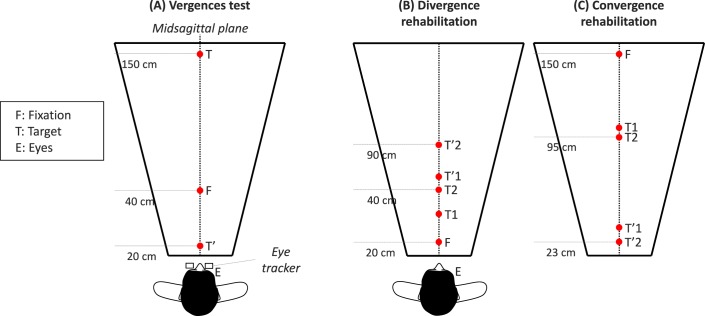
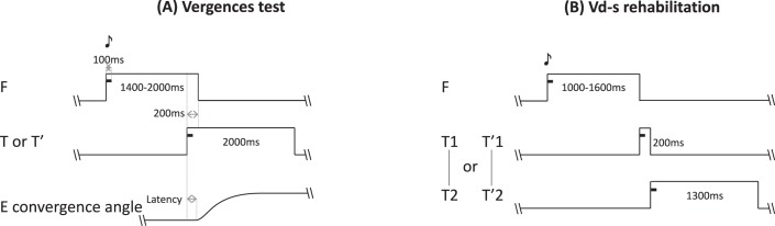
Methods: A total of 19 students (20-27 years old) underwent ophthalmologic, orthoptic examination, and a vergence test coupled with video-oculography. Eight patients were diagnosed with vergence disorders with a high symptomatology score (CISS) and performed a 5-week session of vergence rehabilitation. Vergence and rehabilitation tasks were performed with a trapezoid surface of light emitting diodes (LEDs) and adjacent buzzers (US 8851669). We used a novel Vergence double-step (Vd-s) protocol: the target stepped to a second position before the vergence movement completion. Afterward the vergence test was repeated 1 week and 1 month later.

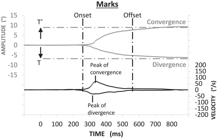
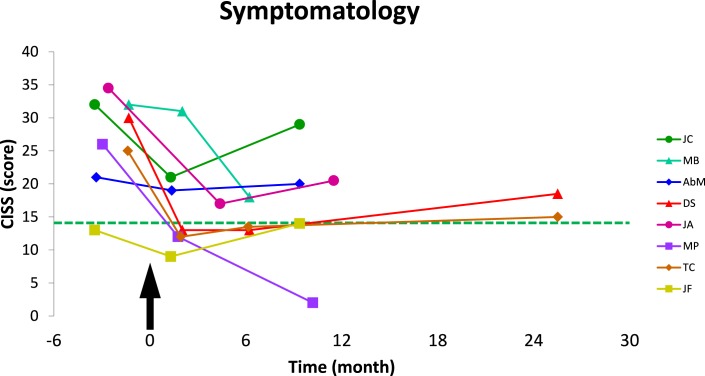
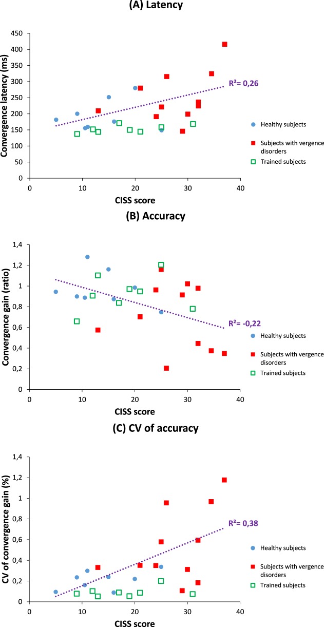
Results: Abnormally increased intertrial variability was observed for many vergence parameters (gain, duration, and speed) for the subjects with vergence disorders. High CISS scores were correlated with variability and increased latency. After the Vd-s, variability of all parameters dropped to normal or better levels. Moreover, the convergence and divergence latency diminished significantly to levels better than normal; benefits were maintained 1 month after completion of Vd-s. CISS scores dropped to normal level, which was maintained up to 1 year.

Conclusions and translational relevance: Intertrial variability is the major marker of vergence disorders. The Vd-s research-based method leads to normalization of vergence properties and lasting removal of symptoms. The efficiency of the method is due to the spatiotemporal parameters of repetitive trials that stimulate neural plasticity.

Keywords: accommodation; binocular vision/stereopsis; double-step task; rehabilitation; vergence.


Figures

Figure 1
Figure 1
Spatial arrangement of targets in the vergence test ([A] subjects are looking successively at different LEDs; from F, the initial fixation LED, to T, the target LED for divergence; or T', the target LED for convergence). Double-step targets used for divergence and convergence rehabilitation ([B, C] from F to T1 or T2, the first target location; then to T'1 or T'2, the final target location). The two types of trials were randomly interleaved.
Figure 2
Figure 2
(A) Temporal arrangement of the vergence test. Each trial starts with the fixation target that appears for a variable period of 1200 to 1800 ms; following this period the target LED lights are on for 2000 ms together with a paired buzzer that lasts only 100 ms; the fixation LED switches off 200 ms after the onset of the target LED (overlap period). (B) Temporal arrangement of the vergence double-step rehabilitation protocol. The trial starts with lighting on the fixation LED for a random period between 1000 and 1600 ms; the fixation LED is accompanied with a buzzer sound for the first 100 ms. Following the fixation period, the target LED lights are on at its first position together with a paired buzzer for 100 ms. After 200 ms the fixation LED switches off and at the same time the LED target steps to it final position (T2 or T'2) where it stays on for 1300 ms; a sound accompanies this second step also for the first 100 ms. A blank period of 300 to 400 ms separates trials.
Figure 3
Figure 3
Method for vergence analysis: convergence and divergence (in gray) were obtained by subtraction of right eye position from the left eye position (LE – RE). The corresponding velocity trace is shown below. Mark of onset and offset of vergence is based on velocity criteria: the time point when the eye velocity respectively exceeded or dropped below 5°/s. The horizontal dotted lines indicate the targets' position.
Figure 4
Figure 4
Follow-up of the symptomatology scores (CISS) of subjects with vergence disorders are plotted over time. Arrow indicates the beginning; that is, the first day of Vd-s rehabilitation protocol. Dotted line shows the mean score of the healthy group. CISS score decreased after rehabilitation, and the results were stable over time.
Figure 5
Figure 5
Individual values of orthoptic tests for the vergence disorders group: dotted lines show the mean values of the healthy group. Following the Vd-s rehabilitation, the near point of convergence decreased, and the convergence amplitude measured with prisms at far or near increased; the results are durable as the orthoptic examination is done 1 month after the end Vd-s rehabilitation.
Figure 6
Figure 6
Trajectories of vergence movements from three healthy subjects ASF (A), AZ (B) and EO (C), and three subjects with vergence disorders JF (D–F), TC (G–I), and MP (J–L). Convergence and divergence traces are superimposed at the top of each figure (in gray); their respective velocity traces are shown at bottom (in black). Vergence traces have small variability, that is, they are reproducible for healthy subjects (A–C). In contrast, variability is high for subjects JF (D) and TC (G), and subject MP shows quasi-paucity of divergence (J). After rehabilitation with the Vd-s protocol: variability decreased for all subjects. For each subject the benefits are stable over 1 week (E, H, K) and 1 month (F, I, L) after the end of rehabilitation.
Figure 7
Figure 7
Convergence latency is correlated positively with symptomatology measured by CISS score (A), and convergence gain is negatively correlated with symptomatology (B), while it is positively correlated with the variability of convergence gain (C). Data from healthy subjects are illustrated in blue circles, and for subjects diagnosed for vergence disorders, with full red squares (before rehabilitation) or empty green squares (after Vd-s rehabilitation); linear correlation model was run by considering only the data from the healthy and vergence disorder groups before rehabilitation. Values of subjects after rehabilitation are shown for illustrating the clear move towards normal levels.

Similar articles

Cited by

References

    1. Montés-Micó R. Prevalence of general dysfunctions in binocular vision. Ann Ophthalmol. 2001; 33: 205–208.
    1. Lavrich JB. Convergence insufficiency and its current treatment. Curr Opin Ophthalmol. 2010; 21: 356–360. - PubMed
    1. Scheiman M,, Cooper J,, Mitchell GL,, et al. A survey of treatment modalities for convergence insufficiency. Optom Vis Sci. 2002; 79: 151–157. - PubMed
    1. Horwood AM,, Toor S,, Riddell PM. Screening for convergence insufficiency using the CISS is not indicated in young adults. Br J Ophthalmol. 2014; 98: 679–683. - PMC - PubMed
    1. Cacho-Martínez P,, García-Muñoz Á,, Ruiz-Cantero MT. Is there any evidence for the validity of diagnostic criteria used for accommodative and nonstrabismic binocular dysfunctions? J Optom. 2014; 7: 2–21. - PMC - PubMed