. 2016 Mar 11;5(2):9.
doi: 10.1167/tvst.5.2.9. eCollection 2016 Mar.

VEGF: From Discovery to Therapy: The Champalimaud Award Lecture

Affiliations

VEGF: From Discovery to Therapy: The Champalimaud Award Lecture

Joan W Miller. Transl Vis Sci Technol. .

Abstract

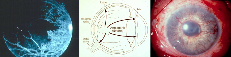
Purpose: Intraocular vascular diseases are leading causes of adult vision loss, and in the mid-1900s, I. C. Michaelson postulated that the retina releases a soluble, diffusible factor that causes abnormal vascular growth and leakage. What became known as "Factor X" eluded investigators for decades.

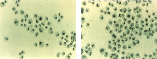
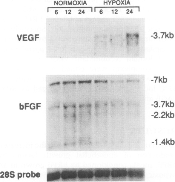
Methods: The field of cancer research, where Judah Folkman pioneered the concept of angiogenesis, provided the inspiration for the work honored by the 2014 Champalimaud Vision Award. Recognizing that tumors recruit their own blood supply to achieve critical mass, Dr Folkman proposed that angiogenic factors could be therapeutic targets in cancer. Napoleone Ferrara identified vascular endothelial growth factor (VEGF) as such an angiogenic agent: stimulated by hypoxic tumor tissue, secreted, and able to induce neovascularization. VEGF also was a candidate for Factor X, and the 2014 Champalimaud Laureates and colleagues worked individually and collaboratively to identify the role of VEGF in ocular disease.

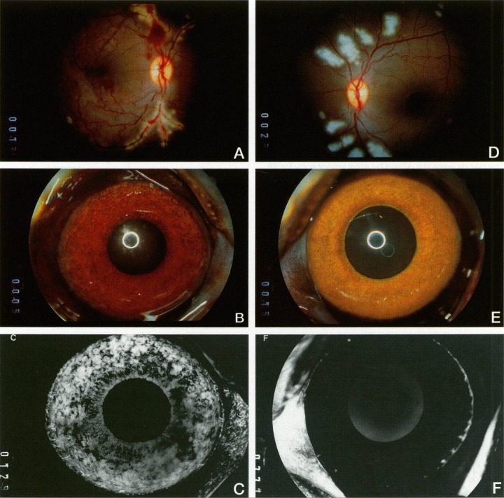
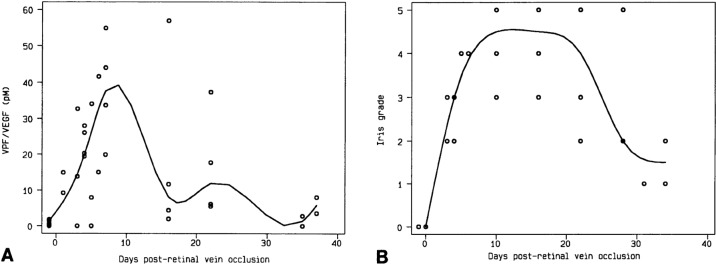
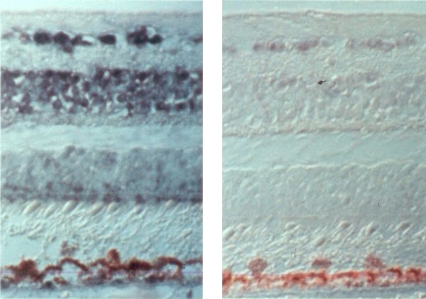
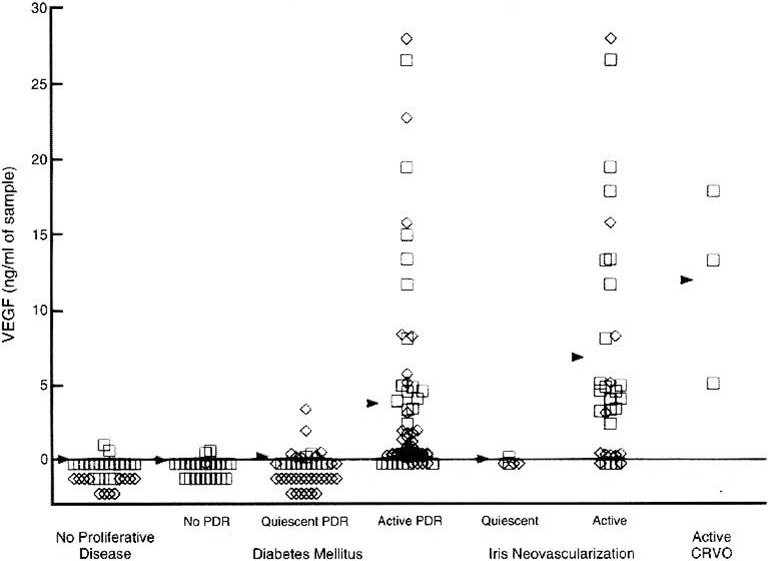
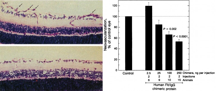
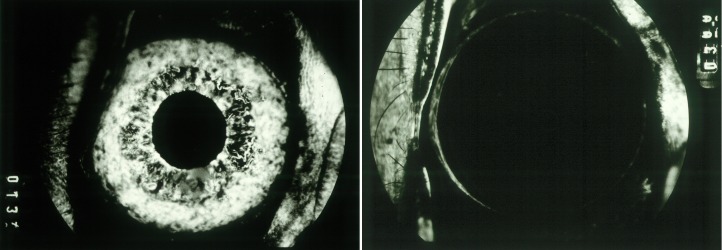
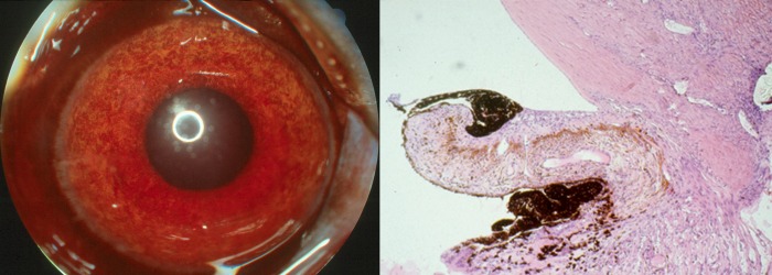
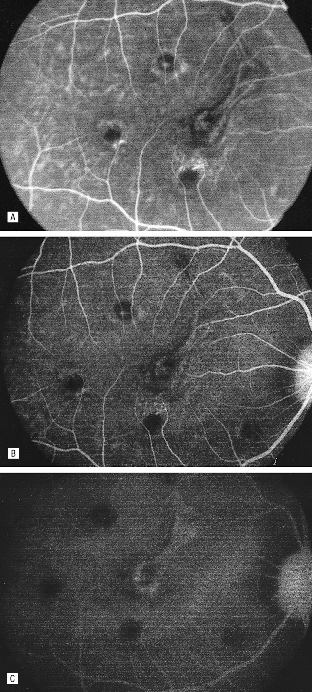
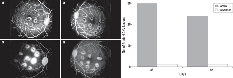
Results: The Champalimaud Laureates correlated VEGF with ocular neovascularization in animal models and in patients. Moreover, they showed that VEGF not only was sufficient, but it also was required to induce neovascularization in normal animal eyes, as VEGF inhibition abolished ocular neovascularization in key animal models.

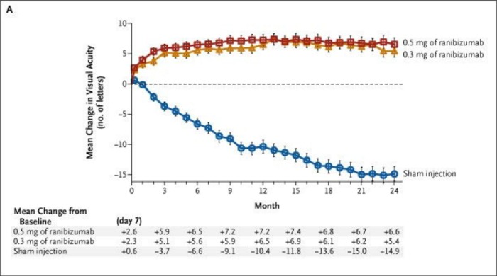
Conclusions: The identification of VEGF as Factor X altered the therapeutic paradigms for age-related macular degeneration (AMD), diabetic retinopathy, retinal vein occlusion, and other retinal disorders.

Translational relevance: The translation of VEGF from discovery to therapy resulted in the most successful applications of antiangiogenic therapy to date. Annually, over one million patients with eye disease are treated with anti-VEGF agents.

Keywords: António Champalimaud Vision Award; Factor X; age-related macular degeneration (AMD); angiogenesis; vascular endothelial growth factor (VEGF).


Figures

Figure 1
Figure 1
Researchers involved in the discovery of VEGF as Factor X and translation to therapy. Top row (left to right): Judah Folkman, Harold Dvorak, Napoleone Ferrara, Evangelos Gragoudas, Donald D'Amico, Lloyd Paul Aiello. Bottom row (left to right): George King, Lois Smith, Eric Pierce, Patricia D'Amore, David Shima, Anthony Adamis, and Joan Miller.
Figure 2
Figure 2
The search for Factor X. Left: Fluorescein angiogram of the retina in a patient with diabetic retinopathy (JWM patient seen at Massachusetts Eye and Ear). Black area represents nonperfused, ischemic retina. Center: Proposed action of unidentified angiogenic factor(s) in ocular neovascularization. Image courtesy of Anthony Adamis, MD; reproduced with permission. Right: Color photo of iris neovascularization in a patient with neovascular glaucoma (JWM patient seen at Massachusetts Eye and Ear).
Figure 3
Figure 3
Left: Human retinal pigmented epithelial (hRPE) cells hybridized in situ with a VEGF sense riboprobe (control), showing nonspecific cellular labeling and low background levels. Right: hRPE hybridized in situ with a VEGF antisense riboprobe, showing strong labeling of all hRPE cells and indicating VEGF expression. Reprinted with permission from Adamis AP, Shima DT, Yeo KT, et al. Synthesis and secretion of vascular permeability factor/vascular endothelial growth factor by human retinal pigment epithelial cells. Biochem Biophys Res Commun. 1993;193:631–638. Copyright 1993 Elsevier.
Figure 4
Figure 4
Northern analysis of total RNA extracted from rRPE cells grown under normoxic and hypoxic conditions for 6, 12, and 24 hours, probed for VEGF (upper), bFGF (middle), and 28S RNA (lower). Reproduced with permission from Shima DT, Adamis AP, Ferrara N, et al. Hypoxic induction of endothelial cell growth factors in retinal cells: identification and characterization of vascular endothelial growth factor (VEGF) as the mitogen. Mol Med. 1995;1:182–193.
Figure 5
Figure 5
Experimental iris neovascularization. (A) Fundus photograph immediately following laser vein occlusion. (B) Color photograph showing new vessels on the surface of the iris, which appear 4 to 7 days after laser vein occlusion. (C) Fluorescein angiography showing iris neovascularization with abundant leakage of fluorescein (grade 3). (D) Fundus photograph immediately following sham laser, aimed adjacent to the retinal vessels and producing retinal injury but preserving normal vasculature. (E) Color photograph of iris 12 days after sham laser, which appears normal. (F) Fluorescein angiography of the iris in (E), showing normal iris vessels with no fluorescein leakage (grade 0). Reproduced with permission from Miller JW, Adamis AP, Shima DT, et al. Vascular endothelial growth factor/vascular permeability factor is temporally and spatially correlated with ocular angiogensis in a primate model. Am J Pathol. 1994;145:574–584. Copyright 1994 Elsevier.
Figure 6
Figure 6
Correlation of VEGF levels in the aqueous (A) and grade of iris neovascularization (B) of four monkey eyes after laser vein occlusion. VEGF levels and neovascularization are represented as a scatterplot with best fit curves. Reproduced with permission from Miller JW, Adamis AP, Shima DT, et al. Vascular endothelial growth factor/vascular permeability factor is temporally and spatially correlated with ocular angiogensis in a primate model. Am J Pathol. 1994;145:574–584. Copyright 1994 Elsevier.
Figure 7
Figure 7
In situ localization of VEGF mRNA in ischemic retinas. Left: Cellular localization of VEGF mRNA expression by hybridization with an antisense VEGF riboprobe 13 days after laser vein occlusion. Right: Sense (control) riboprobe hybridized in 13-day ischemic retina. Adapted with permission from Shima DT, Gougos A, Miller JW, et al. Cloning and mRNA expression of vascular endothelial growth factor in ischemic retinas of Macaca fascicularis. Invest Ophthalmol Vis Sci. 1996;37:1334–1340.
Figure 8
Figure 8
VEGF concentrations in the aqueous (squares) and vitreous (diamonds) of patients undergoing intraocular procedures. Arrowheads indicate mean values. Reproduced with permission from Aiello LP, Avery RL, Arrigg PG, et al. Vascular endothelial growth factor in ocular fluid of patients with diabetic retinopathy and other retinal disorders. N Engl J Med. 1994;331:1480–1487. Copyright 1994 Massachusetts Medical Society.
Figure 9
Figure 9
Inhibition of neovascularization using soluble VEGF receptor-IgG chimeric proteins in a mouse model of retinal ischemia. Left: Retinal neovascularization extending into the vitreous is indicated by arrows in control-treated mice (upper panel), and absent in mice treated with human Flt-IgG chimeric proteins (lower panel). Right: Dose-dependent inhibition of retinal vascularization with human Flt-IgG chimeric proteins. Reproduced with permission from Aiello LP, Pierce EA, Foley ED, et al. Suppression of retinal neovascularization in vivo by inhibition of vascular endothelial growth factor (VEGF) using soluble VEGF-receptor chimeric proteins. Proc Natl Acad Sci U S A. 1995;92:10457–10461. Copyright 1995 National Academy of Sciences.
Figure 10
Figure 10
Inhibition of iris neovascularization using anti-VEGF antibody. Left: Early-phase fluorescein angiogram of an eye treated with anti-gp120 mAb (control), showing new iris vessels filling with fluorescein dye. Right: Early-phase fluorescein angiograms of the contralateral eye, treated with anti-VEGF mAb, showing no neovascularization. Reproduced with permission from Adamis AP, Shima DT, Tolentino MJ, et al. Inhibition of vascular endothelial growth factor prevents retinal ischemia-associated iris neovascularization in a nonhuman primate. Arch Ophthalmol. 1996;114:66–71. Copyright 1996 American Medical Association.
Figure 11
Figure 11
VEGF induces iris neovascularization and neovascular glaucoma. Left: Color iris photograph of an eye that received four 1.25-μg injections of VEGF, showing diffuse iris neovascularization and ectropion uveae (JWM image). Right: Histopathologic examination of the anterior chamber angle of an eye that received ten 1.25-μg injections of VEGF, showing a dense anterior fibrovascular membrane, ectropion uveae, and trabecular meshwork scarring. Reproduced with permission from Tolentino MJ, Miller JW, Gragoudas ES, Chatzistefanou K, Ferrara N, Adamis AP. Vascular endothelial growth factor is sufficient to produce iris neovascularization and neovascular glaucoma in a nonhuman primate. Arch Ophthalmol. 1996;114:964–970. Copyright 1996 American Medical Association.
Figure 12
Figure 12
Fluorescein angiogram of one eye after intravenous injection with fluoresceinated anti-VEGF antibody, demonstrating localization to experimental choroidal neovascularization. Hyperfluorescence noted 1 minute after injection (A), 20 minutes after injection (B), and with leakage noted one hour after injection. Reproduced with permission from Tolentino MJ, Husain D, Theodosiadis P, et al. Angiography of fluoresceinated anti-vascular endothelial growth factor antibody and dextrans in experimental choroidal neovascularization. Arch Ophthalmol. 2000;118:78–84. Copyright 2000 American Medical Association.
Figure 13
Figure 13
VEGF blockade prevents CNV. Left: Fluorescein angiogram of control (A, C) and prevention (B, D) eyes with laser-induced lesions. (A) Early frame of the control eye, which received intravitreal vehicle injection. (B) Early frame of the prevention eye, injected with humanized monoclonal anti-VEGF antibody (rhuFab VEGF). (C) Late frame of the control eye (vehicle). (D) Late frame of the prevention eye (rhuFab VEGF). Late frames demonstrate presence of grade 4 lesions in the control eye but not in the prevention eye. Right: Total number of grade 4 CNV lesions in the control group (shaded bars) versus prevention group (empty bar) 2 weeks (day 35) and 3 weeks (day 42) after laser induction. Injections of anti-VEGF or vehicle preceded laser induction of CNF. Reproduced with permission from Krzystolik MG, Afshari MA, Adamis AP, et al. Prevention of experimental choroidal neovascularization with intravitreal anti-vascular endothelial growth factor antibody fragment. Arch Ophthalmol. 2002;120:338–346. Copyright 2002 American Medical Association.
Figure 14
Figure 14
MARINA 2-year results: Mean changes in visual acuity from baseline to 24 months in patients injected with ranibizumab (0.3 and 0.5 mg monthly) versus sham injections. Reproduced with permission from Rosenfeld PJ, Brown DM, Heier JS, et al. Ranibizumab for neovascular age-related macular degeneration. N Engl J Med. 2006;355:1419–1431. Copyright 1996 Massachusetts Medical Society.
Figure 15
Figure 15
Neovascular AMD showing improved OCT after intravitreal injection of approximately 1.0 mg Avastin. (A) Baseline, (B) 1 week postinjection, (C) 4 weeks postinjection. Reproduced with permission from Rosenfeld PJ, Moshfeghi AA, Puliafito CA. Optical coherence tomography findings after an intravitreal injection of bevacizumab (Avastin) for neovascular age-related macular degeneration. Ophthalmic Surg Lasers Imaging. 2005;36:331–335. Copyright 2005 Slack, Inc.
Figure 16
Figure 16
OCT scans of a patient with RVO (JWM patient seen at Massachusetts Eye and Ear). The intraretinal cystic changes and subretinal fluid (left) completely resolved after treatment with anti-VEGF therapy (right).
Figure 17
Figure 17
VEGF-induced blood-retinal breakdown. Fluorescein angiogram shows dilation, tortuosity, and leakage of temporal retinal vessels, 4 days after a single injection of 1.25 μg VEGF mixed with nonneutralizing antibody. Reproduced with permission from Tolentino MJ, Miller JW, Gragoudas ES, et al. Intravitreous injections of vascular endothelial growth factor produce retinal ischemia and microangiopathy in an adult primate. Ophthalmology. 1996;103:1820–1828. Copyright 1996 Elsevier.
Figure 18
Figure 18
Left: Preretinal neovascularization in peripheral retina. Artery (“a”) and veins (“v”) in the peripheral retinal of an eye injected with VEGF. The artery displays tortuosity and dilation, with microaneurysmal-like formations at the levels of precapillary arterioles and capillaries. In the flat perspective (lower left), saccular neovascular buds obscure the parent veins (paired arrows). Right: Upper right shows a macula of a VEGF-injected eye with grotesquely dilated and saccular capillaries and veins. White arrowhead indicates microaneurysmal-like structure, and black arrow indicates arteriole. FAZ, foveal avascular zone. Lower: Section taken through the microaneurysmal-like structure (indicated by white arrowhead in the upper panel), showing endothelial cell hyperplasia (black arrowhead) and basement membrane material occluding the lumen. Reproduced with permission from Tolentino MJ, McLeod DS, Taomoto M, Otsuji T, Adamis AP, Lutty GA. Pathologic features of vascular endothelial growth factor-induced retinopathy in the nonhuman primate. Am J Ophthalmol. 2002;133:373–385. Copyright 2002 Elsevier.

References

    1. Senger DR,, Galli SJ,, Dvorak AM,, Perruzzi CA,, Harvey VS,, Dvorak HF. Tumor cells secrete a vascular permeability factor that promotes accumulation of ascites fluid. Science. 1983; 219: 983–985. - PubMed
    1. Leung DW,, Cachianes G,, Kuang WJ,, Goeddel DV,, Ferrara N. Vascular endothelial growth factor is a secreted angiogenic mitogen. Science. 1989; 246: 1306–1309. - PubMed
    1. Michaelson IC. The mode of development of the vascular system of the retina with some observations on its significance for certain retinal diseases. Trans Ophthalmol Soc U K. 1948; 68: 137–180.
    1. Gimbrone MA,, Jr,, Leapman SB,, Cotran RS,, Folkman J. Tumor dormancy in vivo by prevention of neovascularization. J Exp Med. 1972; 136: 261–276. - PMC - PubMed
    1. Folkman J. Tumor angiogenesis: therapeutic implications. N Engl J Med. 1971; 285: 1182–1186. - PubMed