. 2015 Jul-Dec;5(2):148-57.
doi: 10.4103/2231-0746.175778.

Perpendicular serial maxillary distraction osteogenesis in cleft lip and palate patients

Affiliations

Perpendicular serial maxillary distraction osteogenesis in cleft lip and palate patients

Leena P Ylikontiola et al. Ann Maxillofac Surg. 2015 Jul-Dec.

Abstract

Background: Cleft lip and palate patients often have a retruded maxilla with a severely narrowed deficient maxillary arch. This report aims to describe the management of severe maxillary retrusion and constriction in cleft lip and palate patients using distraction osteogenesis applied in serial sequence in two directions perpendicular to each other.

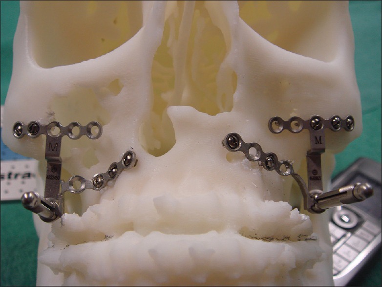
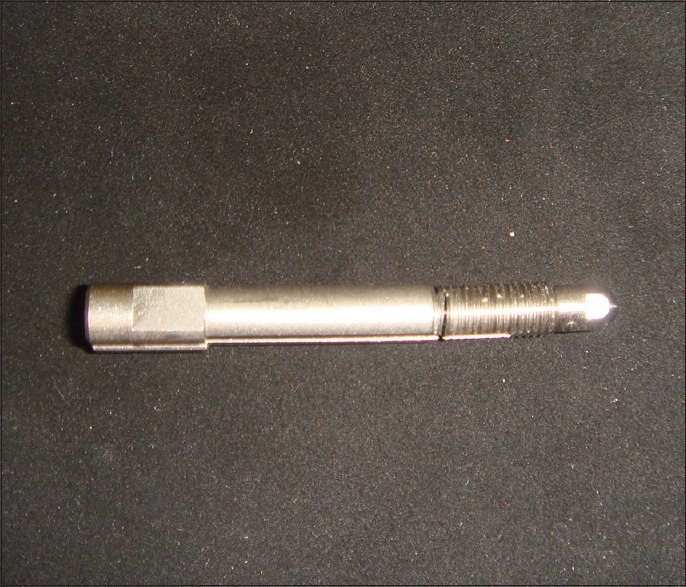
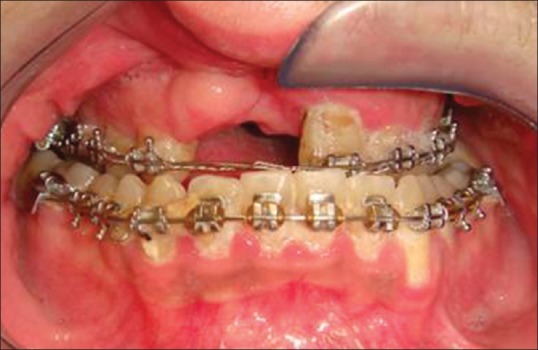
Materials and methods: Two adult male cleft lip and palate patients were treated with maxillary distraction osteogenesis in two stages. In the first stage, surgically assisted rapid palatal expansion with a tooth-borne device was performed to significantly expand the maxillary arch in the transverse dimension. After the teeth were orthodontically aligned, the horizontal distraction of the maxilla was made by two internal maxillary distraction devices.

Results: In the first patient, the maxilla was initially widened by 11 mm and then distracted forward by 20 mm. Despite the breakage of the shaft of one of the two distractors at the end of distraction, a satisfactory occlusion was found at the time of distractor device removal. The maxillary position has remained stable through 8 years of follow-up. In the second patient, the palate was widened by 14 mm and the maxilla was distracted forward by 22 mm. The maxillary position has remained stable through 3 years of follow-up.

Conclusion: Sequential serial distraction of maxilla in two planes perpendicular to each other is a safe and stable approach for the treatment of cleft lip and palate patients with severe transverse and anteroposterior discrepancies.

Keywords: Cleft lip and palate; maxillary distraction osteogenesis; palate expansion.


Figures

Figure 1
Figure 1
Preoperative frontal photograph of case 1
Figure 2
Figure 2
Preoperative lateral photograph of patient
Figure 3
Figure 3
Preoperative photograph of severely malaligned and malpositioned teeth with severe transverse maxillary deficiency
Figure 4
Figure 4
Preoperative left lateral buccal segment view of occlusion
Figure 5
Figure 5
Preoperative occlusal view showing severely constricted maxillary arch
Figure 6
Figure 6
Maxillary tooth-borne device being activated to initiate transverse maxillary distraction
Figure 7
Figure 7
Occlusal radiograph taken at the start of maxillary transverse distraction
Figure 8
Figure 8
Occlusal radiograph taken at the end of maxillary transverse distraction. Note the large diastemma that has been produced in the midline
Figure 9
Figure 9
Maxillary occlusal view photograph showing teeth being aligned in the maxilla following its transverse distraction
Figure 10
Figure 10
Photograph showing retrusive maxilla prepared for advancement
Figure 11
Figure 11
Lateral cephalometric radiograph just prior to maxillary anterior distraction
Figure 12
Figure 12
Maxillary distractor footplates are prebent and fitted and secured with screws to stereolithic skull of patient
Figure 13
Figure 13
Alignment rods are attached to the distractors installed on stereolithic skull to check alignment
Figure 14
Figure 14
Maxillary distractors fitted at time of Le Fort I osteotomy
Figure 15
Figure 15
Postoperative lateral cephalometric radiograph showing maxillary distractors at the beginning of distraction
Figure 16
Figure 16
Maxilla distracted into its new occlusal relationship. The distractors are kept in place for 3 months following the attainment of the final maxillary position as retention devices
Figure 17
Figure 17
Lateral cephalometric radiograph taken after distractor removal and replacement by 4 L-shaped internal fixation plates
Figure 18
Figure 18
Broken distractor rod with a crack in its shaft noted at the time of distractor removal. This breakage seemed to have no influence on the distraction outcome
Figure 19
Figure 19
Postoperative frontal photograph of patient
Figure 20
Figure 20
Postoperative anterior view of occlusion with class I molar relationship and no cross-bite
Figure 21
Figure 21
Postoperative right lateral occlusal view
Figure 22
Figure 22
Postoperative left lateral occlusal view
Figure 23
Figure 23
Preoperative frontal photograph of case 2
Figure 24
Figure 24
Preoperative anterior view of occlusion of constricted and retrusive maxilla after preliminary orthodontic alignment
Figure 25
Figure 25
Right lateral view of buccal segment showing severely retrusive maxilla and complete cross-bite
Figure 26
Figure 26
Left lateral view of buccal segment showing severely retrusive maxilla and complete cross-bite
Figure 27
Figure 27
Anterior view of occlusion after transverse distraction of maxilla increasing the palatal width by 14 mm
Figure 28
Figure 28
Predistraction lateral cephalometric radiograph showing severe maxillary retrusion
Figure 29
Figure 29
Lateral cephalometric radiograph showing maxilla with distraction devices and maxilla partway advanced
Figure 30
Figure 30
Dental implant being placed into generously wide anterior maxillary ridge left by the wake of bone produced by maxillary transverse distraction procedure without the need for bone grafting or augmentation of the ridge
Figure 31
Figure 31
Postretention lateral cephalometric radiograph with distractors having been removed 3 months following the completion of distraction. Internal fixation devices have been applied for long-term retention, and dental implants are in place with anterior incisal alignment normalized
Figure 32
Figure 32
Posttreatment panoramic radiograph showing restored dental implants in position
Figure 33
Figure 33
Postoperative frontal facial photograph of case 2 showing improved facial esthetics with new position of anteriorly distracted maxilla with improved subnasal support
Figure 34
Figure 34
Postoperative anterior view of the occlusion and dental implant restorations
Figure 35
Figure 35
Postoperative right lateral buccal segment view of occlusion with no cross-bite
Figure 36
Figure 36
Postoperative left lateral buccal segment view of occlusion with no cross-bite

References

    1. Harila V, Ylikontiola LP, Palola R, Sándor GK. Maxillary arch dimensions in cleft infants in Northern Finland. Acta Odontol Scand. 2013;71:930–6. - PubMed
    1. Harila V, Ylikontiola LP, Sándor GK. Dental arch relationships assessed by GOSLON Yardstick in children with clefts in Northern Finland. Eur J Paediatr Dent. 2014;15:389–91. - PubMed
    1. Molina F. Distraction osteogenesis for the cleft lip and palate patient. Clin Plast Surg. 2004;31:291–302. - PubMed
    1. Sándor GK, Ylikontiola LP, Serlo W, Pirttiniemi PM, Carmichael RP. Midfacial distraction osteogenesis. Atlas Oral Maxillofac Surg Clin North Am. 2008;16:249–72. - PubMed
    1. Balaji SM. History of craniofacial distraction osteogenesis. Abstracts of the Second Asia Pacific Congress on Distraction Osteogenesis. Bandos, Maldives. 2003:1–9.