A case of laryngeal leiomyosarcoma and review of the literature
- PMID: 26981488
- PMCID: PMC4772578
- DOI: 10.4103/2231-0746.175772
A case of laryngeal leiomyosarcoma and review of the literature
Abstract
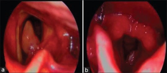
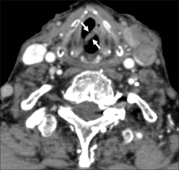
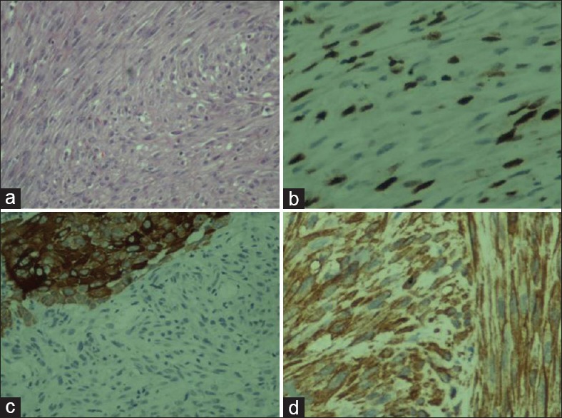
Leiomyosarcoma (LMS) of the larynx is a very rare malignancy that originates from blood vessel, smooth muscle or from the heterotopic mesenchymal tissue in the larynx. The histological diagnosis of LMS depends on the immunohistochemical investigation. The case is here presented of an 82-year-old man with shortness of breath and hoarseness. Indirect laryngoscopy showed a pedunculated large glottic lesion causing airway obstruction. Direct laryngoscopy was performed and biopsies were taken. From the pathological examination, the diagnosis of LMS was made. This case is presented of laryngeal LMS with the clinical, radiological, and histological findings.
Keywords: Glottis; laryngoscopy; larynx; leiomyosarcoma.
Figures
References
-
- Skoulakis CE, Stavroulaki P, Moschotzopoulos P, Paxinos M, Fericean A, Valagiannis DE. Laryngeal leiomyosarcoma: A case report and review of the literature. Eur Arch Otorhinolaryngol. 2006;263:929–34. - PubMed
-
- Tran LM, Mark R, Meier R, Calcaterra TC, Parker RG. Sarcomas of the head and neck. Prognostic factors and treatment strategies. Cancer. 1992;70:169–77. - PubMed
-
- Morera Serna E, Pérez Fernández CA, de la Fuente Jambrina C, Razquin Muñoz J, Pérez Gil MA. Laryngeal leiomyosarcoma. Acta Otorrinolaringol Esp. 2007;58:445–8. - PubMed
Publication types
LinkOut - more resources
Full Text Sources
Other Literature Sources