Review
. 2016 Mar 15;44(3):450-462.
doi: 10.1016/j.immuni.2016.02.015.

Macrophages in Tissue Repair, Regeneration, and Fibrosis

Affiliations
Review

Macrophages in Tissue Repair, Regeneration, and Fibrosis

Thomas A Wynn et al. Immunity. .

Abstract

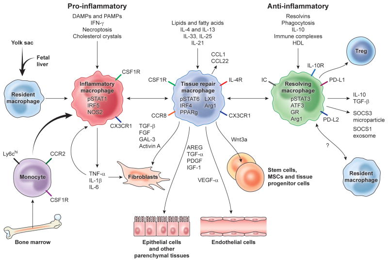
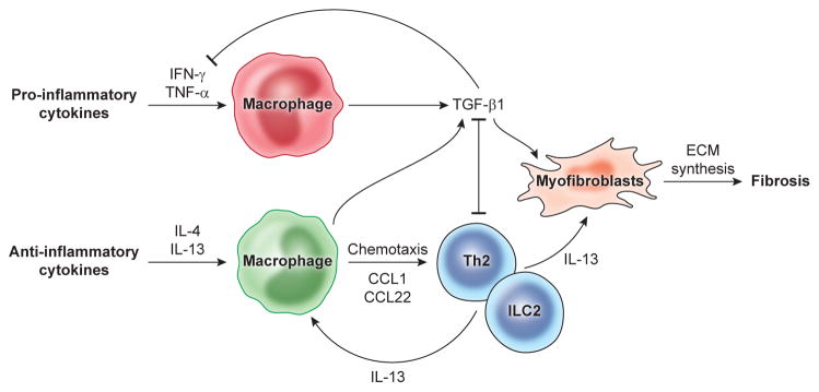
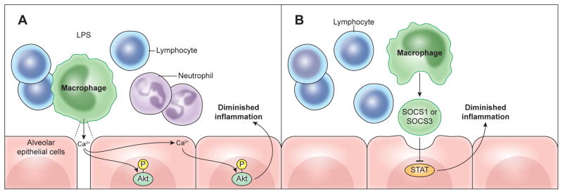
Inflammatory monocytes and tissue-resident macrophages are key regulators of tissue repair, regeneration, and fibrosis. After tissue injury, monocytes and macrophages undergo marked phenotypic and functional changes to play critical roles during the initiation, maintenance, and resolution phases of tissue repair. Disturbances in macrophage function can lead to aberrant repair, such that uncontrolled production of inflammatory mediators and growth factors, deficient generation of anti-inflammatory macrophages, or failed communication between macrophages and epithelial cells, endothelial cells, fibroblasts, and stem or tissue progenitor cells all contribute to a state of persistent injury, and this could lead to the development of pathological fibrosis. In this review, we discuss the mechanisms that instruct macrophages to adopt pro-inflammatory, pro-wound-healing, pro-fibrotic, anti-inflammatory, anti-fibrotic, pro-resolving, and tissue-regenerating phenotypes after injury, and we highlight how some of these mechanisms and macrophage activation states could be exploited therapeutically.


Figures

Fig. 1
Fig. 1. Mechanisms driving major macrophage activation phenotypes in tissue repair, regeneration, and fibrosis
In many tissues the resident tissue macrophage population is derived from the yolk sac and fetal liver during development but are complimented by inflammatory monocytes recruited from the bone marrow following injury. The recruited and resident macrophages undergo marked phenotypic and functional changes in response to DAMPs, PAMPs, growth factors, cytokines, and other mediators released in the local tissue microenvironment, with the dominant phenotypic variants depicted here regulating inflammation, tissue repair, regeneration, and resolution. Macrophages produce a variety of factors that stimulate the proliferation, differentiation, and activation of fibroblasts, epithelial cells, endothelial cells, and stem and progenitor cells that facilitate tissue repair. During the later stages of the repair process, they assume a regulatory pro-resolving phenotype that ensures the tissue damaging inflammatory response is suppressed and normal tissue architecture is restored. If the process is not controlled effectively, persistent inflammation and/or maladaptive repair processes can lead to tissue destructive fibrosis. In some cases, the recruited monocytes seed the tissues and adopt a resident macrophage phenotype, however the mechanisms that restore tissue homeostasis are still under debate. Damage associated molecular patterns (DAMPs), PAMPs (pathogen associated molecular patterns), Regulatory T cells (Treg), interferon-regulatory factor 5 (IRF5), nitric oxide synthase 2 (NOS2), Liver X receptor (LXR), Amphiregulin (AREG), Arginase-1 (Arg1), interferon regulatory factor 4 (IRF4), peroxisome proliferator-activated receptor gamma (PPARγ), fibroblast growth factor (FGF), galectin-3 (GAL-3), transforming growth factor (TGF), Immune complex (IC), glucocorticoid receptor (GR), transcription factor ATF3, silencers of cytokine signaling (SOCS).
Fig. 2
Fig. 2. Cytokine and macrophage-mediated mechanisms of fibrosis
Inflammatory responses that develop following tissue injury are associated with the production of a variety of inflammatory mediators like IFN-γ and TNF-α that promote classical macrophage activation. TGF-β1 is then produced by macrophages as a regulatory feedback mechanism to facilitate the resolution of the pro-inflammatory response. TGF-β1 also triggers fibroblast activation and development of ECM-producing myofibroblasts that facilitate repair and drive fibrosis. The anti-inflammatory cytokine IL-13, produced by a variety of cell types including type 2 innate lymphoid cells, eosinophils, basophils, and CD4+ Th2 cells, also serves as a major driver of tissue repair and fibrosis by inducing TGF-β1, by directly targeting myofibroblast function, and by promoting the development of restorative M(IL-4)-like macrophages that facilitate the recruitment of IL-13-producing leukocytes to sites of tissue injury.
Fig. 3
Fig. 3. Anti-inflammatory signaling by macrophages
Alveolar macrophages transmit key signals to neighboring cells that help facilitate the resolution of inflammation in the lung. A) After LPS inhalation, a specialized population of sessile alveolar macrophages attached to the alveolar wall form gap junction channels with the epithelium, which suppress inflammation by transmitting Ca2+ waves to epithelial cells that result in the phosphorylation of the pro-survival kinase Akt. B) Alveolar macrophages can also dampen inflammation by secreting SOCS1 or SOCS3 proteins in vesicles that are taken up by alveolar epithelial cells. Delivery of SOCS subsequently inhibits epithelial STAT activation, leading to the suppression of pro-inflammatory mediator production.

References

    1. Alexander KA, Flynn R, Lineburg KE, Kuns RD, Teal BE, Olver SD, Lor M, Raffelt NC, Koyama M, Leveque L, et al. CSF-1-dependant donor-derived macrophages mediate chronic graft-versus-host disease. J Clin Invest. 2014;124:4266–4280. - PMC - PubMed
    1. Antoniades CG, Quaglia A, Taams LS, Mitry RR, Hussain M, Abeles R, Possamai LA, Bruce M, McPhail M, Starling C, et al. Source and characterization of hepatic macrophages in acetaminophen-induced acute liver failure in humans. Hepatology. 2012;56:735–746. - PubMed
    1. Aurora AB, Porrello ER, Tan W, Mahmoud AI, Hill JA, Bassel-Duby R, Sadek HA, Olson EN. Macrophages are required for neonatal heart regeneration. J Clin Invest. 2014;124:1382–1392. - PMC - PubMed
    1. Baeck C, Wehr A, Karlmark KR, Heymann F, Vucur M, Gassler N, Huss S, Klussmann S, Eulberg D, Luedde T, et al. Pharmacological inhibition of the chemokine CCL2 (MCP-1) diminishes liver macrophage infiltration and steatohepatitis in chronic hepatic injury. Gut. 2012;61:416–426. - PubMed
    1. Baeck C, Wei X, Bartneck M, Fech V, Heymann F, Gassler N, Hittatiya K, Eulberg D, Luedde T, Trautwein C, Tacke F. Pharmacological inhibition of the chemokine C-C motif chemokine ligand 2 (monocyte chemoattractant protein 1) accelerates liver fibrosis regression by suppressing Ly-6C(+) macrophage infiltration in mice. Hepatology. 2014;59:1060–1072. - PubMed

Publication types