. 2016 Apr 5;55(13):1997-2007.
doi: 10.1021/acs.biochem.5b01330. Epub 2016 Mar 24.

Coumarin Derivatives as Substrate Probes of Mammalian Cytochromes P450 2B4 and 2B6: Assessing the Importance of 7-Alkoxy Chain Length, Halogen Substitution, and Non-Active Site Mutations

Affiliations

Coumarin Derivatives as Substrate Probes of Mammalian Cytochromes P450 2B4 and 2B6: Assessing the Importance of 7-Alkoxy Chain Length, Halogen Substitution, and Non-Active Site Mutations

Jingbao Liu et al. Biochemistry. .

Abstract

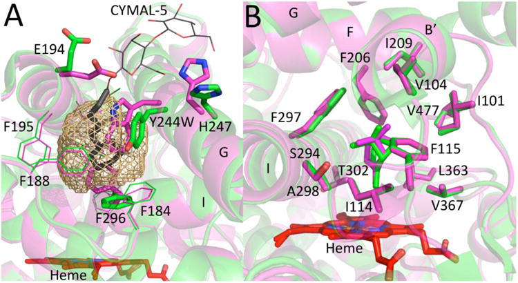
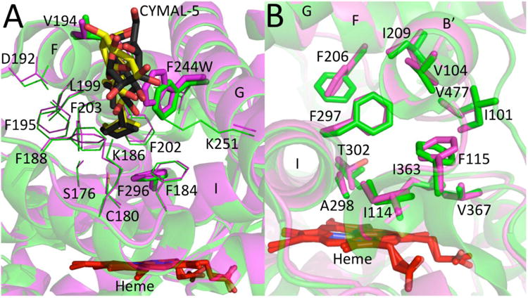
Using a combined structural and biochemical approach, the functional importance of a recently described peripheral pocket bounded by the E-, F-, G-, and I-helices in CYP2B4 and 2B6 was probed. Three series of 4-substituted-7-alkoxycoumarin derivatives with -H, -CH3, or -CF3 at the 4 position of the coumarin core were used initially to monitor functional differences between CYP2B4 and 2B6. 7-Ethoxy-4-(trifluoromethyl)coumarin (7-EFC) displayed the highest catalytic efficiency among these substrates. Mutants were made to alter side-chain polarity (V/E194Q) or bulk (F/Y244W) to alter access to the peripheral pocket. Modest increases in catalytic efficiency of 7-EFC O-deethylation by the mutants were magnified considerably by chlorination or bromination of the substrate ethoxy chain. A structure of CYP2B6 Y244W in complex with (+)-α-pinene was solved at 2.2 Å and showed no CYMAL-5 in the peripheral pocket. A ligand free structure of CYP2B4 F244W was solved at 3.0 Å with CYMAL-5 in the peripheral pocket. In both instances, comparison of the respective wild-type and mutant CYP2B enzymes revealed that CYMAL-5 occupancy of the peripheral pocket had little effect on the topology of active site residue side-chains, despite the fact that the peripheral pocket and active site are located on opposite sides of the I-helix. Analysis of available CYP2B structures suggest that the effect of the amino acid substitutions within the peripheral pocket derive from altered interactions between the F and G helices.


Figures

Figure 1
Figure 1
Observed peripheral pocket in CYP2B enzymes. Cavities found in the A) CYP2B4-paroxetine complex (4JLT) and the B) CYP2B6-(+)-α-pinene complex (4I91) using Mole 2.0 are depicted as surfaces, the protein backbone is shown as a gray ribbon, and the solvent accessible surface of the cavity is in green. In both cases, the active site, labeled A, is physically separated from the peripheral pocket, labeled P, by the I-helix (cyan).
Figure 2
Figure 2
Chemical structure of 4-substituted-7-alkoxycoumarins.
Figure 3
Figure 3
Views of CYP2B6 WT and CYP2B6 Y244W complexes with (+)-α-pinene. A) Overlay of the residues in sticks lining the peripheral pocket (brown mesh) of the CYP2B6-(+)-α-pinene (green) complex and the CYP2B6 Y244W-(+)-α-pinene (magenta) complex. The CYMAL-5 molecule observed in CYP2B6 peripheral pocket is shown in black sticks. For visual purposes, the sugar moiety of CYMAL-5 that extends to the protein surface is shown in thin lines. The side chains of residues 194, 244, and 296 shown in sticks demonstrated significant differences in orientation in the peripheral pocket. B) Overlay of the residues in sticks lining the active-sites of CYP2B6-(+)-α-pinene (green) and CYP2B6-Y244W-(+)-α-pinene (magenta) within a 5 Å radius of the ligand. The heme is shown in red sticks. Respective (+)-α-pinene molecules in the active site and side chain F297 on the I-helix are represented in sticks.
Figure 4
Figure 4
Views of CYP2B4 WT and CYP2B4 F244W structures. A) Overlay of the residues lining the peripheral pockets of ligand-free CYP2B4 (green) and ligand-free CYP2B4 F244W (magenta). CYMAL-5 molecules observed in CYP2B4 and CYP2B4 F244W peripheral pocket are shown in gray and yellow sticks, respectively. The sugar moiety of CYMAL-5 is not clearly visible due to its alternate orientation towards the viewer. Residues show little or no difference in orientation between the two CYP2B4 structures in the presence of CYMAL-5 in the peripheral pocket. The side chain of residues 194, 244, and 296 that demonstrated difference in CYP2B6 complex with and without CYMAL-5 are shown in sticks. B) Overlay of the residues in sticks of the active-sites of ligand-free CYP2B4 (green) and ligand-free CYP2B4-F244W (magenta). The heme is shown in red sticks.
Figure 5
Figure 5
Views comparing orientations of residues 194, 202, 241, and 244 in CYP2B4 and CYP2B6. A) Overlay of the CYP2B4-4-CPI complex (magenta, 1SUO) and the CYP2B4-amlodipine complex (yellow, 3TMZ). B) Overlay of the CYP2B6-4-CPI complex (cyan, 3IBD) and the CYP2B6-amlodipine complex (orange, 3UA5). Residues 194, 202, 241, and 244 are shown in sticks. The phenyl ring of F202 and δ-carbon of I241 are within 4 Å of each other in each structure.

Similar articles

Cited by

References

    1. Rydberg P, Vasanthanathan P, Oostenbrink C, Olsen L. Fast prediction of cytochrome P450 mediated drug metabolism. ChemMedChem. 2009;4:2070–2079. - PubMed
    1. Rydberg P, Olsen L. Predicting drug metabolism by cytochrome P450 2C9: comparison with the 2D6 and 3A4 isoforms. ChemMedChem. 2012;7:1202–1209. - PubMed
    1. Halpert JR. Structure and function of cytochromes P450 2B: from mechanism-based inactivators to X-ray crystal structures and back. Drug Metab Dispos. 2011;39:1113–1121. - PMC - PubMed
    1. Johnson EF, Connick JP, Reed JR, Backes WL, Desai MC, Xu L, Estrada DF, Laurence JS, Scott EE. Correlating structure and function of drug-metabolizing enzymes: progress and ongoing challenges. Drug Metab Dispos. 2014;42:9–22. - PMC - PubMed
    1. Anzenbacher P, Anzenbacherova E, Lange R, Skopalik J, Otyepka M. Active sites of cytochromes P450: What are they like? Acta Chim Slov. 2008;55:63–66.

Publication types

MeSH terms

LinkOut - more resources