

doi: 10.3201/eid2204.150653.
Neisseria meningitidis Serogroup X in Sub-Saharan Africa
- PMID: 26982628
- PMCID: PMC4806945
- DOI: 10.3201/eid2204.150653
Item in Clipboard
Neisseria meningitidis Serogroup X in Sub-Saharan Africa
Emerg Infect Dis.
2016 Apr.
Abstract
The epidemiology of meningococcal disease varies by geography and time. Whole-genome sequencing of Neisseria meningitidis serogroup X isolates from sub-Saharan Africa and Europe showed that serogroup X emergence in sub-Saharan Africa resulted from expansion of particular variants within clonal complex 181. Virulence of these isolates in experimental mouse models was high.
Keywords: Neisseria meningitidis; bacteria; emergence; meningitis; meningococci; serogroup X; sub-Saharan Africa; virulence; whole-genome sequencing.
Figures
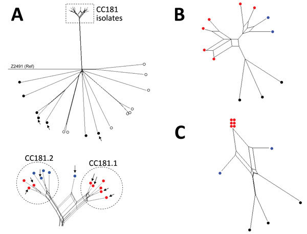
Neighbor-Net SplitsTree graphs generated using SplitsTree4 version 4.13.1 (http://www.splitstree.org ) to visualize trees of Neisseria meningitidis serogroup X isolates. A) All 30 Neisseria meningitidis serogroup X isolates available on BIGSdb (11) were analyzed, including the 11 isolates from this study (8 from sub-Saharan Africa and 3 from France), 9 carriage isolates, 3 invasive isolates from Europe, 1 isolate from the United States, and 6 isolates from sub-Saharan African countries. Open (white) circles indicate carriage isolates; black circles indicate invasive isolates obtained outside of Africa; red circles indicate isolates obtained in sub-Saharan Africa since 2006; blue circles indicate isolates obtained from sub-Saharan Africa during the 1990s. Arrows indicate the 11 isolates obtained during this study. All isolates were compared with reference (Ref) meningococcal strain Z2491. The dashed rectangle indicates the cluster of all the clonal complex (CC) 181 isolates from sub-Saharan Africa (enlarged view at bottom of panel). B) The 11 isolates obtained during this study were compared for iron-acquisition genes (tpbA, tbpB, hpuA, hpuB, lbpA, lbpB, and hmbR). C) Genes of the 11 isolates obtained during this study compared with the 41 genes that differed between all the isolates obtained in Africa since 2006 and the isolate LNP13407 or the isolate LNP14354 (obtained during the 1990s).
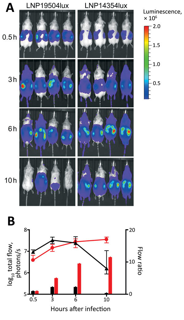
Dynamic imaging showing the multiplication and spread of Neisseria meningitidis in BALB/c transgenic mice expressing the human transferrin. A) Dorsal views of 8 mice (4/group) analyzed for bioluminescence at intervals after infection, as shown on left. Mice were infected by intraperitoneal injection of 5 × 10⁶ CFU of N. meningitidis strain LNP19504lux (derived from an isolate from France) or LNP14354lux (derived from an isolate from Africa). Both strains expressed the luciferase (lux) operon. Photographs are overlaid with color representations of luminescence intensity, measured in total photons per second and indicated on the scales: red, most intense (2.00 × 10⁶ p/s/cm2/steradian); blue, least intense (1.00 × 10⁴ p/s/cm2/steradian). B) Luminescence for LNP19504lux (black) and LNP14354lux (red) was quantified and expressed by using GraphPad PRISM version 4.00 (http://www.graphpad.com/ ). Means ± 95% CIs of total photons per second (lines, left y-axis) were calculated by defining the specific representative region of interest encompassing the entire animal. Signals differed significantly between the 2 groups after 10 h of infection (p = 0.01 by t-test). After 24 h, all mice had survived and signals declined for both isolates but remained detectable for LNP14354lux (not shown). The data are also expressed as flow ratio (total photons per second for each point/total photons at the first point [after 0.5 h of infection]) (bars, right y-axis).
References
-
- World Health Organization. Meningococcal disease control in countries of the African meningitis belt, 2013. Wkly Epidemiol Rec. 2014;89:206–14. - PubMed
-
- Etienne J, Sperber G, Adamou A, Picq JJ. Epidemiological notes: meningococcal meningitis of serogroup X in Niamey (Niger) [in French]. Med Trop (Mars). 1990;50:227–9. - PubMed
Publication types
MeSH terms
Substances
Grants and funding
LinkOut - more resources
Full Text Sources
Other Literature Sources
