Case Reports
. 2016 Mar 16;11(3):e0149619.
doi: 10.1371/journal.pone.0149619. eCollection 2016.

Structural Abnormalities in the Hair of a Patient with a Novel Ribosomopathy

Affiliations
Case Reports

Structural Abnormalities in the Hair of a Patient with a Novel Ribosomopathy

Richard J Alsop et al. PLoS One. .

Abstract

We report the biophysical characterization of hair from a patient with a de novo ribosomopathy. The patient was diagnosed with a mutation on gene RPS23, which codes for a protein which comprises part of the 40S subunit of the ribosome. The patient presents with a number of phenotypes, including hypotonia, autism, extra teeth, elastic skin, and thin/brittle hair. We combined optical microscopy, tensile tests, and X-ray diffraction experiments on hair samples obtained from the scalp of the patient to a multi-scale characterization of the hair from macroscopic to molecular length scales and observe distinct differences in the biophysical properties in the patient's hair when compared to hair from other family members. While no differences were observed in the coiled-coil structure of the keratin proteins or the structure of the intermediate filaments, the patient's hair was 22% thinner, while the Young's modulus remained roughly constant. The X-ray diffraction results give evidence that the amount of lipids in the cell membrane complex is reduced by 20%, which well accounts for the other observations. The pathologies characterized by these techniques may be used to inform the diagnosis of similar de novo mutations in the future.


Conflict of interest statement

Competing Interests: The authors have declared that no competing interests exist.

Figures

Fig 1
Fig 1. Illustration of the structure of hair.
a) Sketch of a human hair showing the three main regions, the medulla, the cuticle, and the cortex. b) Molecular cartoons of hair structures: keratin coiled-coil dimers and lipids in the cell membrane complex. c) A sample 2-dimensional X-ray diffraction image, indicating the assignment of structures to signals, as outlined in the Results. d) Optical microscopy images of the hair surface of all persons involved in this study.
Fig 2
Fig 2. Results of microscopy and hair experiments.
a) The average diameter of the hair samples. Diameters were measured in the centre of the hair fibre. Ten hairs were measured per person and averaged. b) The Young’s modulus of hair from different individuals, measured from tensile tests. The modulus from three hairs were measured and averaged (with the exception of S3). Note that hairs from F were too short to measure with the setup used. The inset in b) shows a typical stress-strain curve. Error bars in both plots represent the standard deviation within the measurements.
Fig 3
Fig 3. Two-dimensional X-ray diffraction images from all hair samples.
The hair strands were oriented with the long axis of the hair parallel with the vertical z-axis. The displayed (qz, q||) range covered length scales from 3 Å up to 250 Å, to study the coiled-coil α-keratin phase, as well as the membrane layer in the cortex [8, 9]. The features observed are common among the individuals in this study, and agree with previous reports.
Fig 4
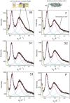
Fig 4. High resolution scans were measured from 0.2 Å−1 < q|| < 3.0 Å−1.
Five peaks describe these scans, with an exponential background: i) a peak at 45 Å (yellow), assigned to keratin bundles and observed in SAXS profiles (Fig 6); ii) A peak at 9.8 Å, assigned to lateral packing of keratin coiled-coils; iii) A lipid peak at 4.5 Å; iv) A peak at 3.5 Å, assigned to amorphous protein; and v) A second lipid peak at 2.3 Å. No significant differences are observed in peak position or width, as listed in Table A in S1 File [8, 9, 19].
Fig 5
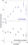
Fig 5. Angular distribution of scattering at the lipid and amorphous position.
Peaks observed at a) Q = 1.40 Å−1 and b) Q = 1.80 Å−1 were integrated as a function of azimuth angle ϕ from the q|| axis, for subject M. While the peak at Q = 1.80 Å−1 is isotropic in ϕ, the peak at Q = 1.40 Å−1 is anisotropically distributed with a maximum in intensity at ϕ = 90°.
Fig 6
Fig 6. Small angle X-ray scattering profiles, covering length scales of 21 Å up to 250 Å.
Peaks at 90 Å, 45 Å, and 27 Å are assigned to the structure of keratin bundles. Cartoons depict the structure of the keratin bundles: A hexagonal packing of keratin dimers for hairs from F, S1, S2, S3, and P, and a hexagonal packing of keratin monomers for M. The difference in packing between M and the others likely comes from the use of hair dyes or perming [4, 9, 20].
Fig 7
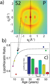
Fig 7. Analysis of diffraction images.
a) A split-image, with the right showing the 2-dimensional pattern from P and the left showing the pattern from S2. Qualitative differences in the relative intensity of the keratin peak at 9.8 Å are visible. The keratin and lipid peaks were integrated from 0 < ϕ < 45° (where ϕ is an angle defined from the q|| axis), and from 0.55 Å−1 < Q < 0.8 Å−1 or 1.0 Å−1 < Q < 2.5 Å−1, respectively. b) The lipid/keratin ratio of intensities is shown as a function of age and c) identified by each person. The dashed-black line in b) is not a fit, but instead a guide derived from past reports on changes in lipid content in hair [14, 32, 33].
Fig 8
Fig 8. A diagram illustrating how the cross-sectional diameter of the hair is obtained.
Hair samples are imaged on the Celestron 10X microscope. Using ImageJ, the intensity profile tangential to the hair axis is obtained. The width of the hair is determined from the width of the intensity profile across the hair.
Fig 9
Fig 9. A description of the stress-strain curves obtained from tensile tests of the hair samples.
1) An initial linear regime is observed, characterized by elastic stretching of the keratin proteins in the hair; ii) A plateau region is observed, when the coiled-coil α-keratin is pulled into a β-sheet configuration; and 3) A second linear regime is observed up to the point of hair fracture [–18, 38, 39].
Fig 10
Fig 10. A diagram illustrating the X-ray diffraction setup for measuring the hair fibres.
The hairs were cut to a length of 3 cm, and ∼50 such hairs were placed on an aluminum mount with a cut-out. The mount was fixed to the X-ray stage with sticky tack.

References

    1. Lewis R. Exome Sequencing Comes to the Clinic. JAMA. 2015;313(13):1301–1303. 10.1001/jama.2015.1389 - DOI - PubMed
    1. Lee H, Deignan JL, Dorrani N, Strom SP, Kantarci S, Quintero-Rivera F, et al. Clinical exome sequencing for genetic identification of rare Mendelian disorders. JAMA. 2014;312(18):1880–1887. 10.1001/jama.2014.14604 - DOI - PMC - PubMed
    1. Soden SE, Saunders CJ, Willig LK, Farrow EG, Smith LD, Petrikin JE, et al. Effectiveness of exome and genome sequencing guided by acuity of illness for diagnosis of neurodevelopmental disorders. Science translational medicine. 2014;6(265):265ra168–265ra168. 10.1126/scitranslmed.3010076 - DOI - PMC - PubMed
    1. Robbins CR. Chemical and Physical Behavior of Human Hair. 5th ed Berlin, Heidelberg: Springer Berlin Heidelberg; 2012. Available from: http://link.springer.com/10.1007/978-3-642-25611-0 - DOI
    1. Astbury W, Sisson WA. X-ray studies of the structure of hair, wool, and related fibres. III. The configuration of the keratin molecule and its orientation in the biological cell. Proceedings of the Royal Society of London Series A, Mathematical and Physical Sciences. 1935;150(871):533–551. 10.1098/rspa.1935.0121 - DOI

Publication types