. 2016 Mar 24;531(7595):523-527.
doi: 10.1038/nature17186. Epub 2016 Mar 16.

The amino acid sensor GCN2 controls gut inflammation by inhibiting inflammasome activation

Affiliations

The amino acid sensor GCN2 controls gut inflammation by inhibiting inflammasome activation

Rajesh Ravindran et al. Nature. .

Abstract

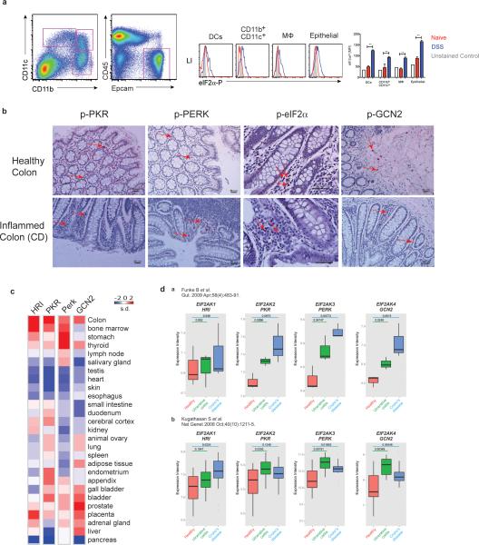
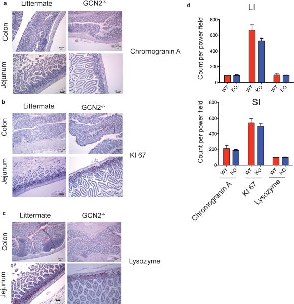
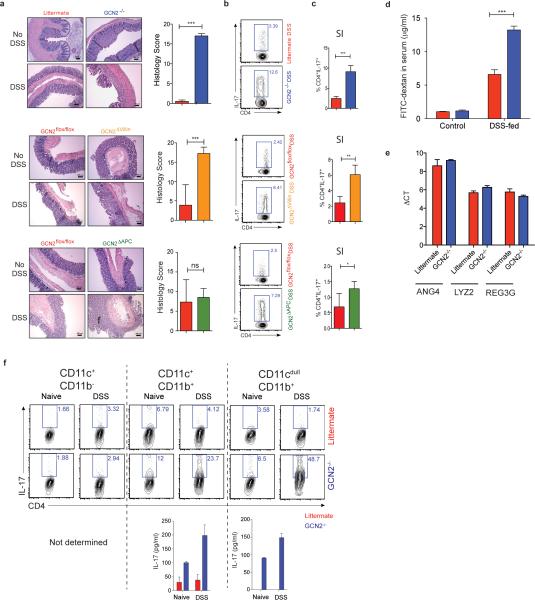
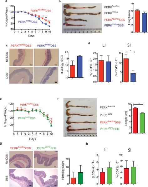
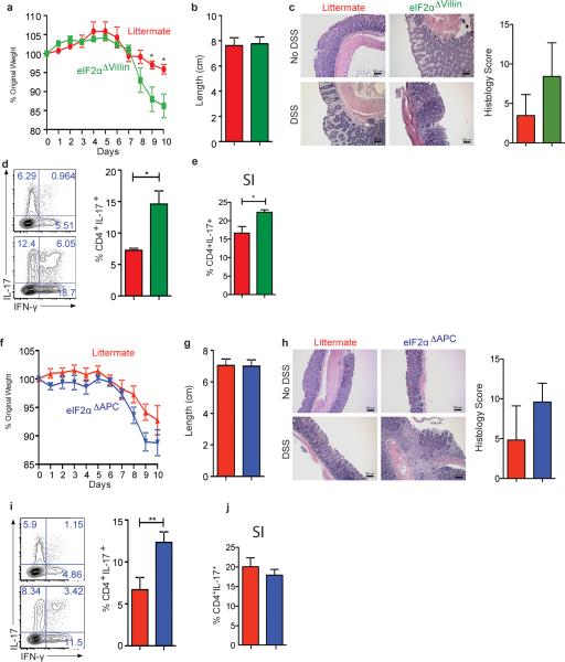
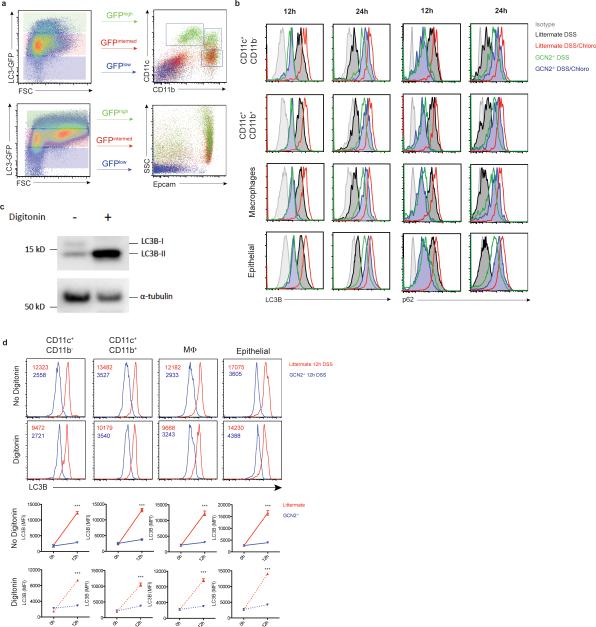
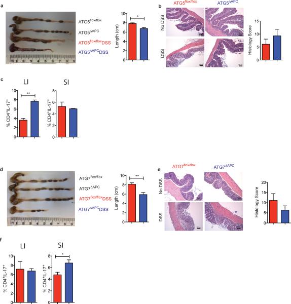
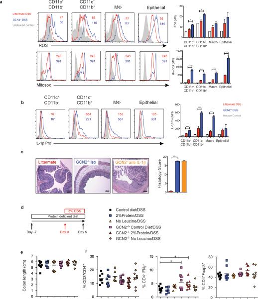
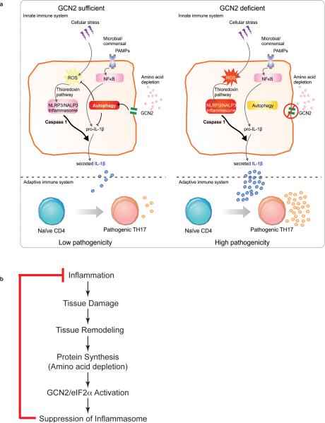
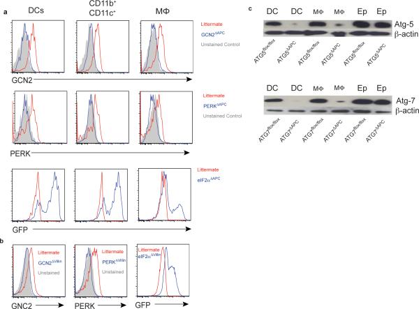
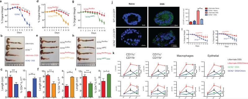
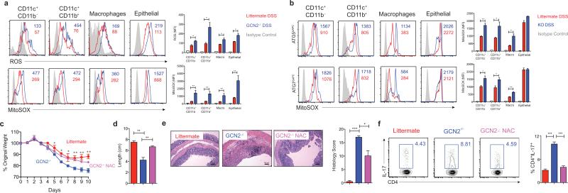
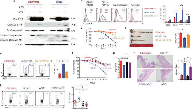
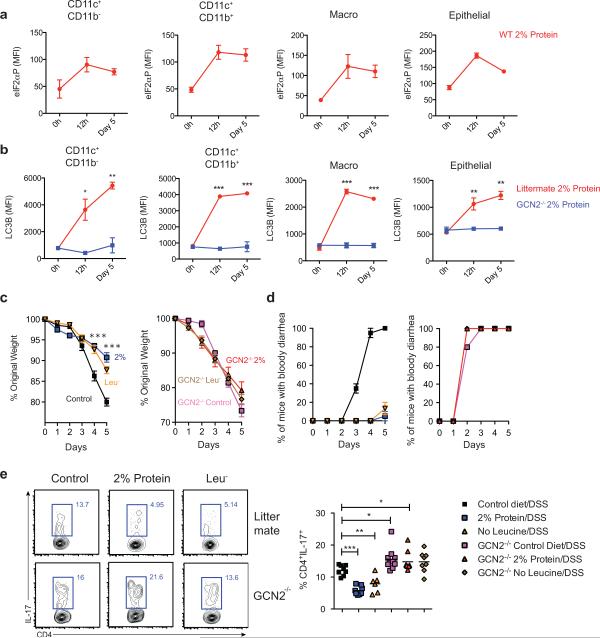
The integrated stress response (ISR) is a homeostatic mechanism by which eukaryotic cells sense and respond to stress-inducing signals, such as amino acid starvation. General controlled non-repressed (GCN2) kinase is a key orchestrator of the ISR, and modulates protein synthesis in response to amino acid starvation. Here we demonstrate in mice that GCN2 controls intestinal inflammation by suppressing inflammasome activation. Enhanced activation of ISR was observed in intestinal antigen presenting cells (APCs) and epithelial cells during amino acid starvation, or intestinal inflammation. Genetic deletion of Gcn2 (also known as Eif2ka4) in CD11c(+) APCs or intestinal epithelial cells resulted in enhanced intestinal inflammation and T helper 17 cell (TH17) responses, owing to enhanced inflammasome activation and interleukin (IL)-1β production. This was caused by reduced autophagy in Gcn2(-/-) intestinal APCs and epithelial cells, leading to increased reactive oxygen species (ROS), a potent activator of inflammasomes. Thus, conditional ablation of Atg5 or Atg7 in intestinal APCs resulted in enhanced ROS and TH17 responses. Furthermore, in vivo blockade of ROS and IL-1β resulted in inhibition of TH17 responses and reduced inflammation in Gcn2(-/-) mice. Importantly, acute amino acid starvation suppressed intestinal inflammation via a mechanism dependent on GCN2. These results reveal a mechanism that couples amino acid sensing with control of intestinal inflammation via GCN2.


Figures

Extended data Fig 1
Extended data Fig 1. eIF2α kinases are expressed in human and murine gut cells
(a) Analysis of p-eIF2α expression in APCs and epithelial cells in large intestine of naïve and 2% DSS treated mice by flow cytometer. (b) Comparison of immunohistological analysis of phosphorylated PKR, PERK, eIF2α and GNC2 in healthy and inflamed human colon tissue (n=1). (c) Expression levels of HRI, PKR, PERK and GCN2 in human organs quantified based on information from a public database (www.ebi.ac.uk). (d) Expression intensity of various eIF2α kinases plotted from known published microarray data from colonic biopsies of patients with either Ulcerative colitis or Crohn's compared to healthy controls. Data are from one experiment representative of three separate experiments. *P < 0.05; **P < 0.005, ***P < 0.0005. Error bars indicate mean ±SEM.
Extended data Fig 2
Extended data Fig 2. GCN2 deficiency does not affect the proliferation or differentiation of IECs
Immunohistology analysis (a-c) and quantification (d) of colons and ileums from GCN2−/− and wild type (littermate) mice for (a) chromagranin A, (b) Ki67 and (c) Lysozme. Data are from one experiment representative of three separate experiments (n=3). *P < 0.05; **P < 0.005, ***P < 0.0005. Error bars indicate mean ±SEM.
Extended data Fig 3
Extended data Fig 3. GCN2 expression protects mice from DSS-induced colitis
(a) H&E staining of colon sections before and after DSS in Wildtype vs GCN2−/− mice, GCN2flox/flox vs GCN2ΔVillin and GCN2flox/flox vs GCN2ΔAPC. IL-17 levels in (b) large intestineal and (c) small intestinal CD4+ T cells measured by flow cytometry; (d) GCN2−/− mice show increased intestinal permeability following DSS treatment evidenced by higher levels of FITC-dextran in the serum. (e) Expression of antimicrobial defensins in wild type and GCN2−/− mice via qPCR. (f) IL-17 production by flow cytometry and ELISA of OTII-CD4 T cells after culturing with different large intestinal APC subsets. Data are from one experiment representative of three separate experiments (n=4-5). *P < 0.05; **P < 0.005, ***P < 0.0005. Error bars indicate mean ±SEM.
Extended data Fig 4
Extended data Fig 4. PERK expression in epithelial cells plays only minor role in controlling mucosal homeostasis following DSS challenge
(a-d) The body weight (a), colon length (b), histology by H&E (c) and Th17 responses (d) both in the colon (LI) and small intestine (SI) of PERKΔvillin and control wild type littermates treated with DSS. (e-h) The body weight (e), colon length (f), histology by H&E and histology score (g) and Th17 responses (h) both in the colon (LI) and small intestine (SI) of PERKΔAPC and control wild type littermates treated with DSS. (Data are representative of two separate experiments (n=5). Data are representative of two separate experiments. *P < 0.05; **P < 0.005, ***P < 0.0005. Error bars indicate mean ±SEM.
Extended data Fig 5
Extended data Fig 5. eIF2α expression in epithelial cells and antigen presenting cells controls weight loss and partially Th17 responses following DSS challenge
(a-e) The body weight (a), colon length (b), histology by H&E and histology score (c) and Th17 responses in both the colon (LI) (d) and small intestine (SI) (e) of eIF2α Δvillin and control wild type littermates treated with DSS. (f-j) The body weight (f), colon length (g), histology by H&E and histology score (h) and Th17 responses in both the colon (LI) (i) and small intestine (SI) (j) of eIF2α ΔAPC and control wild type littermates treated with DSS. Data are representative of three separate experiments (n=5). *P < 0.05; **P < 0.005, ***P < 0.0005. Error bars indicate mean ±SEM.
Extended data Fig 6
Extended data Fig 6. Intestinal APCs and epithelial cells reveal high expression of LC3
(a) Expression of LC3-GFP in APC subsets and epithelial cells of naive LC3-GFP mice by flow cytometry (n=3). Data is from a single experiment. (b) Kinetic Mean fluorescence Intensity (MFI) comparison of LC3B and p62 expression with and without chloroquine on individual APC subsets and epithelial cells by flow cytometry after 12 and 24 h of single DSS administration (c) Western blot detection of LC3-I and II on lamina propria APCs before and after digitonin (d) LC3B staining of individual APCs and epithelial cells 12 hrs after they were treated with DSS. The portion of the lamina propria cells were subjected to digitonin before intracellular staining with the LC3B antibody. Data are representative of two separate experiments (n=4-5). *P < 0.05; **P < 0.005, ***P < 0.0005. Error bars indicate mean ±SEM.
Extended data Fig 7
Extended data Fig 7. ATG5 and ATG7 expression in antigen presenting cells partially protects mice from DSS challenge
(a-c) Colon length (a), histology by H&E and histology score (b) and Th17 responses (c) in both the colon (LI) and small intestine (SI) of Atg5ΔAPC and control wild type littermates treated with DSS. (d-f) Colon length (d), histology by H&E and histology score (e) and Th17responses (f) in both the colon (LI) and small intestine (SI) of Atg7ΔAPC and control wild type littermates treated with DSS Data are representative of three separate experiments (n=5). Data are representative of two separate experiments (n=5). *P < 0.05; **P < 0.005, ***P < 0.0005. Error bars indicate mean ±SEM.
Extended data Fig 8
Extended data Fig 8. GCN2 induced autophagy protects the intestinal tissue from the effects of excess oxidation and inflammation
GCN2−/− and littermate wild type mice were treated with 2% DSS in the drinking water for 5 days. (a) MFIs of ROS and mitochondrial ROS (MitoSox) in individual APC subsets and epithelial cells of the small intestine isolated from wild type, and GCN2−/− were analyzed by flow cytometry (n=5). (b) MFIs of pro-IL-1β in individual APC subsets and epithelial cells of the small intestine isolated from wild type or GCN2−/− mice (Day 5 post DSS) were analyzed by flow cytometry (n=3). (c) Histology analysis of GCN2 mice that were treated with neutralizing antibody. (d) Effects of low protein diet on DSS induced colitis (e) Colon length. (f) Frequencies of CD4+, CD4+IFNγ+ and CD4+Foxp3+ T cells. Data are from one experiment representative of two or three separate experiments. *P < 0.05; **P < 0.005, ***P < 0.0005. Error bars indicate mean ±SEM.
Extended data Fig 9
Extended data Fig 9. Mechanisms by which GCN2 contributes to protection in the gut against acute colitis
(a) Amino acid starvation induced by an inflamed colon activates GCN2 that triggers autophagy which is important in inhibiting oxidative stress and pro-IL-1β levels of IL-1β dictate the magnitude of IL-17A producing CD4 T cells in the colon. (b) A hypothetical model for the evolutionary significance of coupling amino acid starvation with control of inflammation.
Extended data Fig 10
Extended data Fig 10. Mouse phenotyping by flow cytometry and western blot
In addition to molecular genotyping via tail DNA we analyzed the protein levels in various mucosal subsets by flow cytometry in various APC (a) and epithelial (b) specific conditional knockouts (c) Western blot to show selective depletion in APC populations in the Atg5flox/flox CD11c cre (Atg5ΔAPC) and the Atg7flox/flox CD11c cre (Atg7ΔAPC).
Figure 1
Figure 1. GCN2 activation in APCs and epithelial cells suppresses intestinal inflammation by a mechanism dependent on autophagy
GCN2 deficiency leads to loss of body weight, colon shortening and enhanced production of IL-17 by colonic CD4+ T cells (a-c). Expression of GCN2 in epithelial cells (GCN2Δvillin) (d-f) or APCs (GCN2ΔAPC) (g-i) protects mice from DSS induced colitis. (j). LC3-GFP expression and the GFP punctae counts in the crypts (3D) from GCN2−/− LC3-GFP and LC3-GFP mice before and 12 hrs after DSS (k) Mean fluorescence Intensity (MFI) comparison of LC3B and p62 expression with and without chloroquine on APC subsets and epithelial cells by flow cytometry before and following 3% DSS administration. (l) Comparison of body weights of Atg5ΔAPC and Atg7ΔAPC mice to littermate controls subjected to acute 2% DSS-induced colitis. Data are representative of three separate experiments (n=5). *P < 0.05; **P < 0.005, ***P < 0.0005. Error bars indicate mean ±SEM., two-tailed unpaired student's t-test.
Figure 2
Figure 2. GCN2 suppresses ROS activity and intestinal inflammation
(a) Histograms and MFI quantification of ROS and mitochondrial ROS in colonic APC subsets and epithelial cells isolated from wild type and GCN2−/− mice after 5 days of DSS. (b) Atg5ΔAPC and the Atg7ΔAPC compared to littermate before and after 5 days of DSS. NAC protects the GCN2−/− mice from weight loss (c), colon length (d), decreased pathology (e) and reduced colonic Th17 frequencies (f), induced by DSS. Data are representative of three experiments (n=5). *P < 0.05; **P < 0.005, ***P < 0.0005. Error bars indicate mean ±SEM.
Figure 3
Figure 3. Enhanced intestinal inflammation in GCN2−/− mice is dependent on inflammasome activation
(a) Western blot analysis of pro-IL-1β, pro-caspase, cleaved IL-1β (p17) and cleaved caspase (p20) in lysate and culture supernatants of BMDC cultures of wildtype and GCN2−/− treated with LPS alone or LPS + Adenosine tri phosphate (ATP) (Potassium efflux agent that triggers inflammasomes) under amino acid starvation condition. (b) Quantification of MFIs of pro-IL-1β in colonic APC subsets in GCN2−/− and littermates following DSS. (c-e) Body weight (c), colon length, and colonic Th17 responses (e) in DSS treated GCN2−/− mice treated with anti-IL-1β antibody or isotype control. (f-i) Comparison of body weight (f), colon length (g), histology (h) and Th17 frequencies between littermates, GCN2−/−, ASC−/− and GCN2−/− ASC−/− mice subjected to DSS induced colitis. Data are from one experiment representative of three separate experiments n=(4-5). *P < 0.05; **P < 0.005, ***P < 0.0005. Error bars indicate mean ±SEM.
Figure 4
Figure 4. Dietary restriction of amino acids can partially protect against DSS induced colitis
(a) Expression of p-eIF2α on APC subsets and epithelial cells from wild type mice on 2% protein diet (b) Kinetics of LC3B-GFP expression after 2% protein diet in colonic APC subsets and epithelial cells isolated from GCN2−/− LC3-GFP and LC3-GFP mice. (C) Mice on modified protein diet are protected from DSS induced colitis. (c) Weight loss (d) percent of animals having bloody diarrhea and (e) colonic Th17 responses, in 3% DSS induced wild type or GCN2−/− mice that were on protein modified diets (2% protein diet or leucine deficient diet) compared to control diet (16%). Data are from of two separate experiments that were then pooled. *P < 0.05; **P < 0.005, ***P < 0.0005. Error bars indicate mean ±SEM.

Comment in

References

    1. Zhou R, Yazdi AS, Menu P, Tschopp J. A role for mitochondria in NLRP3 inflammasome activation. Nature. 2011;469:221–225. doi:10.1038/nature09663. - PubMed
    1. Kawai T, Akira S. Toll-like receptors and their crosstalk with other innate receptors in infection and immunity. Immunity. 2011;34:637–650. doi:10.1016/j.immuni.2011.05.006. - PubMed
    1. Pulendran B. The Varieties of Immunological Experience:Of Pathogens, Stress, and Dendritic Cells. Annual Review of Immunology. 2015 Apr;33 - PubMed
    1. Donnelly N, Gorman AM, Gupta S, Samali A. The eIF2alpha kinases: their structures and functions. Cellular and molecular life sciences : CMLS. 2013;70:3493–3511. doi:10.1007/s00018-012-1252-6. - PMC - PubMed
    1. Han AP, et al. Heme-regulated eIF2alpha kinase (HRI) is required for translational regulation and survival of erythroid precursors in iron deficiency. EMBO J. 2001;20:6909–6918. doi:10.1093/emboj/20.23.6909. - PMC - PubMed

Publication types

MeSH terms