. 2016 Mar 24;531(7595):471-475.
doi: 10.1038/nature17157. Epub 2016 Mar 16.

Deletions linked to TP53 loss drive cancer through p53-independent mechanisms

Affiliations

Deletions linked to TP53 loss drive cancer through p53-independent mechanisms

Yu Liu et al. Nature. .

Abstract

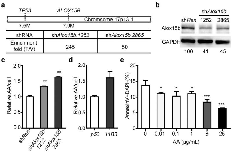
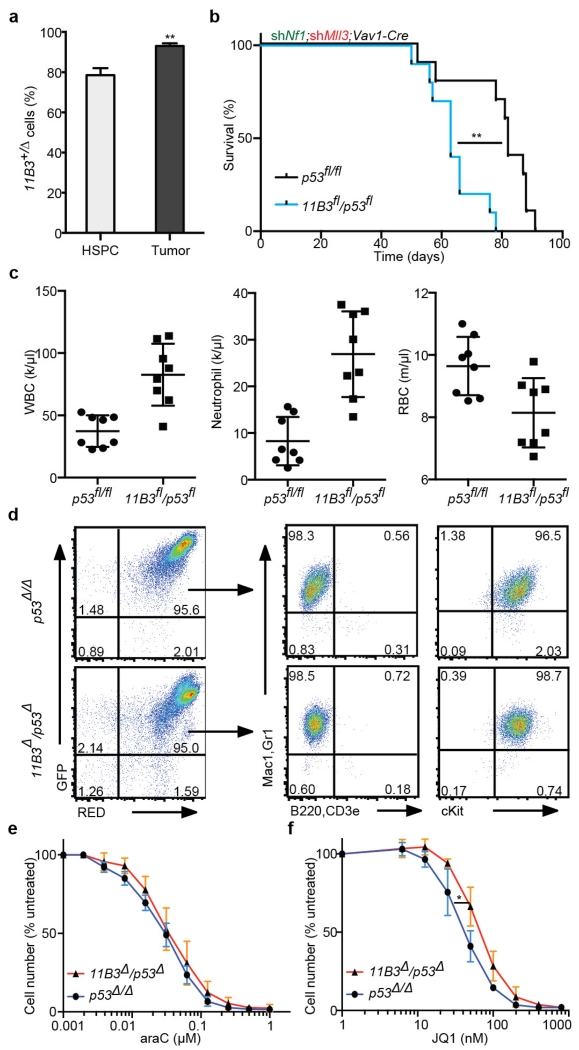
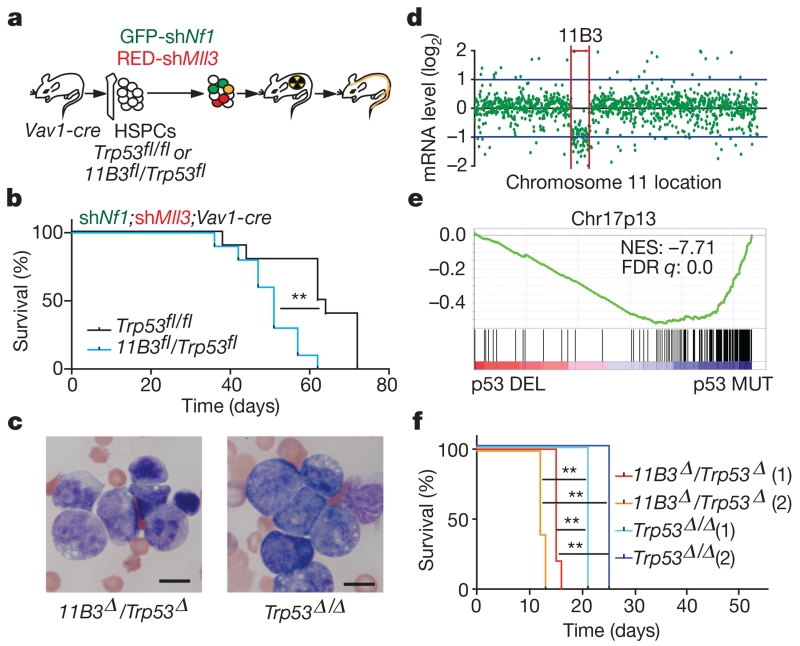
Mutations disabling the TP53 tumour suppressor gene represent the most frequent events in human cancer and typically occur through a two-hit mechanism involving a missense mutation in one allele and a 'loss of heterozygosity' deletion encompassing the other. While TP53 missense mutations can also contribute gain-of-function activities that impact tumour progression, it remains unclear whether the deletion event, which frequently includes many genes, impacts tumorigenesis beyond TP53 loss alone. Here we show that somatic heterozygous deletion of mouse chromosome 11B3, a 4-megabase region syntenic to human 17p13.1, produces a greater effect on lymphoma and leukaemia development than Trp53 deletion. Mechanistically, the effect of 11B3 loss on tumorigenesis involves co-deleted genes such as Eif5a and Alox15b (also known as Alox8), the suppression of which cooperates with Trp53 loss to produce more aggressive disease. Our results imply that the selective advantage produced by human chromosome 17p deletion reflects the combined impact of TP53 loss and the reduced dosage of linked tumour suppressor genes.


Figures

Extended Data Figure 1
Extended Data Figure 1. The frequency and prognostic impact of chromosome 17p deletion with the copy number loss identified by GISTIC
a, The ratio of chromosome-copy-number-altered cases within TP53-mutated cases compared to those within wild-type cases. TP53 mutations were statistically correlated with 17p loss (P < 0.001) but also other copy number events (P < 0.001). b, Peaks of copy number loss identified by the GISTIC algorithm in NHL or AML. x axis, GISTIC q value; y axis, chromosome. q < 0.25 is considered as significant. c, Overall survival of human diffuse large B-cell lymphoma (DLBCL) patients with chromosome 17p deletion is significantly shortened compared to those with no 17p copy number variants, as annotated from the Gene Expression Omnibus GSE34171 series. *P < 0.05 (log-rank test).
Extended Data Figure 2
Extended Data Figure 2. Generation of a chromosome 11B3 conditional knockout mouse
a, Top, strategy to introduce 5′ HPRT gene and loxP site telomeric to Sco1 on chromosome 11B3 with MICER clone MHPN91j22. Bottom, Southern blot demonstrating correct targeting of the derived ES cells. st, single-targeted allele; wt, wild-type. Blue arrowheads denote loxP sites. b, Top, strategy to introduce 3′ HPRT gene and loxP site centromeric to Alox12 on chromosome 11B3 with MICER clone MHPP248j19. Bottom, Southern blot demonstrating correct targeting of the derived ES cells. dt, double-targeted allele. c, Top, diagram showing the expected PCR results and drug-resistance phenotypes of doubly targeted ES cells harbouring loxP sites in cis versus in trans. Gr, G418 (neomycin) resistance; Pr, puromycin resistance; Hr, HAT resistance. df, deleted allele; dp, duplicated allele. Green bar indicates the PCR product location and length. Bottom, PCR results show different ES cell clones generated in a and b.
Extended Data Figure 3
Extended Data Figure 3. 11B3 recombination and lymphomagenesis in Eu-Myc model
a, The extent of 11B3 deletion in peripheral blood cells, as determined by semi-quantitative PCR, in 11B3fl/+ mice crossed to Cd19-cre (left), Mx1-cre (middle) or Vav1-cre (right). Genomic DNA from 11B3+/Δ ES cells was mixed with 11B3fl/+ cells at different ratios (5% or 20%) as a standard. For Mx1-cre, 6–8-week-old mice were treated with polyinosinic:polycytidylic acid (poly(I:C)) (15 mg kg−1 every other day, 7 times) by intraperitoneal injection. b, Partial 11B3 deletion in Vav1-cre;11B3fl/+ pre-B cells as determined by semi-quantitative PCR, indicating incomplete recombination. c, Complete Trp53fl/+ recombination in Vav1-cre;Trp53fl/+ pre-B cells as determined by PCR. d, Tumour-free survival of Eμ-Myc;Vav1-cre;Trp53fl/+ (n = 9), Eμ-Myc;Vav1-cre;11B3fl/+ (n = 12) and Eμ-Myc;Vav1-cre (n = 6) mice shows that 11B3-deleted tumours have longer tumour latency than Trp53-loss-only controls. ***P < 0.001 (log-rank test).
Extended Data Figure 4
Extended Data Figure 4. Charaterization of 11B3-deleted lymphoma
a, Immunophenotypes of B220+ Eμ-Myc lymphomas generated from Vav1-cre;p53fl/+ or Vav1-cre;11B3fl/+. 11B3-deleted lymphomas were either IgMIgD or IgM+IgD+ while all the Trp53-null lymphomas were IgMIgD. b, Haematoxylin and eosin (H&E) stainings of lymph node, spleen and liver of moribund, lymphoma-bearing mice originating from Eμ-Myc;Vav1-cre;11B3fl/+ or Eμ-Myc;Vav1-cre;Trp53fl/+ genotypes. Scale bar, 50 μm. c, 11B3-deleted lymphoma cells isolated from enlarged lymph nodes are more resistant to chemotherapy drugs 4-hydroxycyclophosphamide (left) and vincristine (right), by in vitro drug sensitivity assay. Shown are representative results of three 11B3Δ/Trp53Δframeshift (11B3) or Trp53Δ/Δ (p53) lymphoma cell lines assayed in quadruplicate. *P < 0.05 (Student’s two-tailed t-test). d, e, No functional p53 was detected in various 11B3-deleted tumours as determined by western blotting of p53 and RT–PCR analysis of p21 induction after 4-h ADR treatment. Eμ-Myc;Arf−/− (Trp53+/+) lymphomas were used as a positive control and p21 levels were normalized to untreated cells. Tumours shown in d were identified as missense (tumour 711) or frameshift mutations (tumour 723), while those in e had no detectable mutation. In total eight tumours were analysed. f, The scope of p53 mutations detected in chromosome 11B3-deleted lymphoma cells as determined by sequencing (n = 12). DBD, DNA-binding domain; FS, frameshift mutation; INS, insertion mutation; MS, missense mutation; TAD, transactivation domain; TET, tetramerization domain.
Extended Data Figure 5
Extended Data Figure 5. Tumours in mice heterozygous for Trp53 mutations lose heterozygosity by duplicating the mutant Trp53 allele
a, No chromosome 11B3 deletion was detected in various Trp53 heterozygous mutants. Relative allele copy number of various chromosome 11B3 genes, as determined by qPCR analysis of genomic DNA from Eμ-Myc lymphomas derived from germline mice harbouring the following additional alleles: Vav1-cre;Trp53fl/+(exon 2–10 flanked), Trp53+/− (exon 2–6 deleted), Vav1-cre;Trp53LSL-R270H/+ or Vav1-cre;Trp53LSL-R172H/+. Rpa3 on chromosome 6 was used as an endogenous normalization control. b, SNP analysis of tumour or normal tissue (tail) genomic DNA harvested from mice in a, indicating that uniparental disomy occurred during Trp53 LOH, in that C57BL/6 (B6)-derived wild-type Trp53 allele is replaced by 129-derived Trp53 mutant allele. Note that all Trp53-engineered alleles retain 129-derived SNPs; the germline wild-type Trp53 allele is C57BL/6-derived. c, Cartoon summary of the results from a and b.
Extended Data Figure 6
Extended Data Figure 6. A Trp53 shRNA induces equivalent knockdown in cells with one or two alleles of the Trp53 gene
Pre-B cells were isolated from Trp53+/+ or Trp53+/− bone marrow, and then transduced with GFP-linked Trp53 shRNA (shp53). GFP+ cells were sorted out by fluorescence-activated cell sorting, and treated with control wild-type pre-B cells in the present of vehicle or 1 μg ml−1 ADR for 4 h. p53 and p21 levels were detected by western blotting and RT–qPCR, respectively. Shown is the representative result of three independent experiments.
Extended Data Figure 7
Extended Data Figure 7. In the Eμ-Myc model, Trp53 and Eif5a cooperate in tumorigenesis
Two-colour assay for the cooperation of Trp53 and Eif5a deficiencies on lymphoma genesis. Eμ-Myc HSPCs retrovirally co-transduced with GFP- (shEif5a or shRen) and mCherry- linked shRNAs (shRen, shp53) were transplanted into sublethally-irradiated syngeneic recipients (n = 5 per group). a, b, The resulting tumours were analysed by flow cytometry (a) and the percentage of GFP+ mCherry+ lymphoma cells in each configuration was quantified (b). Error bars represent s.d. *P < 0.05, ***P < 0.001 (two tailed t-test).
Extended Data Figure 8
Extended Data Figure 8. Alox15b deficiency promotes tumorigenesis and increases AA levels
a, Enrichment fold of shAlox15b.1252 and shAlox15b.2865 in resulting tumours (Fig. 3i, j) compared to those in initiating shRNA libraries. b, Knockdown efficiency of shAlox15b.1252 and shAlox15b.2865 compared to control shRen in NIH3T3 cells, as detected by western blotting and quantitated by ImageJ. c, Relative levels of AA per cell are increased with Alox15b shRNAs as measured by liquid chromatography–mass spectrometry (LC-MS). NIH3T3 cells were transduced with shRen or shAlox15b. n = 3. **P < 0.01 (unpaired two tailed t-test). d, Relative levels of AA per cell in 11B3Δ/Trp53Δframeshift (11B3) lymphoma cells are higher than control cells with Trp53Δ/Δ (p53) as measured by LC-MS. n = 2. P = 0.056 (unpaired two tailed t-test). e, In vitro AA treatments reduce apoptosis, as measured by annexin V staining of pre-B cells after 20 h treatment of indicated concentration of AA. n = 4. *P < 0.05; ***P < 0.001 (unpaired two tailed t-test).
Extended Data Figure 9
Extended Data Figure 9. 11B3 deletion accelerates leukaemogenesis beyond Trp53 loss alone and decreases sensitivity to the BET-protein inhibitor JQ-1
a, The percentage of 11B3 deletion as determined by qPCR in premalignant c-Kit+ HSPCs (n = 2) and resulting leukaemia cells (tumour; n = 4). **P < 0.01 (unpaired two-tailed t-test). b, Overall survival of recipient mice transplanted with HSPCs from Vav1-cre;11B3fl/Trp53fl or Vav1-cre;Tr53fl/fl co-transduced with both Nf1 and Mll3 shRNAs. **P < 0.01 (log-rank test). c, Complete blood cell counts of recipient mice indicate that there are more total white blood cells (WBCs) and neutrophils, and fewer red blood cells in Vav1-cre;11B3fl/Trp53fl mice compared with the Vav1-cre;Trp53fl/fl control group at 8 weeks post-transplantation. (Note that two mice from each group died before analysis and were not included.) d, Flow cytometry analysis of GFP+mCherry+ leukaemic cells in the bone marrow of moribund mice in a shows that leukaemia cells are myeloid cells in origin and contain both shNf1 and shMll3. e, f, In vitro drug sensitivity of leukaemia cells to araC (e) and the BET-bromodomain inhibitor JQ-1 (f). Shown are representative results of three 11B3Δ/Trp53Δ and two Trp53Δ/Δ leukaemia cell lines assayed in quadruplicate. *P < 0.05 (Student’s two-tailed t-test).
Figure 1
Figure 1. The frequency, scope, and prognostic value of chromosome 17p alterations in human cancers
a, The nature of TP53-containing chromosome 17p alterations in pan-human cancer (left), non-Hodgkin lymphomas (NHL; middle) and AML (right). DEL, deletion; MUT, mutations; WT, wild-type. b, The extent of chromosome 17p deletions in NHL (44 cases; top) and AML (25 cases; bottom) data sets, irrespective of TP53 deletion, as determined by single nucleotide polymorphism (SNP) array analysis. Each line represents one patient. Red bar indicates the significant copy number loss (q < 0.25) analysed by GISTIC algorithm. c, Overall survival of complex-karyotype AML patients containing both 17p deletion and TP53 mutation as compared to those without any TP53 alteration (top; P = 0.076), those with homozygous (Homo) TP53 point mutations (bottom; P = 0.059), or those with 17p deletion without any detectable TP53 mutation (bottom; P = 0.03). All are log-rank test.
Figure 2
Figure 2. A mouse model of human 17p13.1 deletion accelerates lymphoma development
a, The synteny of human chromosome (Chr) 17p13.1 and mouse chromosome 11B3 (from Sco1 to Alox12b, ~4 Mb), with several representative genes listed. Blue arrowheads denote loxP sites. b, Conditional, 11B3-deletion strategy with PCR analysis (corresponding to the green bar) showing the desired deletion. c, Tumour-free survival of mice with the indicated genotype (log-rank test). d, e, The extent of 11B3 deletion in non-tumorigenic pre-B cells (Vav1-cre;11B3fl/+) and in lymphomas arising in Eμ-Myc;Vav1-cre;11B3fl/+ mice was determined by semi-quantitative PCR using mixed genomic DNA from 11B3+/Δ ES cells to 11B3fl/+ cells at different ratios (10% or 20%) (d), and qPCR (e; two-tailed t-test). Sample names correspond to the mouse identifier. Error bars represent standard deviation (s.d.). f, Copy number profile of mouse chromosome 11 as determined by low-pass whole genome sequencing of 11B3-deleted lymphoma cells obtained from c. Red arrows highlight the 11B3 region. g, Resulting Trp53 status upon LOH from tumours arising from heterozygous chromosome 11B3 deletion (left) or those originating from heterozygous Trp53 deletion (middle) or point mutation (right). UPD, uniparental disomy. ***P < 0.001.
Figure 3
Figure 3. 11B3 deletion can accelerate lymphomagenesis through p53-independent mechanisms
a, Kaplan–Meier lymphoma-free survival curve of recipient mice receiving Vav1-cre;11B3fl/+ or Vav1-cre; 11B3+/+, transduced simultaneously with a mouse Myc cDNA and an shRNA against Trp53 (shp53). n = 8 per genotype. **P < 0.01 (log-rank test). b, The expression levels of Trp53 and p21 after ADR treatment of resulting lymphoma cells as detected by immunoblotting and RT–qPCR, respectively. p21 levels were normalized to untreated Trp53+/+ lymphomas. Shown are representative results (n = 8 per cohort). c, qPCR analysis to determine the percentage of chromosome 11B3 deletion in pre-B cells (transduced cells before transplantation; n = 3) and resulting lymphomas (n = 5) from the experiment in a. ***P < 0.001 (two-tailed t-test).
Figure 4
Figure 4. 11B3 encodes multiple genes whose attenuation cooperates with Trp53 loss to drive lymphoma
a, Kaplan–Meier lymphoma-free survival of recipient mice transplanted with Eμ-Myc HSPCs with various GFP-linked tandem shRNAs. shp53 indicates shTrp53. n = 10 per group. b, Annexin V staining of Eμ-Myc pre-B cells transduced with the indicated tandem shRNAs constructs. n = 3. Result represents at least two independent experiments. PI, propidium iodide. c, d, In vivo shRNA screen to identify potential tumour suppressors on chromosome 17p13 (c) and resulting tumour-free survival curve of mice receiving HPSCs transduced with shRNA pools (d). Hiseq, high-throughput sequencing; TSG, tumour suppressor gene. e, f, Kaplan–Meier tumour-free survival curve of recipient mice transplanted with pre-B cells co-infected with Myc and the indicated shRNAs (e; n = 10), or infected with Myc-linked tandem shRNAs (f; n = 6). a, df, Log-rank test. b, Unpaired two-tailed t-test, error bars represent s.d. *P < 0.05; **P < 0.01; ***P < 0.001.
Figure 5
Figure 5. Chromosome 11B3 deletion acts beyond Trp53 loss to drive myeloid leukaemia
a, AML generation by HSPC isolation (Vav1-cre; Trp53fl/fl or Vav1-cre;11B3fl/Trp53fl) co-transduction with a GFP-linked Nf1 shRNA and an mCherry-linked Mll3 shRNA, and transplantation into sublethally irradiated recipients (n = 10 per group). b, Post-transplant, leukaemia-free survival **P < 0.01 (log-rank test). c, Blood smear of moribund mice (b) that is representative of all animals analysed in each genotype (n = 4). d, RNA-seq comparison of chromosome 11 gene-expression levels between the 11B3Δ/Trp53Δ and Trp53Δ/Δ leukaemia generated in b (n = 4). e, Gene set enrichment analysis of genes on chromosome 17p13 in chromosome 17p-deleted human AML compared to those with TP53 mutations but without 17p deletions. FDR, false discovery rate; NES, normalized enrichment score. f, Kaplan–Meier survival curve of secondary transplants from two independent primary leukaemias of each genotype in a, b. n = 5 in each cohort. **P < 0.01 (log-rank test).

Comment in

References

    1. Hanahan D, Weinberg RA. Hallmarks of cancer: the next generation. Cell. 2011;144:646–674. - PubMed
    1. Zender L, et al. An oncogenomics-based in vivo RNAi screen identifies tumor suppressors in liver cancer. Cell. 2008;135:852–864. - PMC - PubMed
    1. Xue W, et al. A cluster of cooperating tumor-suppressor gene candidates in chromosomal deletions. Proc. Natl Acad. Sci. USA. 2012;109:8212–8217. - PMC - PubMed
    1. Solimini NL, et al. Recurrent hemizygous deletions in cancers may optimize proliferative potential. Science. 2012;337:104–109. - PMC - PubMed
    1. Davoli T, et al. Cumulative haploinsufficiency and triplosensitivity drive aneuploidy patterns and shape the cancer genome. Cell. 2013;155:948–962. - PMC - PubMed

Publication types

MeSH terms

Substances

Associated data