. 2016 Mar 17:6:23076.
doi: 10.1038/srep23076.

Intra-articular injection of synovial mesenchymal stem cells improves cartilage repair in a mouse injury model

Affiliations

Intra-articular injection of synovial mesenchymal stem cells improves cartilage repair in a mouse injury model

J Mak et al. Sci Rep. .

Abstract

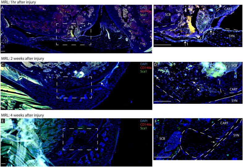
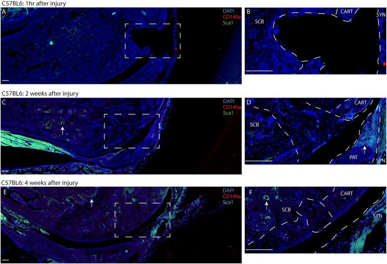
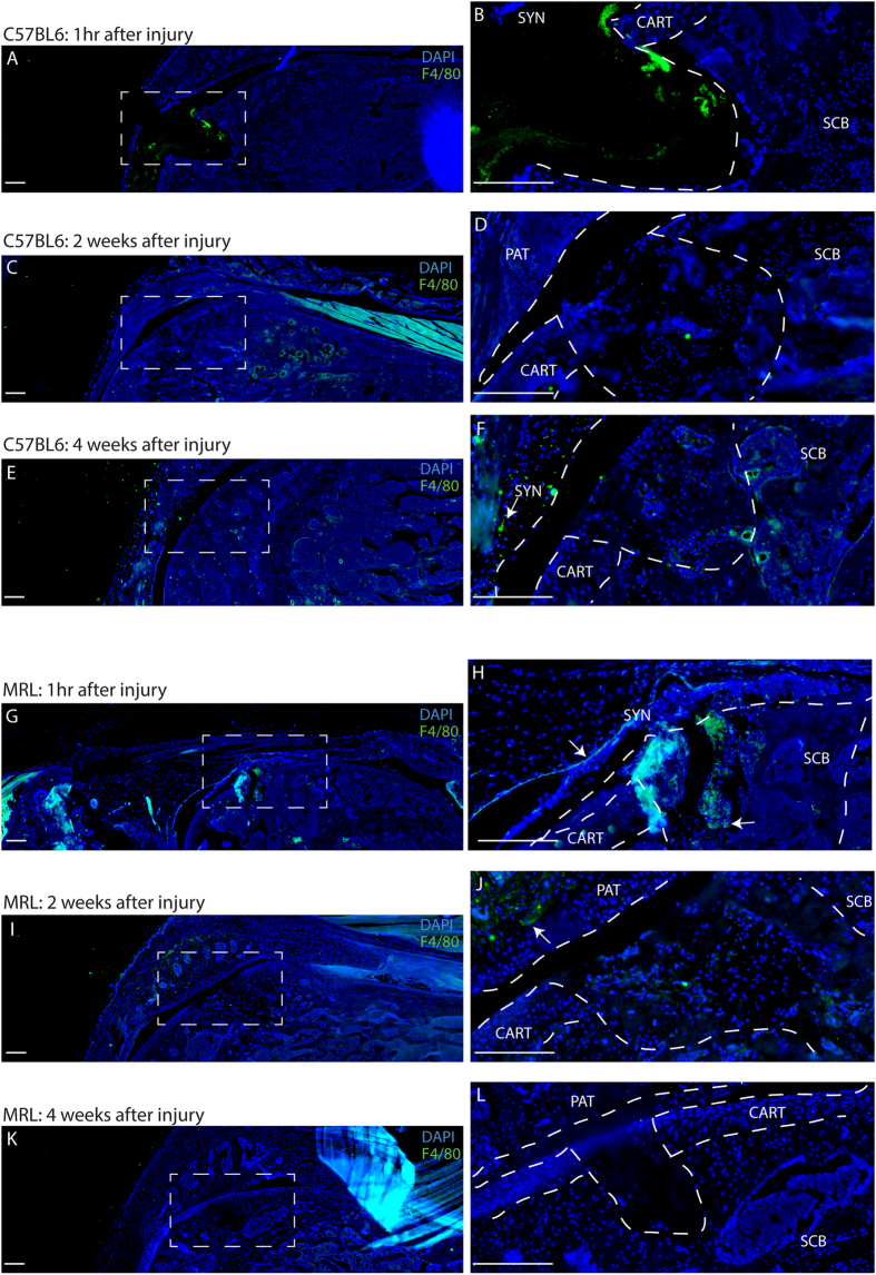
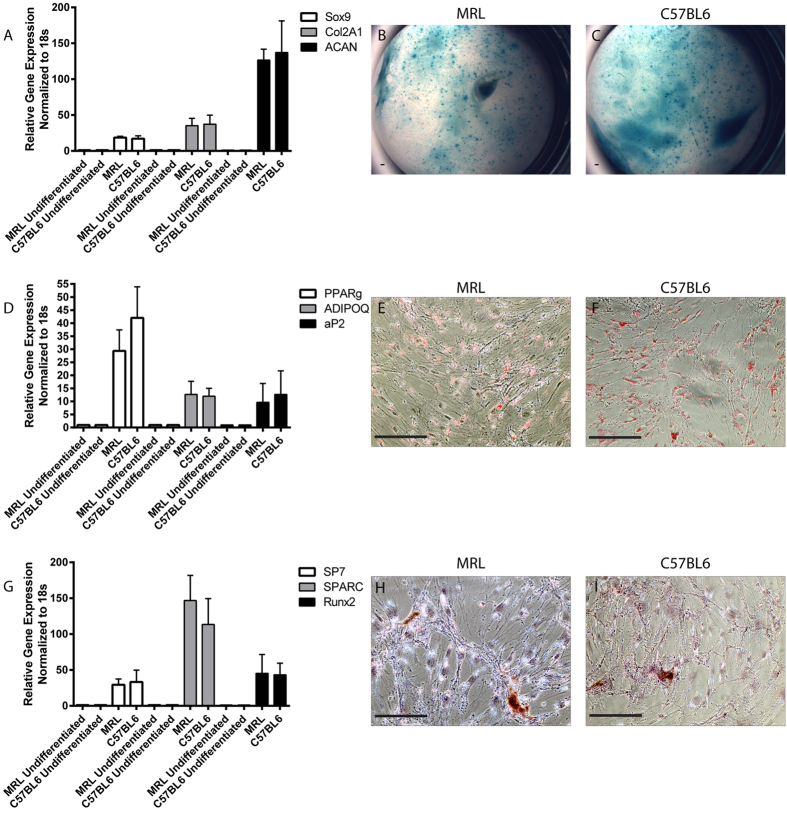
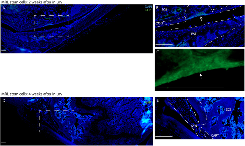
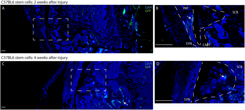
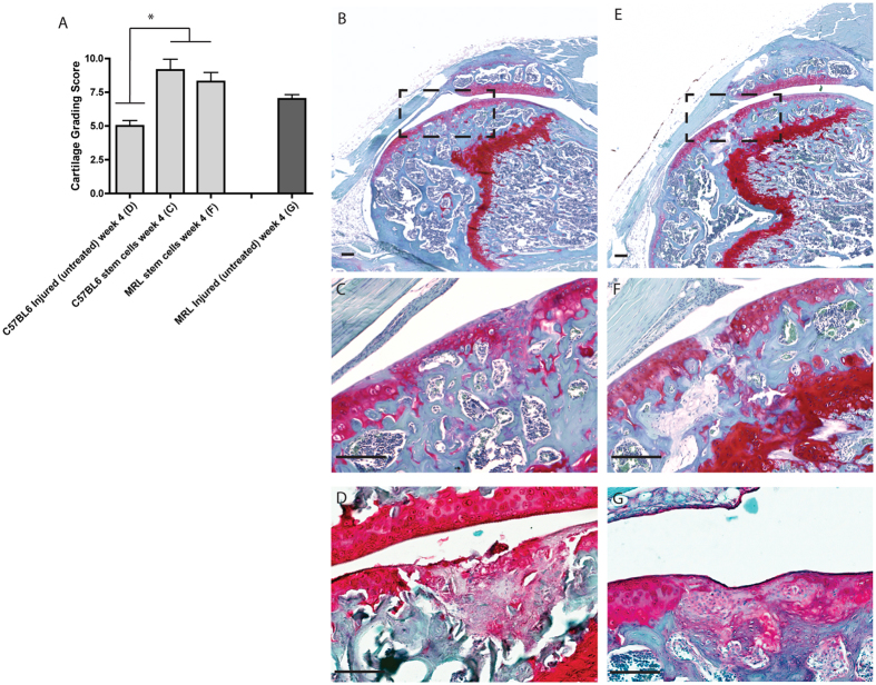
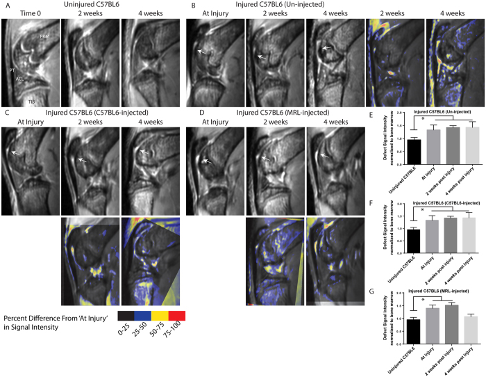
Controversy remains whether articular cartilage has an endogenous stem/progenitor cell population, since its poor healing capacity after injury can lead to diseases such as osteoarthritis. In the joint environment there are mesenchymal stem/progenitor cells (MSCs) in the synovial membrane and synovial fluid that can differentiate into cartilage, but it is still under debate if these cells contribute to cartilage repair in vivo. In this study, we isolated a Sca-1 positive, chondrogenesis capable population of mouse synovial MSCs from C57BL6 and MRL/MpJ "super-healer" strains. Intra-articular injection of Sca-1 + GFP + synovial cells from C57BL6 or MRL/MpJ into C57BL6 mice following cartilage injury led to increased cartilage repair by 4 weeks after injury. GFP expression was detected in the injury site at 2 weeks, but not 4 weeks after injury. These results suggest that synovial stem/progenitor cells, regardless of strain background, have beneficial effects when injected into an injured joint. MSCs derived from MRL/MpJ mice did not promote an increased repair capacity compared to MSCs derived from non-healing C57BL6 controls; however, MRL/MpJ MSCs were observed within the defect area at the time points examined, while C57BL6 MSCs were not.


Figures

Figure 1
Figure 1. Characterization of Endogenous MRL MSCs after Cartilage Injury.
Immediately after joint injury Sca-1+CD140a+ cells can be observed within the defect and the adjacent synovium (arrow B) as well as sub-chondral bone. At 2 weeks after injury, Sca-1+ cells can be observed in the defect, but are no longer staining positive for CD140a (arrows D). By 4 weeks after injury, very few Sca-1+ cells can be found in or around the defect area (arrow F). Scale bars = 200 μm. SCB = sub-chondral bone, CART = cartilage, SYN = synovium.
Figure 2
Figure 2. Characterization of Endogenous C57BL6 MSCs after Cartilage Injury.
Immediately after joint injury neither Sca-1+ nor CD140a+ cells can be observed around the defect area or in the adjacent synovium or sub-chondral bone. At 2 weeks after injury Sca-1+ cells can be observed in sub-chondral bone, and appear to be associated with the vasculature (arrow C). At this time-point Sca-1+ cells can also be seen in the patella (arrow D). By 4 weeks after injury, no Sca-1+ cells can be found in or around the defect area, however, Sca-1+ cells can still be observed in the sub-chondral bone, associated with blood vessels (arrow E,F). Scale bars = 200 μm. SCB = sub-chondral bone, CART = cartilage, SYN = synovium, PAT = patella.
Figure 3
Figure 3. Macrophage identification in C57BL6 and MRL joints after Cartilage Injury.
In C57BL6 mice, no F4/80 positive macrophages are observed in the defect area until 4 weeks after cartilage injury (arrow). In MRL mice, F4/80 positive macrophages are observed in the defect and synovium immediately after injury (arrow) and F4/80 positive cells decrease in weeks 2 and 4 after injury. Scale bars = 200 μm. SCB = sub-chondral bone, CART = cartilage, SYN = synovium, PAT = patella.
Figure 4
Figure 4. Characterization of MSCs from MRL and C57BL6 mice In Vitro.
Synovial MSCs were harvested from MRL and C57BL6 mice and examined for chondrogenic (A–C), adipogenic (D–F) and osteogenic (G–I) potential using qRT-PCR for specific markers (A,D,G) and histological stains (B,C,E,F,H,I). No differences in multipotent differentiation capacity was observed between MSCs derived from the two strains. Scale bars = 100 μm.
Figure 5
Figure 5. Exogenous delivery of MRL MSCs contribute to Cartilage Repair.
MRL synovial GFP+ MSCs migrate to the defect within 2 weeks after injury (A) and directly contribute to cartilage repair (arrows B,C). By 4 weeks after injury, no GFP can be detected at the injury site. Scale bars = 200 μm. SCB = sub-chondral bone, CART = cartilage, SYN = synovium, PAT = patella.
Figure 6
Figure 6. Exogenous delivery of C57BL6 MSCs in a mouse cartilage injury model.
C57BL6 synovial GFP+ MSCs were not found in the defect within 2 weeks after injury (A,B), but were observed with the patella and adjacent synovium (arrow B). By 4 weeks after injury, GFP positive cells were observed in the defect site (arrow D), and below the defect in the sub-chondral bone (arrow C). Scale bars = 200 μm. SCB = sub-chondral bone, CART = cartilage, SYN = synovium, PAT = patella.
Figure 7
Figure 7. Quantification of Cartilage Injury Repair.
Cartilage injuries were graded in C57BL6 mice, MRL mice, and C57BL6 mice that received an injection of either MRL or C57BL6 MSCs (A). No difference was observed between MRL and C57BL6 MSC injected groups, but both were significantly better than injured-untreated C57BL6 mice. Representative images are shown of knees injured and injected with C57BL6 (B,C) or MRL (D,E) MSCs, or C56BL6 (D) and MRL (G) knee that have been injured but not injected with MSCs at the 4 week timpoint. Scale bars = 100 μm (B,D); = 150 μm (C,E).
Figure 8
Figure 8. Repeated Measures MRI Tracking of Cartilage Injury Repair Over Time.
MRI was used to follow repair in vivo over 4 weeks in: control joints without injury (A), C57BL6 injured mice without MSC injection (B), C57BL6 injected with C57BL6-derived MSCs (C) and C57BL6 injected with MRL-derived MSCs (D). Arrows indicate the position of the defect. Images at 2 or 4 weeks after injury were registered (using the defect site) to the images collected at the ‘At Injury’ time-point and pseudo-coloured to demonstrate difference in signal intensity between the two time-points. Signal intensity of the defect was quantified at injury, 2 weeks and 4 weeks post injury for mice that were un-injected (E), injected with C57BL6 MSCs (F), or MRL MSCs (G). A significant reduction in signal intensity was only observed 4 weeks after injection with MRL MSCs. FMR = Femur, TIB = Tibia, ACL = Anterior Cruciate Ligament, PT = Patellar Tendon.

References

    1. Pittenger M. F. et al. Multilineage Potential of Adult Human Mesenchymal Stem Cells. Science. 284, 143–147 (1999). - PubMed
    1. Zuk P. A. et al. Human Adipose Tissue Is a Source of Multipotent Stem Cells. Mol. Biol. Cell. 13, 4279–4295 (2002). - PMC - PubMed
    1. De Bari C., De Accio F. D., Tylzanowski P. & Luyten F. P. Multipotent Mesenchymal Stem Cells From Adult Human Synovial Membrane. Arthritis Rheum. 44, 1928–1942 (2001). - PubMed
    1. Dennis E., Bruder S. P., Lennon D. P. & Caplan A. I. In Vitro Differentiation of Bone and Hypertrophic Cartilage from Periosteal-Derived Cells. Exp Cell Res. 195, 492–503 (1991). - PubMed
    1. Johnson K. et al. A Stem Cell–Based Approach to Cartilage Repair. Science. 336, 717–721 (2012). - PubMed

Publication types

MeSH terms