Clinical-Morphological Features and Outcomes of Lupus Podocytopathy
- PMID: 26983707
- PMCID: PMC4822663
- DOI: 10.2215/CJN.06720615
Clinical-Morphological Features and Outcomes of Lupus Podocytopathy
Abstract
Background and objectives: Lupus podocytopathy, which is characterized by diffuse foot process effacement without peripheral capillary wall immune deposits and glomerular proliferation, has been described in SLE patients with nephrotic syndrome in case reports and small series. This study aimed to better characterize the incidence, clinical-morphologic features, and outcomes of such patients from a large Chinese cohort.
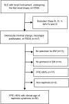
Design, setting, participants, & measurements: Lupus podocytopathy was identified from 3750 biopsies of SLE patients obtained from 2000 to 2013 that showed mild glomerular histology in patients with a clinical sign of nephrotic syndrome. The biopsy results were divided into three groups: glomerular minimal change, mesangial proliferation, and FSGS.
Results: Fifty (1.33%) cases were identified as lupus podocytopathy and included minimal change in 13 cases, mesangial proliferation in 28 cases, and FSGS in nine cases. Extensive foot process effacement appeared in all the biopsies and mesangial electron-dense deposits were present in 47 biopsies. All patients demonstrated nephrotic syndrome, and the median proteinuria was 5.72 g/24 h (interquartile range [IQR], 3.82, 6.92). Seventeen (34%) cases presented with AKI. Forty-seven (94%) patients achieved remission after immunosuppressive therapy for a median time of 4 weeks (IQR, 2, 8). Compared with the patients with minimal change and mesangial proliferation, patients with FSGS showed significantly higher incidence of AKI and severe tubule-interstitial injury and a much lower complete remission rate. During follow-up of a median of 62 (IQR, 36, 84) months, renal relapses occurred in 28 (59.6%) patients. No patient died or developed ESRD.
Conclusions: The findings from this cohort study suggest that lupus podocytopathy may represent a special entity of lupus nephritis with distinct clinical-morphologic features. The differences in AKI incidence, tubular injury severity, and response to treatment between the patients with minimal change/mesangial proliferation and those with FSGS patterns indicate two different subtypes of lupus podocytopathy.
Keywords: acute kidney injury; biopsy; follow-up studies; glomerulosclerosis, focal segmental; humans; kidney failure, chronic; lupus nephritis; nephrotic syndrome; pathology; proteinuria.
Copyright © 2016 by the American Society of Nephrology.
Figures


Comment in
-
Lupus Podocytopathy: A Distinct Entity.Clin J Am Soc Nephrol. 2016 Apr 7;11(4):547-8. doi: 10.2215/CJN.01880216. Epub 2016 Mar 16. Clin J Am Soc Nephrol. 2016. PMID: 26983708 Free PMC article. No abstract available.
References
-
- Baldwin DS, Gluck MC, Lowenstein J, Gallo GR: Lupus nephritis. Clinical course as related to morphologic forms and their transitions. Am J Med 62: 12–30, 1977 - PubMed
-
- Matsumura N, Dohi K, Shiiki H, Morita H, Yamada H, Fujimoto J, Kanauchi M, Hanatani M, Ishikawa H: Three cases presenting with systemic lupus erythematosus and minimal change nephrotic syndrome] [in Japanese]. Nihon Jinzo Gakkai Shi 31: 991–999, 1989 - PubMed
-
- Okai T, Soejima A, Suzuki M, Yomogida S, Nakabayashi K, Kitamoto K, Nagasawa T: A case report of lupus nephritis associated with minimal change nephrotic syndrome–comparison of various histological types of 67 cases with lupus nephritis [in Japanese]. Nihon Jinzo Gakkai Shi 34: 835–840, 1992 - PubMed
-
- Makino H, Haramoto T, Shikata K, Ogura T, Ota Z: Minimal-change nephrotic syndrome associated with systemic lupus erythematosus. Am J Nephrol 15: 439–441, 1995 - PubMed
Publication types
MeSH terms
LinkOut - more resources
Full Text Sources
Other Literature Sources
Medical

