. 2016 Mar 17:6:23165.
doi: 10.1038/srep23165.

A new hand-held microfluidic cytometer for evaluating irradiation damage by analysis of the damaged cells distribution

Affiliations

A new hand-held microfluidic cytometer for evaluating irradiation damage by analysis of the damaged cells distribution

Junsheng Wang et al. Sci Rep. .

Abstract

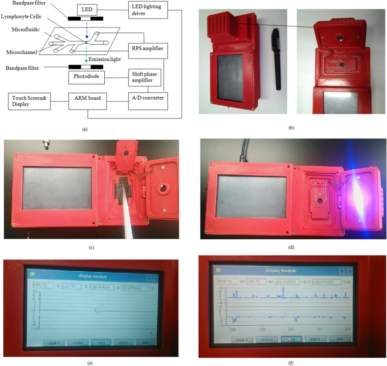
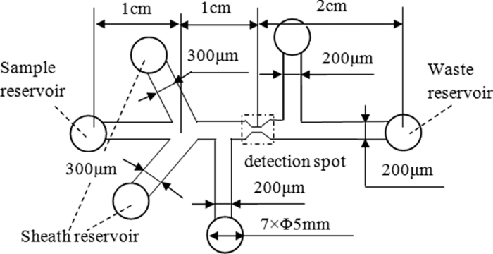
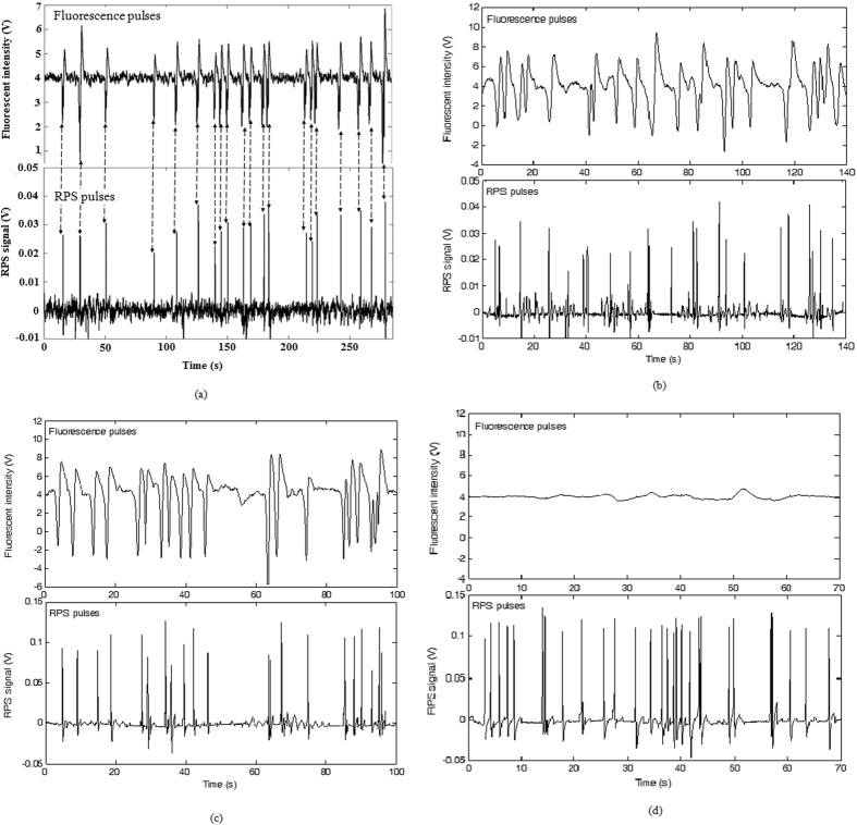
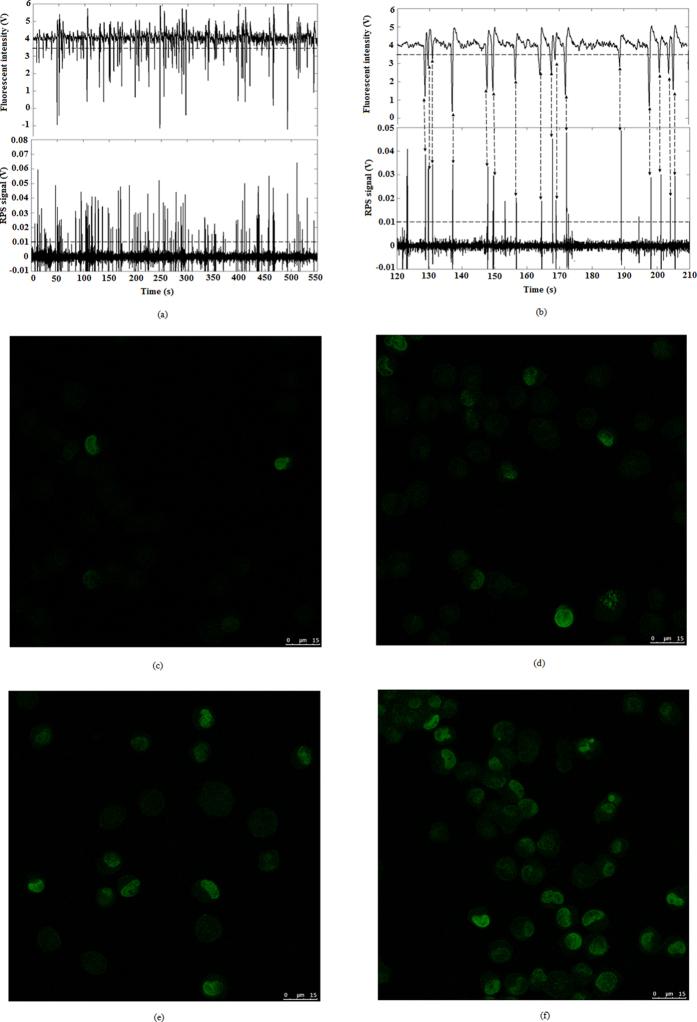
Space radiation brings uneven damages to cells. The detection of the distribution of cell damage plays a very important role in radiation medicine and the related research. In this paper, a new hand-held microfluidic flow cytometer was developed to evaluate the degree of radiation damage of cells. The device we propose overcomes the shortcomings (e.g., large volume and high cost) of commercial flow cytometers and can evaluate the radiation damage of cells accurately and quickly with potential for onsite applications. The distribution of radiation-damaged cells is analyzed by a simultaneous detection of immunofluorescence intensity of γ-H2AX and resistance pulse sensor (RPS) signal. The γ-H2AX fluorescence intensity provides information of the degree of radiation damage in cells. The ratio of the number of cells with γ-H2AX fluorescence signals to the total numbers of cells detected by RPS indicates the percentage of the cells that are damaged by radiation. The comparison experiment between the developed hand-held microfluidic flow cytometer and a commercial confocal microscope indicates a consistent and comparable detection performance.


Figures

Figure 1
Figure 1
(a) Schematic diagram of the microfluidic flow cytometer with fluorescence and RPS detection. (bf )Pictures of the hand-held microfluidic flow cytometer.
Figure 2
Figure 2. Diagram of structure and dimensions of the microfluidic chip used in this study.
Figure 3
Figure 3
RPS and fluorescence signals of individual particles (a) (8.3 μm, fluorescent particle, 0.18% intensity, Dragon Green); (b) (5.8 μm, fluorescent particle, FICP-50-2); (c) (8.3 μm, fluorescent particle, 0.85% intensity, Dragon Green); (d) (10 μm, non-fluorescent particle, PPX-100-10).
Figure 4
Figure 4
Typical signals of individual lymphocyte cells (a) radiated under 32 J/m2; (b) enlarged view of Fig. 4(a) from 120 s to 210 s. The actual confocal images of the γ-H2AX fluorescent marker in imaged cell radiated for different time under 32 J/m2 (c) for 1.25 minutes; (d) for 2.5 minutes; (e) for 5 minutes; (f) for 10 minutes.
Figure 5
Figure 5. Comparison experiments between a commercial confocal microscope and the developed microfluidic cytometer.
(a) Fluorescence intensity distribution for 100 cells after being radiated under 32 J/m2; (b) the ratio of the number of radiation damaged cells to the total number of cells in the sample at different radiation doses.

References

    1. Bai X. M. et al. Efficient annealing of radiation damage near grain boundaries via interstitial emission. Science. 327, 1631 (2010). - PubMed
    1. Davis F., Il’yasova D., Rankin K., McCarthy B. & Bigner D. D. Medical diagnostic radiation exposures and risk of gliomas. Radiat Res. 175, 790 (2011). - PMC - PubMed
    1. Einstein A. J. Effects of radiation exposure from cardiac imaging: how good are the data? J Am Coll Cardiol. 59, 553 (2012). - PMC - PubMed
    1. Horneck G., Klaus, D. M. & Mancinelli R. L. Space microbiology. Microbiol Mol Biol Rev. 74, 121 (2010). - PMC - PubMed
    1. Seymour C. B. & Mothersill C. Radiation-induced bystander effects-implications for cancer. Nat. Rev. Cancer. 4, 158 (2004). - PubMed

Publication types

MeSH terms

LinkOut - more resources