Review
. 2016 Apr;36(3):449-57.
doi: 10.1007/s10571-016-0345-4. Epub 2016 Mar 17.

Ciliary Extracellular Vesicles: Txt Msg Organelles

Affiliations
Review

Ciliary Extracellular Vesicles: Txt Msg Organelles

Juan Wang et al. Cell Mol Neurobiol. 2016 Apr.

Abstract

Cilia are sensory organelles that protrude from cell surfaces to monitor the surrounding environment. In addition to its role as sensory receiver, the cilium also releases extracellular vesicles (EVs). The release of sub-micron sized EVs is a conserved form of intercellular communication used by all three kingdoms of life. These extracellular organelles play important roles in both short and long range signaling between donor and target cells and may coordinate systemic responses within an organism in normal and diseased states. EV shedding from ciliated cells and EV-cilia interactions are evolutionarily conserved phenomena, yet remarkably little is known about the relationship between the cilia and EVs and the fundamental biology of EVs. Studies in the model organisms Chlamydomonas and Caenorhabditis elegans have begun to shed light on ciliary EVs. Chlamydomonas EVs are shed from tips of flagella and are bioactive. Caenorhabditis elegans EVs are shed and released by ciliated sensory neurons in an intraflagellar transport-dependent manner. Caenorhabditis elegans EVs play a role in modulating animal-to-animal communication, and this EV bioactivity is dependent on EV cargo content. Some ciliary pathologies, or ciliopathies, are associated with abnormal EV shedding or with abnormal cilia-EV interactions. Until the 21st century, both cilia and EVs were ignored as vestigial or cellular junk. As research interest in these two organelles continues to gain momentum, we envision a new field of cell biology emerging. Here, we propose that the cilium is a dedicated organelle for EV biogenesis and EV reception. We will also discuss possible mechanisms by which EVs exert bioactivity and explain how what is learned in model organisms regarding EV biogenesis and function may provide insight to human ciliopathies.

Keywords: C. elegans; Cilia; Ectosome; Exosome; Extracellular vesicles; Microvesicle.


Figures
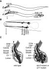
Fig. 1
Fig. 1
Caenorhabditis elegans EV-releasing neurons (EVNs). Caenorhabditis elegans cilia are located on distal dendritic endings of sensory neurons. There are 60 and 108 sensory ciliated neurons in C. elegans hermaphrodite and male, respectively. However, only six IL2 neurons in hermaphrodites and 27 neurons in males shed and release EVs into environment. a Names and anatomical position of the EVNs in the hermaphrodite and male. The number of each EVN type is in parenthesis. b EVN sensory organs display common ultrastructure features. Each EVN has a sister neuron, their cilia and ciliary bases are isolated in a lumen formed by two glial cells, the sheath cell, and the socket cell. The lumen is continuous with a cuticular pore, from which the EVN cilium protrudes into environment directly while the sister cilium does not. Only the male-specific cephalic sensory organ components are shown. c A model of the cephalic sensory organ based on electron tomography reproduced from (Wang et al. 2014). The cephalic sensillum contains CEM and CEP cilia, CEM-derived EVs, and the lumen formed by sheath and socket cell. The CEM cilium sheds EVs into the lumen that may be released through the cuticular pore to environment. In a klp-6 (an EVN-specific ciliary kinesin) or a cil-7 (a myristoylated coiled-coil protein) mutant, EVs accumulate in the cephalic lumen as diagnosed by transmission electron microscopy and PKD-2::GFP EVs are not released outside (Maguire et al. ; Wang et al. 2014). Mutation in either klp-6 or cil-7 disrupts EVN-mediated sensory functions (Color figure online)
Fig. 2
Fig. 2
Model depicting the cilium as an EV donor (a) and EV acceptor (b). Cilia are highly compartmentalized organelles with an microtubule axonemal skeleton (gray lines) (Blacque and Sanders 2014). The ciliary membrane has different domains that are enriched with certain proteins. The IFT machinery and other ciliary complexes contribute to ciliary compartmentalization. The IFT machinery is required for some aspect of EV biogenesis (Wang et al. 2014). Therefore, we propose that a ciliary compartmentalization might be used for sorting of ciliary EV cargoes. EVs may be released by different ciliary regions including the base (green) or from the tip (purple) of the ciliary membrane. b Cilia are sensory organelles that may interact with EVs (gray bubbles). EVs may originate from neighbors or from cells at long distances. EVs may interact with the ciliary membrane (blue) or may fuse with the ciliary membrane (gray dashes) to promote signal transduction (green arrow). In a disease state, aberrant EVs (red) may trigger a pathological signal (b′) or abnormal cilia (red) may fail to transduce an EV-induced signal (b″) (Color figure online)

Comment in

  • Correspondence.
    Rubowitz A. Rubowitz A. Retina. 2016 Aug;36(8):e78-9. doi: 10.1097/IAE.0000000000001160. Retina. 2016. PMID: 27388737 No abstract available.

References

    1. Andres M, Turiegano E, Gopfert MC, Canal I, Torroja L (2014) The extracellular matrix protein artichoke is required for integrity of ciliated mechanosensory and chemosensory organs in Drosophila embryos. Genetics 196:1091–1102 - PMC - PubMed
    1. Bae YK, Barr MM (2008) Sensory roles of neuronal cilia: cilia development, morphogenesis, and function in C. elegans. Front Biosci 13:5959–5974 - PMC - PubMed
    1. Bae YK, Qin H, Knobel KM, Hu J, Rosenbaum JL, Barr MM (2006) General and cell-type specific mechanisms target TRPP2/PKD-2 to cilia. Development 133:3859–3870 - PubMed
    1. Bae YK, Lyman-Gingerich J, Barr MM, Knobel KM (2008) Identification of genes involved in the ciliary trafficking of C. elegans PKD-2. Dev Dyn 237:2021–2029 - PMC - PubMed
    1. Baldari CT, Rosenbaum J (2010) Intraflagellar transport: it’s not just for cilia anymore. Curr Opin Cell Biol 22:75–80 - PMC - PubMed