. 2016 Jun;44(6):2076-89.
doi: 10.1007/s10439-016-1592-8. Epub 2016 Mar 16.

Understanding the Role of ECM Protein Composition and Geometric Micropatterning for Engineering Human Skeletal Muscle

Affiliations

Understanding the Role of ECM Protein Composition and Geometric Micropatterning for Engineering Human Skeletal Muscle

Rebecca M Duffy et al. Ann Biomed Eng. 2016 Jun.

Abstract

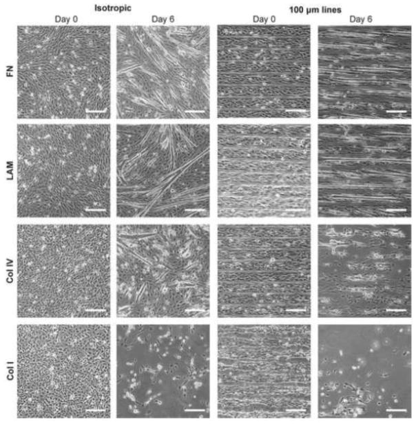
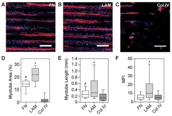
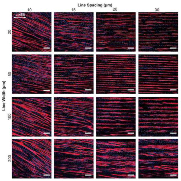
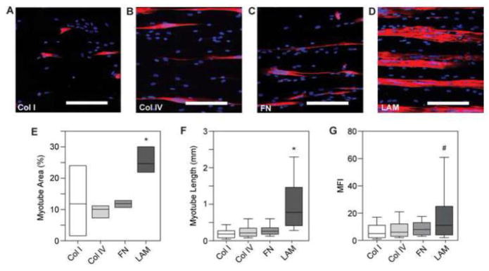
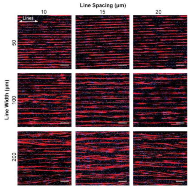
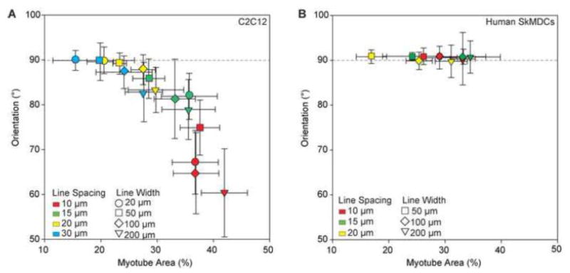
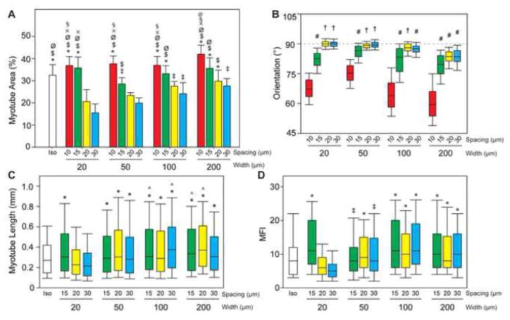
Skeletal muscle lost through trauma or disease has proven difficult to regenerate due to the challenge of differentiating human myoblasts into aligned, contractile tissue. To address this, we investigated microenvironmental cues that drive myoblast differentiation into aligned myotubes for potential applications in skeletal muscle repair, organ-on-chip disease models and actuators for soft robotics. We used a 2D in vitro system to systematically evaluate the role of extracellular matrix (ECM) protein composition and geometric patterning for controlling the formation of highly aligned myotubes. Specifically, we analyzed myotubes differentiated from murine C2C12 cells and human skeletal muscle derived cells (SkMDCs) on micropatterned lines of laminin compared to fibronectin, collagen type I, and collagen type IV. Results showed that laminin supported significantly greater myotube formation from both cells types, resulting in greater than twofold increase in myotube area on these surfaces compared to the other ECM proteins. Species specific differences revealed that human SkMDCs uniaxially aligned over a wide range of micropatterned line dimensions, while C2C12s required specific line widths and spacings to do the same. Future work will incorporate these results to engineer aligned human skeletal muscle tissue in 2D for in vitro applications in disease modeling, drug discovery and toxicity screening.

Keywords: Extracellular matrix; Microcontact printing; Skeletal muscle; Tissue engineering.


Figures

Figure 1
Figure 1
Phase contrast images of C2C12 cells differentiated on FN, LAM, Col IV, and Col I. Samples were differentiated on isotropically coated coverslips and 100×20 micropatterns. After 6 days of differentiation, cells begin to delaminate from μCP lines of Col IV and Col I and isotropically coated Col I. However, myotubes were able to form on both μCP or isotropically coated LAM and FN. Scale bars 200 μm.
Figure 2
Figure 2
C2C12 myotubes differentiated for 6 days on 100×20 lines of (A) FN (B) LAM and (C) Col IV show increased myotube formation on LAM and delamination of myotubes from Col IV after staining for MHC and nuclei. (D) Myotubes formed on LAM lines had significantly higher MFI than those differentiated on FN or Col IV. (E) Myotubes differentiated on LAM were significantly longer than those differentiated on FN or Col IV, and myotubes differentiated on FN were also significantly longer than myotubes that formed on Col IV. (F) Percent area of myotubes formed on LAM lines was also significantly higher than on FN or Col IV, and significantly more percent area myotubes formed on FN than on Col IV. * p < 0.05 compared to FN and Col I. # p < 0.05 compared to Col IV. Scale bars 200 μm. Blue – DAPI; Red – MHC.
Figure 3
Figure 3
Representative images of C2C12 cells differentiated on 16 different line patterns of LAM. Myotubes deviate from aligning with the designated pattern when geometric spacing is too narrow (<15 μm), or when the lines are too wide (200 μm). White arrows represent direction of μCP lines. Scale bars 200 μm. Blue – DAPI; Red – MHC.
Figure 4
Figure 4
(A) Percent area myotubes is higher on 10 and 15 μm spacing conditions because myotubes have more patterned area of LAM on which to form. (B) The orientation of lines is marked at 90°. Myotubes maintain alignment with the patterned ECM when the line spacing is 20 or 30 μm, and line width is < 200 μm. (C) Longer myotubes formed on from patterned lines >20 μm wide. (D) Myotubes formed on wider lines were the result of fusion of significantly more myoblasts as measured by MFI. Length and MFI were not quantified for line spacings of 10 μm as myotubes were not restricted to form uniaxially along the pattern. † p< 0.05 compared to 10 and 15 μm spacings and all widths; # p< 0.05 compared to 10 μm spacings; * p< 0.05 compared to 20×20 and 20×30; ^ p< 0.05 compared to isotropic; ‡ p< 0.05 than 20×30; $ p< 0.05 compared to 50×30; Ø p<0.05 compared to 50×20 and 100×30; § p< 0.05 compared to 50×15 and 200×30; x p< 0.05 compared to 100×20; @ p< 0.05 compared to 100×15 and 200×20.
Figure 5
Figure 5
(A–D) Representative images of MHC and DAPI staining of human myotube formation on 100×20 lines. (E) Quantifying the percent area of myotube formation for human skeletal muscle cells shows significantly higher myotube area on LAM compared to Col I, Col IV, and FN. Myotube formation on Col I is highly variable. (F) Human myotubes formed on LAM lines are significantly longer than myotubes formed on Col I, Col IV, and FN. (G) Human myotubes formed on LAM lines also have significantly higher MFI than those formed on Col I and Col IV lines. * p <0.05 compared to Col I, Col IV, and FN. # p < 0.05 compared to Col I and Col IV. Scale bars 200 μm. Blue – DAPI; Red – MHC.
Figure 6
Figure 6
Representative images of human myotube formation after 6 days of differentiation on 9 line patterns of LAM as well as an isotropic control. Myotubes do not appear to deviate from line patterns with narrow line spacings (<20 μm) to the extent that C2C12 mouse myoblasts do. Scale bars 200 μm. White arrows represent orientation of LAM lines. Blue – DAPI; Red - MHC
Figure 7
Figure 7
(A) Percent area myotubes for each LAM line pattern and isotropic control (not normalized for percent area of patterned LAM) shows an increase in myotube formation as the line width increases, as expected. (B) With 90° representing parallel orientation to the μCP LAM, there was no significant difference in orientation on patterned lines. (C) There is no statistical difference in myotube length or (D) MFI for human myotubes grown on 9 different line width and spacing conditions of LAM. * p< 0.05 compared to 50×20; # p < 0.05 compared to 50×10, 50×15, and 100×20; ‡ p< 0.05 compared to all other conditions.
Figure 8
Figure 8
(A) Scatterplot of percent area myotubes against the orientation of myotubes for each pattern shows a trend of increasing deviation from alignment with the patterned lines as percent area of myotubes increases. There is a tradeoff for increasing the amount of myotube formation and maintaining uniaxial organization of myotubes. (B) Scatterplot with human myotube orientation and percent area shows there is not a decrease in alignment with LAM lines (90°) with an increase in percent area myotubes and decrease in line spacing, as is observed with C2C12 myotubes. Error bars represent standard deviation.

References

    1. Alford PW, Feinberg AW, Sheehy SP, Parker KK. Biohybrid thin films for measuring contractility in engineered cardiovascular muscle. Biomaterials. 2010;31:3613–3621. - PMC - PubMed
    1. Alford PW, Nesmith AP, Seywerd JN, Grosberg A, Parker KK. Vascular smooth muscle contractility depends on cell shape. Integrative Biology. 2011;3:1063–1070. - PMC - PubMed
    1. Altomare L, Riehle M, Gadegaard N, Tanzi M, Fare S. Microcontact printing of fibronectin on a biodegradable polymeric surface for skeletal muscle cell orientation. International Journal of Artificial Organs. 2010;33:535–543. - PubMed
    1. Bajaj P, Reddy B, Jr, Millet L, Wei C, Zorlutuna P, Bao G, Bashir R. Patterning the differentiation of C2C12 skeletal myoblasts. Integr Biol (Camb) 3:897–909. - PubMed
    1. Boonen KJ, Rosaria-Chak KY, Baaijens FP, van der Schaft DW, Post MJ. Essential environmental cues from the satellite cell niche: optimizing proliferation and differentiation. Am J Physiol Cell Physiol. 2009;296:C1338–1345. - PubMed

Publication types

Substances

LinkOut - more resources