. 2016 May 13;291(20):10916-33.
doi: 10.1074/jbc.M116.715573. Epub 2016 Mar 16.

Amidase Activity of AmiC Controls Cell Separation and Stem Peptide Release and Is Enhanced by NlpD in Neisseria gonorrhoeae

Affiliations

Amidase Activity of AmiC Controls Cell Separation and Stem Peptide Release and Is Enhanced by NlpD in Neisseria gonorrhoeae

Jonathan D Lenz et al. J Biol Chem. .

Abstract

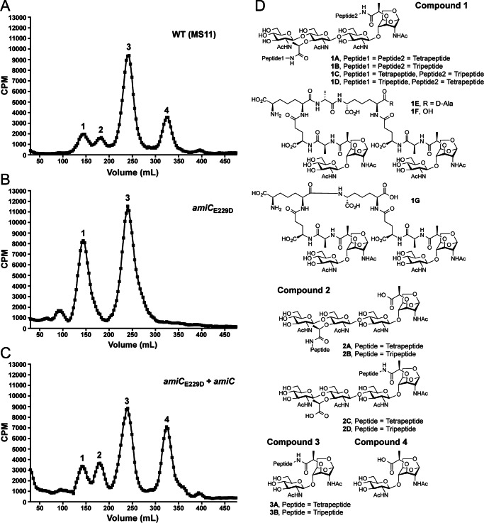
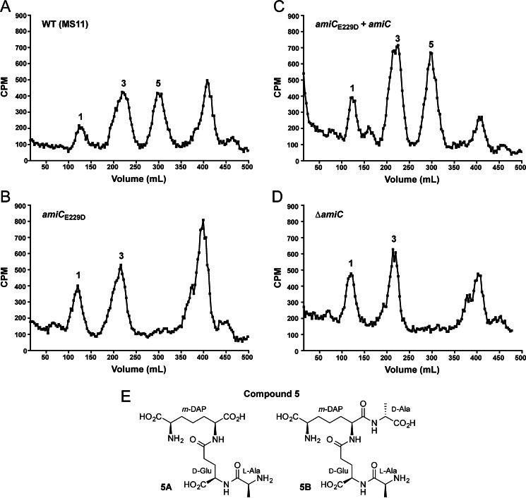
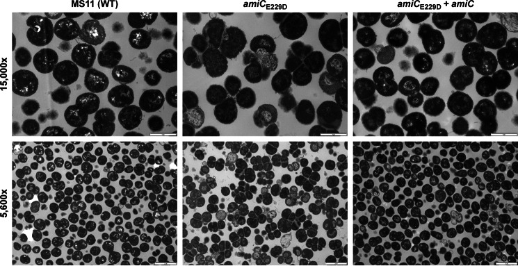
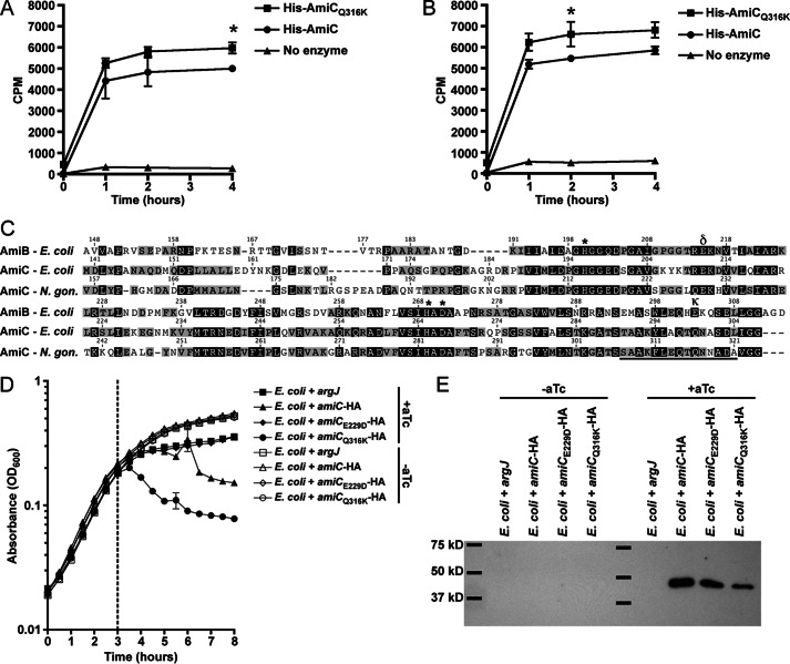
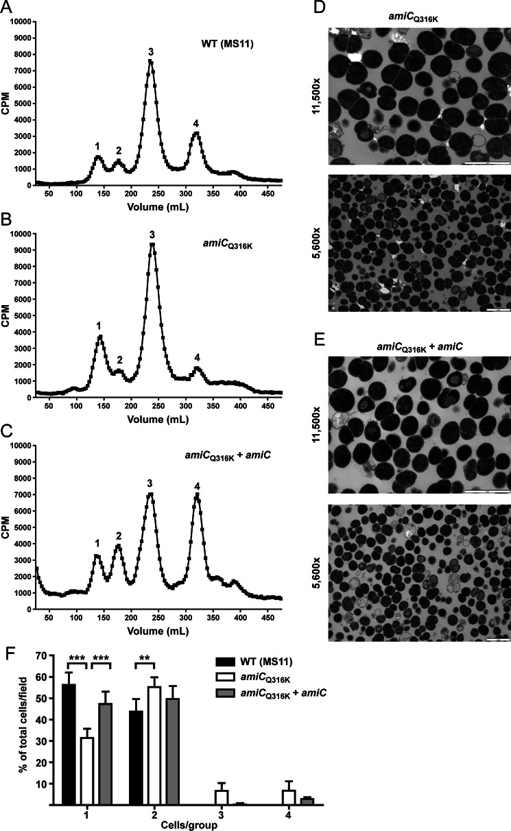
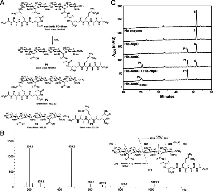
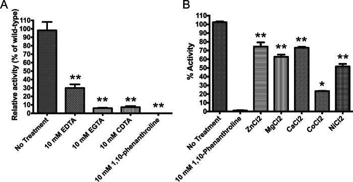
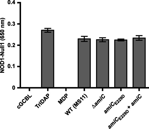
The human-restricted pathogen Neisseria gonorrhoeae encodes a single N-acetylmuramyl-l-alanine amidase involved in cell separation (AmiC), as compared with three largely redundant cell separation amidases found in Escherichia coli (AmiA, AmiB, and AmiC). Deletion of amiC from N. gonorrhoeae results in severely impaired cell separation and altered peptidoglycan (PG) fragment release, but little else is known about how AmiC functions in gonococci. Here, we demonstrated that gonococcal AmiC can act on macromolecular PG to liberate cross-linked and non-cross-linked peptides indicative of amidase activity, and we provided the first evidence that a cell separation amidase can utilize a small synthetic PG fragment as substrate (GlcNAc-MurNAc(pentapeptide)-GlcNAc-MurNAc(pentapeptide)). An investigation of two residues in the active site of AmiC revealed that Glu-229 is critical for both normal cell separation and the release of PG fragments by gonococci during growth. In contrast, Gln-316 has an autoinhibitory role, and its mutation to lysine resulted in an AmiC with increased enzymatic activity on macromolecular PG and on the synthetic PG derivative. Curiously, the same Q316K mutation that increased AmiC activity also resulted in cell separation and PG fragment release defects, indicating that activation state is not the only factor determining normal AmiC activity. In addition to displaying high basal activity on PG, gonococcal AmiC can utilize metal ions other than the zinc cofactor typically used by cell separation amidases, potentially protecting its ability to function in zinc-limiting environments. Thus gonococcal AmiC has distinct differences from related enzymes, and these studies revealed parameters for how AmiC functions in cell separation and PG fragment release.

Keywords: Gram-negative bacteria; NOD-like receptor (NLR); Neisseria gonorrhoeae; amidase; cell separation; cell wall; infectious disease; pathogen-associated molecular pattern (PAMP); peptidoglycan.


Figures

FIGURE 1.
FIGURE 1.
An amiCE229D mutant releases larger [3H]glucosamine-labeled peptidoglycan fragments relative to the wild type and no disaccharide. A–C, N. gonorrhoeae strains wild type (MS11) (A), amiCE229D (KX503) (B), and amiCE229D+amiC (JL537) (C) were metabolically pulse-chase-labeled with [3H]glucosamine, and the released peptidoglycan fragments were fractionated by size-exclusion chromatography. Traces are representative of two independent labeling and chromatography analyses. D, chemical structures of potential variants for released PG fragments found within the numbered peaks in A–C and heretofore referenced as compound families 1–4. Pictured chain lengths for compound families 1 and 2 are based upon their presence in whole sacculi, but the released proportions of compounds 1A–1G and 2A–2D are unknown. Compounds 3A and 3B are released at an ∼1:3 ratio.
FIGURE 2.
FIGURE 2.
An amiCE229D mutant does not release some [3H]DAP-labeled peptide fragments, similar to a ΔamiC strain. N. gonorrhoeae strains wild type (MS11) (A), amiCE229D (KX503) (B), amiCE229D+amiC (JL537) (C), and ΔamiC (DG005) (D) were metabolically pulse-chase-labeled with [3H]DAP, and the released peptidoglycan fragments were fractionated by size-exclusion chromatography. Structures for the compounds corresponding to the indicated fragment peaks are found in Fig. 1D (compound families 1-4) or shown here (E) (compound family 5). Traces are representative of two independent labeling and chromatography analyses.
FIGURE 3.
FIGURE 3.
amiCE229D mutation results in a defect in cell separation. Thin-section electron microscopy of wild-type (MS11), amiCE229D (KX503), and amiCE229D+amiC (JL537) strains are shown. Images are representative of multiple fields taken from each sample at each magnification, and each strain was grown, processed, and imaged a minimum of two independent times. Scale bar, 1 μm at ×15,000 and 2 μm at ×5,600.
FIGURE 4.
FIGURE 4.
amiCQ316K mutation has increased enzymatic and lytic activity, whereas amiCE229D mutation protects E. coli from lytic activity. A and B, [3H]glucosamine-labeled whole sacculi from N. gonorrhoeae (A) or an isogenic pacA mutant that does not acetylate its peptidoglycan (B) were digested with purified His-AmiC or His-AmiCQ316K at 37 °C, and samples were taken at 0, 1, 2, and 4 h. Error bars represent S.D. from duplicate reactions. Differences between His-AmiC and His-AmiCQ316K were determined by Student's t test (*, p < 0.05). Assays in A and B are representative of five independent experiments. C, protein sequence alignment of AmiB and AmiC from E. coli and AmiC from N. gonorrhoeae using MUSCLE (partial sequence displayed). Underlines indicate an α-helix known to block activity in E. coli AmiB; δ symbol indicates site of Glu-229 and κ indicates site of Gln-316 in N. gonorrhoeae; *, indicates other putative Zn-coordinating active-site residues. D, E. coli TAM1 with aTc-inducible, C-terminally HA-tagged versions of AmiC (WT), AmiCE229D, AmiCQ316K, or an untagged metabolic enzyme (argJ) were grown to mid-log phase and induced with aTc, and lysis was monitored for 5 h. Error bars represent mean ± S.D. of triplicate wells. The assay is representative of three independent experiments. E, immunoblot against the HA tag showing that AmiC and all variants are made in E. coli during lysis assays. E. coli were grown as described in D except that samples were removed for immunoblot analysis just prior to and 30 min following aTc addition. Immunoblot is representative of two independent growth experiments.
FIGURE 5.
FIGURE 5.
amiCQ316K mutation causes intermediate PG fragment release and cell separation phenotypes. A–C, N. gonorrhoeae strains wild type (MS11) (A), amiCQ316K (JL535) (B), and amiCQ316K+amiC (JL536) (C) were metabolically pulse-chase-labeled with [3H]glucosamine, and the released peptidoglycan fragments were fractionated by size-exclusion chromatography. Structures for the compounds corresponding to the indicated fragment peaks are found in the legend for Fig. 1D (compound families 14). D and E, thin-section electron microscopy of amiCQ316K (JL535) (D) and amiCQ316K+amiC (JL536) (E) strains. Images are representative of multiple fields taken for each sample at each magnification. Scale bar = 2 μm. F, quantification of thin-section electron microscopy. Cells presenting as individuals (1), diplococci (2), clusters of three (3), or clusters of four (4) were enumerated from a minimum of five fields taken of the wild-type, amiCQ316K, and amiCQ316K+amiC strains (total of >3000 cells). For each group, bars represent the mean ± S.D. of the percentage of cells in a given group within a field. Differences were determined by one-way analysis of variance (***, p < 0.001; **, p < 0.01).
FIGURE 6.
FIGURE 6.
AmiC is capable of cleaving a synthetic PG substrate. A, structure of synthetic PG substrate and four possible products of reaction with AmiC (P1–P4). Partial amidase reaction would give a tetrasaccharide-pentapeptide P1 and/or P2 (predicted mass of 1502.62) along with pentapeptide P4 (predicted mass of 532.25). The complete amidase reaction would give tetrasaccharide P3 and pentapeptide P4. B, collision-induced spectrum of AmiC tetrasaccharide-peptide product and fragmentation. The m/z values in bold (682, 822, and 1025) are unique fragment ions and can only be formed in P1, not P2. C, reverse-phase HPLC analysis of digestions of PG dimer using His-AmiC, His-NlpD, His-AmiCQ316K, or a combination of enzymes. S, synthetic dimer substrate; P1, tetrasaccharide-pentapeptide; P4, pentapeptide. Products were confirmed by mass spectrometry. HPLC analysis is representative of two independent experiments on a synthetic dimer under identical conditions used for independent LC-MS analysis of reactions (Table 2).
FIGURE 7.
FIGURE 7.
AmiC requires metal cations for activity. A, treatment of AmiC-CTD with metal chelators results in nearly complete ablation of activity. The most marked effect was with the zinc-specific chelator 1,10-phenanthroline. B, addition of metal ions rescue AmiC-CTD activity. Following treatment of AmiC-CTD with 1,10-phenanthroline, the chelator was removed by dialysis, and 10 mm metal salts were added to the protein. The addition of ZnCl2, MgCl2, and CaCl2 resulted in 70–80% restoration of protein activity. Error bars represent mean ± S.D. of triplicate reactions. (**, p < 0.01; *, p < 0.05).
FIGURE 8.
FIGURE 8.
Reducing the peptide and disaccharide release does not reduce signaling through hNOD1. HEK-Blue cells overexpressing hNOD1 and carrying a reporter driven by NF-κB and AP-1 were exposed to supernatant from wild-type (MS11), amiCE229D (KX503), amiCE229D+amiC (JL537), and ΔamiC (DG005) strains. For each assay, each strain was grown three independent times, and supernatants from each of these replicates were assayed in triplicate wells alongside positive and negative controls on the same passage of HEK-Blue cells. All exposures on hNOD1 cells were mirrored on the parental Null1 strain grown in parallel, and values from Null1 cells were subtracted as background from the values obtained for hNOD1 cells. Error bars of controls represent the mean ± S.D. of triplicate technical replicates, and error bars of experimental samples represent the means ± S.E. of the value obtained for the three biological replicates after averaging the triplicate technical replicate wells for each. TriDAP, l-Ala-γ-d-Glu-mDAP (hNOD1 agonist, 10 μg/ml); MDP, muramyl dipeptide (hNOD2 agonist, 10 μg/ml).

References

    1. Vollmer W. (2008) Structural variation in the glycan strands of bacterial peptidoglycan. FEMS Microbiol. Rev. 32, 287–306 - PubMed
    1. Centers for Disease Control and Prevention (2013) Antibiotic Resistance Threats in the United States, 2013, http://www.cdc.gov/drugresistance/threat-report-2013/
    1. Gregg C. R., Melly M. A., Hellerqvist C. G., Coniglio J. G., and McGee Z. A. (1981) Toxic activity of purified lipopolysaccharide of Neisseria gonorrhoeae for human fallopian tube mucosa. J. Infect. Dis. 143, 432–439 - PubMed
    1. Massari P., Henneke P., Ho Y., Latz E., Golenbock D. T., and Wetzler L. M. (2002) Cutting edge: immune stimulation by neisserial porins is toll-like receptor 2 and MyD88 dependent. J. Immunol. 168, 1533–1537 - PubMed
    1. Melly M. A., McGee Z. A., and Rosenthal R. S. (1984) Ability of monomeric peptidoglycan fragments from Neisseria gonorrhoeae to damage human fallopian tube mucosa. J. Infect. Dis. 149, 378–386 - PubMed

Publication types

MeSH terms

LinkOut - more resources