. 2016 Apr 29;90(10):5163-5175.
doi: 10.1128/JVI.00036-16. Print 2016 May 15.

Porcine Reproductive and Respiratory Syndrome Virus Utilizes Nanotubes for Intercellular Spread

Affiliations

Porcine Reproductive and Respiratory Syndrome Virus Utilizes Nanotubes for Intercellular Spread

Rui Guo et al. J Virol. .

Abstract

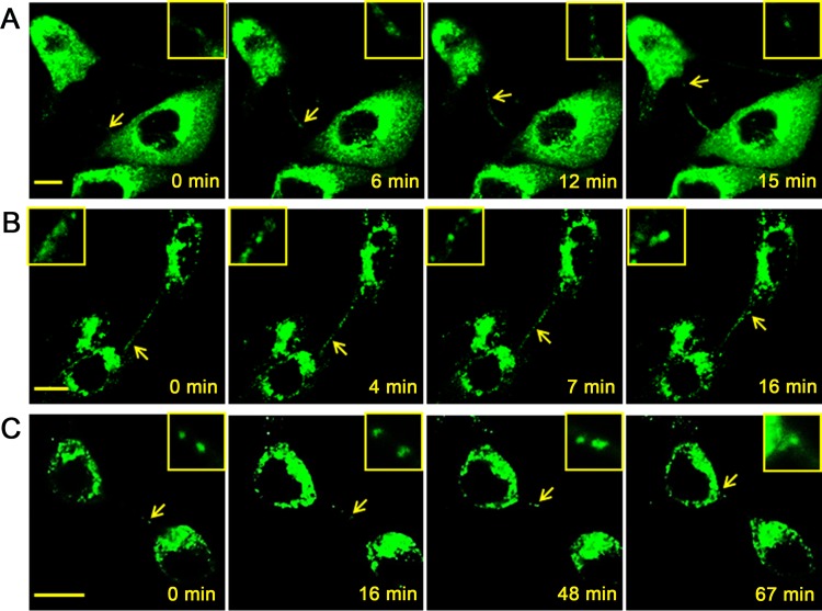
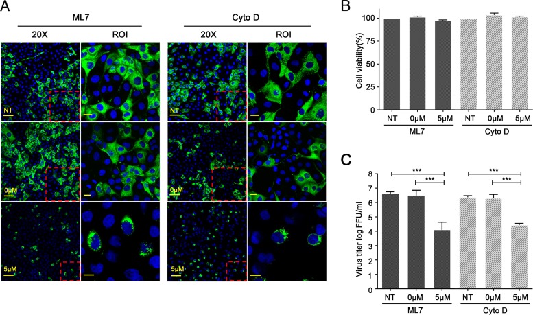
Intercellular nanotube connections have been identified as an alternative pathway for cellular spreading of certain viruses. In cells infected with porcine reproductive and respiratory syndrome virus (PRRSV), nanotubes were observed connecting two distant cells with contiguous membranes, with the core infectious viral machinery (viral RNA, certain replicases, and certain structural proteins) present in/on the intercellular nanotubes. Live-cell movies tracked the intercellular transport of a recombinant PRRSV that expressed green fluorescent protein (GFP)-tagged nsp2. In MARC-145 cells expressing PRRSV receptors, GFP-nsp2 moved from one cell to another through nanotubes in the presence of virus-neutralizing antibodies. Intercellular transport of viral proteins did not require the PRRSV receptor as it was observed in receptor-negative HEK-293T cells after transfection with an infectious clone of GFP-PRRSV. In addition, GFP-nsp2 was detected in HEK-293T cells cocultured with recombinant PRRSV-infected MARC-145 cells. The intercellular nanotubes contained filamentous actin (F-actin) with myosin-associated motor proteins. The F-actin and myosin IIA were identified as coprecipitates with PRRSV nsp1β, nsp2, nsp2TF, nsp4, nsp7-nsp8, GP5, and N proteins. Drugs inhibiting actin polymerization or myosin IIA activation prevented nanotube formation and viral clusters in virus-infected cells. These data lead us to propose that PRRSV utilizes the host cell cytoskeletal machinery inside nanotubes for efficient cell-to-cell spread. This form of virus transport represents an alternative pathway for virus spread, which is resistant to the host humoral immune response.

Importance: Extracellular virus particles transmit infection between organisms, but within infected hosts intercellular infection can be spread by additional mechanisms. In this study, we describe an alternative pathway for intercellular transmission of PRRSV in which the virus uses nanotube connections to transport infectious viral RNA, certain replicases, and certain structural proteins to neighboring cells. This process involves interaction of viral proteins with cytoskeletal proteins that form the nanotube connections. Intercellular viral spread through nanotubes allows the virus to escape the neutralizing antibody response and may contribute to the pathogenesis of viral infections. The development of strategies that interfere with this process could be critical in preventing the spread of viral infection.


Figures

FIG 1
FIG 1
Detection of intercellular nanotubes containing PRRSV GP5 and N protein. (A to D) MARC-145 cells were infected by PRRSV strain SD95-21 at an MOI of 0.1 and fixed at 12 hpi. (E and F) Porcine alveolar macrophages were infected at an MOI of 1 with PRRSV and fixed at 12 hpi. The fixed cells were immunostained for GP5 (green; A, B, and E) or N protein (green; C, D, and F) together with cytoskeleton protein of F-actin (red; A and C) or myosin IIA (red; B, D, E, and F). Images were taken by a confocal microscope (LSM 880; Zeiss). Scale bars, 10 μm (merged images) and 5 μm (zoomed images, right column).
FIG 2
FIG 2
Intercellular nanotubes contain viral proteins in PRRSV-infected cells. MARC-145 cells were infected by PRRSV strain SD95-21 at an MOI of 0.1 and fixed at 12 hpi. (A to N) The fixed cells were immunostained for nsp1β (A and H), nsp2TF (B and I), nsp2 (C and J), nsp4 (D and K), nsp7 (E and L), nsp8 (F and M), or GP4 (G and N) together with F-actin (A to G) or myosin IIA (H to N). Viral proteins were labeled with green fluorescence, and cytoskeleton proteins were labeled with red fluorescence. (O to Q) Double staining of nsp2 with GP4 (O and Q) or GP5 (P). The PRRSV nsp2 was labeled with green fluorescence, and GP4 or GP5 was labeled with red fluorescence. Pictures were taken by a confocal microscope (LSM 880; Zeiss). Scale bars, 10 μm (merged images) and 5 μm (zoomed images, right column).
FIG 2
FIG 2
Intercellular nanotubes contain viral proteins in PRRSV-infected cells. MARC-145 cells were infected by PRRSV strain SD95-21 at an MOI of 0.1 and fixed at 12 hpi. (A to N) The fixed cells were immunostained for nsp1β (A and H), nsp2TF (B and I), nsp2 (C and J), nsp4 (D and K), nsp7 (E and L), nsp8 (F and M), or GP4 (G and N) together with F-actin (A to G) or myosin IIA (H to N). Viral proteins were labeled with green fluorescence, and cytoskeleton proteins were labeled with red fluorescence. (O to Q) Double staining of nsp2 with GP4 (O and Q) or GP5 (P). The PRRSV nsp2 was labeled with green fluorescence, and GP4 or GP5 was labeled with red fluorescence. Pictures were taken by a confocal microscope (LSM 880; Zeiss). Scale bars, 10 μm (merged images) and 5 μm (zoomed images, right column).
FIG 3
FIG 3
Detection of PRRSV RNA in the intercellular nanotube connections. MARC-145 cells were infected by the PRRSV SD95-21 strain at an MOI of 0.1 and fixed at 12 hpi. (A) Viral RNA was detected by fluorescence in situ hybridization of RNA using a CAL594-labeled PRRSV N gene RNA FISH probe (red). (B) PRRSV N protein was immunostained using anti-N MAb SDOW17 (green). (C) Cell nucleus is stained with DAPI (blue). (D) The colocalized foci are readily visible in the nanotubes (yellow). Images were taken by a confocal microscope (LSM 880; Zeiss). Scale bar, 20 μm.
FIG 4
FIG 4
Intercellular spreading of PRRSV infection in cells with the presence of virus-neutralizing antibody. (A) Standard virus-neutralizing assay. Immune serum from PRRSV-infected pigs was serially diluted and incubated with the virus for 1 h; the virus-antibody complex was then added on the MARC-145 cells. At 12 hpi, cells were stained with anti-N MAb, and virus foci were counted under the fluorescence microscope. The percentage of viral growth reduction was calculated in comparison to the result from cells treated with negative-control serum. (B) Cell-to-cell spreading of the viruses under the existence of virus-neutralizing antibody. MARC-145 cells were infected with PRRSV SD95-21. At 3 hpi, the 1:4-diluted immune serum or negative-control serum was added onto the infected cells. At 6, 12, 24, or 36 hpi, cell culture supernatant was harvested, and cells were fixed and stained with anti-N MAb. Images were taken by a confocal microscope (LSM 880; Zeiss). Scale bar, 50 μm. (C) Virus titers in the harvested cell culture supernatant from the experiment described in panel B. Virus titer was determined by counting the virus foci under the fluorescence microscope, and the result was interpreted as the number of fluorescent focus units (FFU) per ml. NS, negative-control serum from a noninfected pig; PS, immune serum from a PRRSV-infected pig.
FIG 5
FIG 5
Live-cell images demonstrating intercellular transport of PRRSV GFP-tagged nsp2 through nanotubes. (A) GFP-PRRSV-infected cells maintained in culture medium containing PRRSV-neutralizing antibody. (B) GFP-PRRSV-infected cells maintained in regular cultural medium (no PRRSV-neutralizing antibody). (C) HEK-293T cells were transfected with a PRRSV full-length cDNA infectious clone, pCMV-SD95-21-GFP. At 24 h postinfection (A and B) or transfection (C), cells were analyzed using a live-cell image system of a confocal microscope (LSM 880; Zeiss). In both infected MARC-145 cells and transfected HEK-293T cells, the dot-like GFP-nsp2 proteins were visualized as moving through an intercellular nanotube connection (arrows) into the cytoplasm of a neighboring cell. Insets show a zoomed area of interest that contains the GFP-nsp2 proteins. The specific nanotube shown between MARC-145 cells is about 20 μm in length, while the specific nanotube shown between HEK-293T cells is about 12 μm in length. Scale bar, 10 μm.
FIG 6
FIG 6
Intercellular transport of the GFP-nsp2 protein in cocultured MARC-145 and HEK-293T cells. (A) GFP-PRRSV-infected MARC-145 cells were fixed and stained with anti-nsp2 MAb 140-68 and anti-SV40 T antigen pAb sc-20800. (B) GFP-PRRSV-infected HEK-293T cells were fixed and stained with anti-nsp2 MAb 140-68 and anti-SV40 T antigen pAb sc-20800. (C) GFP-PRRSV-infected MARC-145 cells were trypsinized at 12 hpi and mixed with naive HEK-293T cells. At 36 h postcultivation, cells were fixed and stained with anti-nsp2 MAb 140-68 and anti-SV40 T antigen pAb sc-20800. The nsp2 was labeled with green fluorescence, and SV40 large T antigen was labeled with red fluorescence. Pictures were taken by a confocal microscope (LSM 880; Zeiss). Scale bar, 10 μm.
FIG 7
FIG 7
Coprecipitation of PRRSV proteins with cytoskeleton proteins F-actin and myosin IIA. (A to C) PRRSV-infected (+) MARC-145 cell lysates or mock-infected (−) cell lysates were used for immunoprecipitation using PRRSV protein-specific MAbs as indicated on the top of each panel. Anti-mouse IgG (α-mIgG) was used as a control. The immunoprecipitated proteins were separated by 8 to 16% Tris-glycine gradient gels (A) and immunoblotted by anti-myosin IIA pAb and anti-F-actin MAb (B and C). (D to L) PRRSV-infected MARC-145 cell lysates (+) or mock-infected (−) cell lysates were used for immunoprecipitation using anti-myosin IIA pAb. The immunoprecipitated proteins were separated by 8 to 16% Tris-glycine gradient gels and immunoblotted by PRRSV protein-specific MAbs as indicated on the bottom of each panel. IP, immunoprecipitation; WB, Western blotting. Arrows in panels A to C indicate F-actin (42 kDa) and nonmuscle myosin heavy chain IIA (215 kDa). Arrows in Panels D, E, G, H, and J indicate the target protein, as shown at the bottom of each panel.
FIG 8
FIG 8
Inhibitors of F-actin and myosin IIA affect the intercellular spread of PRRSV. (A) Confluent MARC-145 cell monolayers were pretreated with ML7 or cytochalasin D (Cyto D) compound at a concentration of 5 μM or 0 μM. Nontreated cells (NT) were used as a control. After a 30-min incubation at 37°C, the cells were infected at an MOI of 0.1 with PRRSV. After 1 h of incubation, the infected cells were washed with PBS, and fresh medium containing cytochalasin D or ML7 compound was added. At 24 hpi, cells were fixed and immunostained with PRRSV N protein-specific MAb. Images in columns 1 and 3 were taken using a 20× objective lens (scale bar, 50 μm). The inset at the bottom right corner of each image shows a ×40 zoomed region of interest (ROI). Scale bar for the inset, 10 μm. (B) Effect of cytochalasin D or ML7 on the viability of the MARC-145 cells compared to the viability of DMSO-treated (0 μM) or nontreated (NT) control cells. Graphs show the results (mean and standard deviation) of a representative experiment (n = 3). (C) Viral growth inhibition efficiency of the myosin IIA inhibitor ML7 or F-actin inhibitor cytochalasin D. Virus titration was conducted using cell culture supernatant collected from the experiment in shown in panel A. Virus titer was determined by counting the virus foci under the fluorescence microscope, and the result was interpreted as fluorescent focus units (FFU) per milliliter. Statistical significance between the groups was determined by a one-way analysis of variance test using GraphPad InStat Prism (version 5.0), and a P value of < 0.01 (***) was considered statistically significant.

References

    1. Marsh M, Helenius A. 1989. Virus entry into animal-cells. Adv Virus Res 36:107–151. doi: 10.1016/S0065-3527(08)60583-7. - DOI - PMC - PubMed
    1. Zhong P, Agosto LM, Munro JB, Mothes W. 2013. Cell-to-cell transmission of viruses. Curr Opin Virol 3:44–50. doi: 10.1016/j.coviro.2012.11.004. - DOI - PMC - PubMed
    1. Mothes W, Sherer NM, Jin J, Zhong P. 2010. Virus cell-to-cell transmission. J Virol 84:8360–8368. doi: 10.1128/JVI.00443-10. - DOI - PMC - PubMed
    1. Sattentau Q. 2008. Avoiding the void: cell-to-cell spread of human viruses. Nat Rev Microbiol 6:815–826. doi: 10.1038/nrmicro1972. - DOI - PubMed
    1. Xu WF, Santini PA, Sullivan JS, He B, Shan MM, Ball SC, Dyer WB, Ketas TJ, Chadburn A, Cohen-Gould L, Knowles DM, Chiu A, Sanders RW, Chen K, Cerutti A. 2009. HIV-1 evades virus-specific IgG2 and IgA responses by targeting systemic and intestinal B cells via long-range intercellular conduits. Nat Immunol 10:1008–U1106. doi: 10.1038/ni.1753. - DOI - PMC - PubMed

Publication types

MeSH terms

LinkOut - more resources