Review
. 2016 Mar 16;89(6):1131-1156.
doi: 10.1016/j.neuron.2016.02.017.

Unifying Views of Autism Spectrum Disorders: A Consideration of Autoregulatory Feedback Loops

Affiliations
Review

Unifying Views of Autism Spectrum Disorders: A Consideration of Autoregulatory Feedback Loops

Caitlin Mullins et al. Neuron. .

Abstract

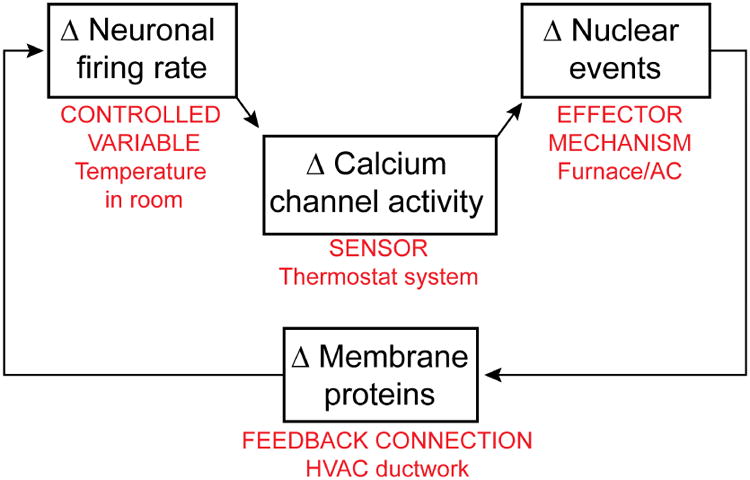
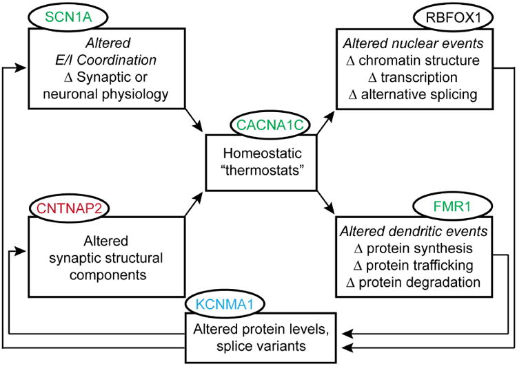
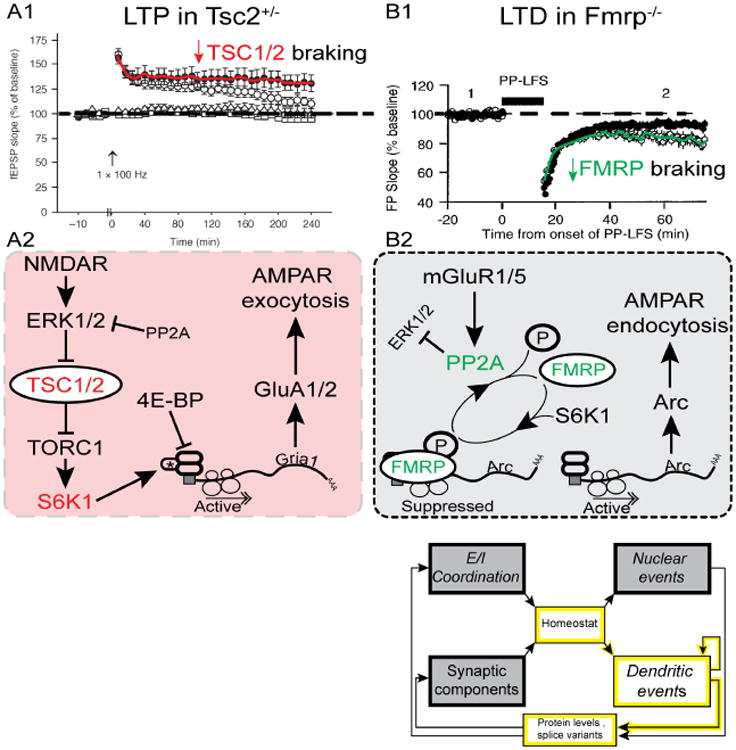
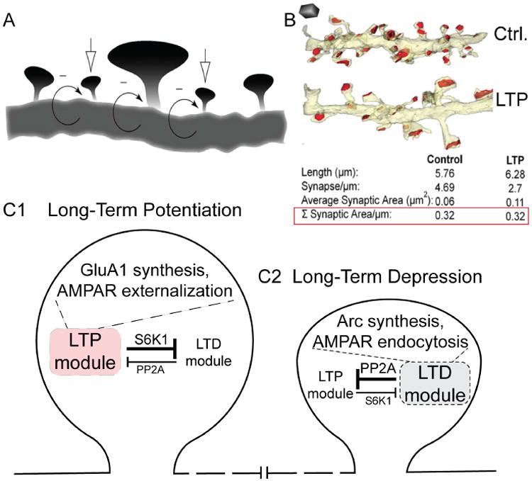
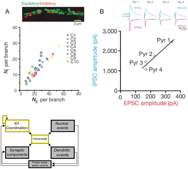
Understanding the mechanisms underlying autism spectrum disorders (ASDs) is a challenging goal. Here we review recent progress on several fronts, including genetics, proteomics, biochemistry, and electrophysiology, that raise motivation for forming a viable pathophysiological hypothesis. In place of a traditionally unidirectional progression, we put forward a framework that extends homeostatic hypotheses by explicitly emphasizing autoregulatory feedback loops and known synaptic biology. The regulated biological feature can be neuronal electrical activity, the collective strength of synapses onto a dendritic branch, the local concentration of a signaling molecule, or the relative strengths of synaptic excitation and inhibition. The sensor of the biological variable (which we have termed the homeostat) engages mechanisms that operate as negative feedback elements to keep the biological variable tightly confined. We categorize known ASD-associated gene products according to their roles in such feedback loops and provide detailed commentary for exemplar genes within each module.


Figures

Fig 1
Fig 1. Ca2+ regulation of gene expression in neuronal development & homeostasis
A canonical homeostatic feedback loop, proposed by Abbott, Marder et al. (LeMasson et al., 1993; Marder et al., 1996; Siegel et al., 1994), hypothesizes that Δ firing → Δ Ca2+ → Δ gene expression → Δ membrane channel proteins regulating neuronal excitability. This feedback loop is taken as key to how neuronal firing is kept away from extremes. The components of the feedback loop can be likened to elements of a temperature control system (labeled in red).
Fig 2
Fig 2. Homeostatic feedback through a multi-pathway loop
Expanding upon Figure 1 to incorporate alterations in both nuclear and dendritic events that arise during a homeostatic response. These effector mechanisms operate via feedback to alter synaptic or neuronal physiology and synaptic structure. ASD-associated mutations have been identified in genes associated with each component of this feedback loop (ovals); dysfunction in any one section can affect the operation of the entire loop to impact function at a variety of levels. This schematic representation of autoregulation appears in simplified versions in later figures (3, 4, and 6), highlighting the specific modules being discussed.
Fig. 3
Fig. 3. Canonical Wnt/β-catenin signaling pathway and ASD-related protein players
Hepatocyte growth factor (HGF) activation of the MET receptor tyrosine kinase releases β-catenin (CTNNB1) from surface cadherin molecules. β-catenin in the cytoplasm is bound by proteins that make up a destruction complex (DC). The DC marks β-catenin for proteasomal degradation. Wnt2 signaling via binding with a Frizzled receptor (FZD9) and its co-receptor LRP5/6 activates dishevelled (DVL), which saves β-catenin from degradation by inhibiting the DC. When DVL is active, β-catenin enters the nucleus, where it activates gene transcription by binding with the TCF/LEF1 family of transcription factors. Gene names are shown in bold if associated with ASD and present in Table 1 (utilizing the same color-coding scheme). The icon, simplified from Fig. 2, highlights the specific modules directly involved in the pathway illustrated by this figure (and utilized in the same manner for Figs. 4 and 6).
Fig. 4
Fig. 4. Linking L-type channels to nuclear events
Calcium entering through CaV1.2 channels (CACNA1C) binds to calmodulin (CALM1, CaM), and triggers activation of α2/β-CaMKII (CAMK2A/B), and calcineurin (PPP3CC, CaN). The aggregation of α2/β-CaMKII at postsynaptic sites may contribute to synaptic tagging, a key step in long-lasting LTP (Frey and Morris, 1997; Hudmon et al., 2005; Redondo et al., 2010). γCaMKII (CAMK2G) is phosphorylated by α2/β-CaMKII to trap the Ca2+/CaM signal in place, and dephosphorylated at a different residue by CaN. This dephosphorylation event exposes a nuclear localization sequence on γCaMKII and triggers translocation, shuttling Ca2+/CaM into the nucleus. Nuclear Ca2+/CaM activates CaMKK (CAMKK1/2) and CaMKIV (CAMK4), which phosphorylates CREB, activating CRE-mediated gene transcription. CaMKIV also phosphorylates CREB binding protein (CBP, CREBBP), further favoring transcription, and activates alternative splicing, via splicing factors such as Rbfox1 (RBFOX1). In this scheme, both βCaMKII and CaN act in close proximity to CaV1 channels, while γCaMKII appears to operate as a carrier, not a kinase. Gene names are shown in bold if associated with ASD and present in Table 1 (utilizing the same color-coding scheme).
Fig. 5
Fig. 5. Contrasting roles of TSC1/2 and FMRP
A1, LTP is exaggerated in Tsc2+/- mice. From (Ehninger et al., 2008). A2, Activation of NMDARs leads to the inhibition of TSC1/2, which disinhibits TORC1. Activation of TORC1 leads to the translation of proteins important for LTP, including GluA1/2 subunits. AMPAR exocytosis supports LTP. Loss of TSC1/2 (as seen in tuberous sclerosis) would lead to excessive TORC1 activation, and exaggerated LTP (experimentally supported by A1). B1, LTD is exaggerated in FMRP KO mice. From (Huber et al., 2002)(Copyright 2002 National Academy of Sciences, USA). B2, Putative signaling pathway linking metabotropic GluR action to local translation of Arc and LTD. Activation of mGluR1/5 activates PP2A, which dephosphorylates FMRP. This releases mRNA transcripts from FMRP repression, and allows the local translation of LTD-relevant proteins. Translation of Arc supports readout of low spine activity by β-CaMKII, Arc-dependent AMPAR endocytosis, and LTD (Okuno et al., 2012). Loss of FMRP (as seen in FXS), leads to decreased inhibition of translation, and increased LTD (see panel B1).
Fig. 6
Fig. 6. Competing LTD and LTP processes
A, LTP and LTD are illustrated here as occurring within the same dendritic compartment, at neighboring spines. From (Rabinowitch and Segev, 2008). B, Following LTP-induction protocol, some spines increase in size, without the overall synaptic area changing. From (Bourne and Harris, 2012). C1,C2, LTP and LTD are seen as competing processes within single spines. PP2A activation is critical for the LTD module but inhibits the LTP module, whereas S6K1 is critical for the LTP module but inhibits the LTD module. Mutual inhibition supports a winner-take-all scenario. When the action of S6K1 predominates, the LTP module overrides LTD, and GluA1 synthesis and AMPAR externalization takes place (C1; Fig. 5A2). When the action of PP2A predominates, the LTD module prevails over LTP, and Arc synthesis and AMPAR endocytosis takes place (C2; Fig. 5B2). For clarity, this model focuses on simple aspects of multiple mutually inhibitory interactions between the FMRP and TSC1/2 pathways (Ashley and Warren, 1995; Bian et al., 2015; Darnell and Klann, 2013).
Fig. 7
Fig. 7. E:I coordination in neuronal, circuit, and pathophysiological contexts
A, Coordination (but not equality) of the numbers of excitatory and inhibitory synapses per dendritic branch (NE, NI respectively). NI is ∼0.5× NE. From (Liu, 2004). B, Coordination (but not equality) of the collective strengths of E and I transmission, illustrated for 4 pyramidal neurons driven by the same optogenetic stimulation of layer IV feedforward inputs. Inset shows recordings of EPSCs (red) and IPSCs (blue) with |driving force| of ∼70 mV. |IPSC| is ∼7×|EPSC|, reflecting a ∼7-fold ratio of synaptic conductances. From (Xue et al., 2014).

References

    1. Aguilar-Valles A, Matta-Camacho E, Khoutorsky A, Gkogkas C, Nader K, Lacaille JC, Sonenberg N. Inhibition of Group I Metabotropic Glutamate Receptors Reverses Autistic-Like Phenotypes Caused by Deficiency of the Translation Repressor eIF4E Binding Protein 2. J Neurosci. 2015;35:11125–11132. - PMC - PubMed
    1. Alessi DR, Gomez N, Moorhead G, Lewis T, Keyse SM, Cohen P. Inactivation of p42 MAP kinase by protein phosphatase 2A and a protein tyrosine phosphatase, but not CL100, in various cell lines. Curr Biol. 1995;5:283–295. - PubMed
    1. Alvarez Retuerto AI, Cantor RM, Gleeson JG, Ustaszewska A, Schackwitz WS, Pennacchio LA, Geschwind DH. Association of common variants in the Joubert syndrome gene (AHI1) with autism. Hum Mol Genet. 2008;17:3887–3896. - PMC - PubMed
    1. Amir RE, Van den Veyver IB, Wan M, Tran CQ, Francke U, Zoghbi HY. Rett syndrome is caused by mutations in X-linked MECP2, encoding methyl-CpG-binding protein 2. Nature genetics. 1999;23:185–188. - PubMed
    1. Anderson DJ, Jan YN. The determination of the neuronal phenotype. In: Cowan WM, Jessell TM, Zipursky SL, editors. Molecular and Cellular Approaches to Neural Development. New York: Oxford University Press; 1997. pp. 26–63.

Publication types

MeSH terms