. 2016 Mar 18:6:22772.
doi: 10.1038/srep22772.

The fertilization-induced zinc spark is a novel biomarker of mouse embryo quality and early development

Affiliations

The fertilization-induced zinc spark is a novel biomarker of mouse embryo quality and early development

Nan Zhang et al. Sci Rep. .

Abstract

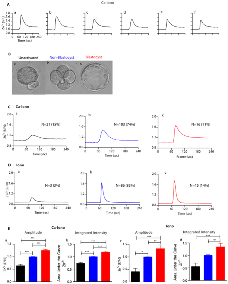
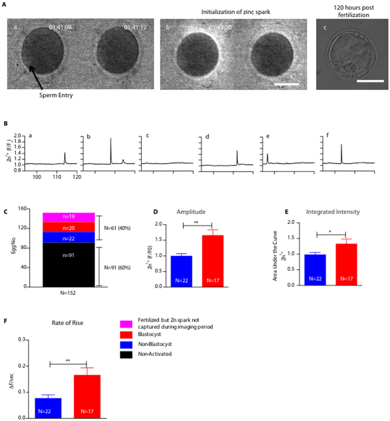
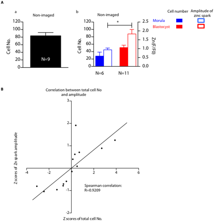
Upon activation, mammalian eggs release billions of zinc ions in an exocytotic event termed the "zinc spark." The zinc spark is dependent on and occurs coordinately with intracellular calcium transients, which are tightly associated with embryonic development. Thus, we hypothesized that the zinc spark represents an early extracellular physicochemical marker of the developmental potential of the zygote. To test this hypothesis, we monitored zinc exocytosis in individual mouse eggs following parthenogenetic activation or in vitro fertilization (IVF) and tracked their development. Retrospective analysis of zinc spark profiles revealed that parthenotes and zygotes that developed into blastocysts released more zinc than those that failed to develop. Prospective selection of embryos based on their zinc spark profile significantly improved developmental outcomes and more than doubled the percentage of embryos that reached the blastocyst stage. Moreover, the zinc spark profile was also associated with embryo quality as the total cell number in the resulting morulae and blastocysts positively correlated with the zinc spark amplitude (R = 0.9209). Zinc sparks can thus serve as an early biomarker of zygote quality in mouse model.


Figures

Figure 1
Figure 1. Distinct Ionomycin-induced zinc spark profiles are correlated with egg activation and blastocyst formation.
(A) Representative time traces of normalized zinc fluorescence (F/F0) following activation by 5 μM Ca-Iono. F0 was calculated by averaging the first five fluorescence measurements for each egg prior to zinc spark. All six eggs (a-f) were collected from the same individual mouse. (B) Representative images of unactivated eggs (a, black color), non-blastocysts (b, blue color) and blastocysts (c, red color). (C) Representative profiles of Ca-Iono induced zinc sparks in the eggs that showed differential developmental outcomes: (a) unactivated, (b) non-blastocyst, and (c) blastocyst. (D) Representative profiles of Iono induced zinc sparks in the eggs that showed differential developmental outcomes: (a) unactivated, (b) non-blastocyst, and (c) blastocyst. (E) Amplitude (a,c) and integrated intensity (b,d) of the Ca-Iono (a,b) or Iono (c,d) induced zinc sparks in unactivated eggs (black), non-blastocysts (blue) and blastocysts (red). The non-blastocyst group was normalized to one and values for the other groups are presented relative to that. (*p < 0.05; ** p < 0.01; ***p < 0.001).
Figure 2
Figure 2. Larger zinc spark release during IVF is associated with embryos that develop to the blastocyst stage.
(A) (a) Representative images of a MII egg being binding a single sperm. Asterisks and an arrow denote the location where the sperm entered the egg; (b) the egg released zinc shortly after the sperm entry; (c) image of the same fertilized eggs 120 hours post zinc imaging. (B) Representative time traces of normalized zinc sparks (F/F0) in mouse eggs upon fertilization. (C) Statistical summary of the outcomes of the imaged eggs. (D) Comparison of amplitude of zinc sparks in the non-blastocyst and blastocyst embryos. (E) Comparison of the integrated intensity of zinc sparks in the non-blastocyst and blastocyst embryos; (F) Comparison of the rate of rise of zinc sparks in non-blastocyst and blastocyst embryos. (*p < 0.05; **p < 0.01; ***p < 0.001).
Figure 3
Figure 3. Zinc sparks are highly correlated with the total cell number of late stage pre-implantation embryos.
(A) (a) Cell numbers of the embryos that were not imaged during fertilization (non-imaged); (b) amplitude of zinc sparks (empty bars) and total cell numbers (solid bars) in the embryos that arrested at morula stage (blue) or reached blastocyst stage (red) 120 hours post zinc imaging. (B) Z scores, which is calculated as (x-xbar)/SD, of total cell number of embryos and the z scores of the zinc spark amplitude in the imaged embryos from (A) are highly correlated. (*p < 0.05, ***p < 0.001).

References

    1. Bernhardt M. L., Kim A. M., O’Halloran T. V. & Woodruff T. K. Zinc requirement during meiosis I-meiosis II transition in mouse oocytes is independent of the MOS-MAPK pathway. Biology of reproduction 84, 526–536, doi: 10.1095/biolreprod.110.086488 (2011). - DOI - PMC - PubMed
    1. Bernhardt M. L., Kong B. Y., Kim A. M., O’Halloran T. V. & Woodruff T. K. A zinc-dependent mechanism regulates meiotic progression in mammalian oocytes. Biology of reproduction 86, 114, doi: 10.1095/biolreprod.111.097253 (2012). - DOI - PMC - PubMed
    1. Kim A. M. et al. Zinc sparks are triggered by fertilization and facilitate cell cycle resumption in mammalian eggs. ACS Chem. Biol. 6, 716–723 (2011). - PMC - PubMed
    1. Kim A. M., Vogt S., O’Halloran T. V. & Woodruff T. K. Zinc availability regulates exit from meiosis in maturing mammalian oocytes. Nature chemical biology 6, 674–681, doi: 10.1038/nchembio.419 (2010). - DOI - PMC - PubMed
    1. Kong B. Y., Bernhardt M. L., Kim A. M., O’Halloran T. V. & Woodruff T. K. Zinc maintains prophase I arrest in mouse oocytes through regulation of the MOS-MAPK pathway. Biology of reproduction 87, 11, 11–12, doi: 10.1095/biolreprod.112.099390 (2012). - DOI - PMC - PubMed

Publication types