. 2016 May;5(5):683-93.
doi: 10.5966/sctm.2015-0231. Epub 2016 Mar 17.

Systemic Mesenchymal Stromal Cell Transplantation Prevents Functional Bone Loss in a Mouse Model of Age-Related Osteoporosis

Affiliations

Systemic Mesenchymal Stromal Cell Transplantation Prevents Functional Bone Loss in a Mouse Model of Age-Related Osteoporosis

Jeffrey Kiernan et al. Stem Cells Transl Med. 2016 May.

Abstract

Age-related osteoporosis is driven by defects in the tissue-resident mesenchymal stromal cells (MSCs), a heterogeneous population of musculoskeletal progenitors that includes skeletal stem cells. MSC decline leads to reduced bone formation, causing loss of bone volume and the breakdown of bony microarchitecture crucial to trabecular strength. Furthermore, the low-turnover state precipitated by MSC loss leads to low-quality bone that is unable to perform remodeling-mediated maintenance--replacing old damaged bone with new healthy tissue. Using minimally expanded exogenous MSCs injected systemically into a mouse model of human age-related osteoporosis, we show long-term engraftment and markedly increased bone formation. This led to improved bone quality and turnover and, importantly, sustained microarchitectural competence. These data establish proof of concept that MSC transplantation may be used to prevent or treat human age-related osteoporosis.

Significance: This study shows that a single dose of minimally expanded mesenchymal stromal cells (MSCs) injected systemically into a mouse model of human age-related osteoporosis display long-term engraftment and prevent the decline in bone formation, bone quality, and microarchitectural competence. This work adds to a growing body of evidence suggesting that the decline of MSCs associated with age-related osteoporosis is a major transformative event in the progression of the disease. Furthermore, it establishes proof of concept that MSC transplantation may be a viable therapeutic strategy to treat or prevent human age-related osteoporosis.

Keywords: Mesenchymal stem cell; Osteoporosis; Sca-1; Stem cell transplantation; Tissue-specific stem cells.


Figures

Figure 1.
Figure 1.
Isolation and differentiation capacity of donor MSCs. (A): A workflow diagram showing WT MSC isolation. (B): Single cell derived colony derived from passage 1 donor MSCs (×4 magnification, Crystal Violet stain). (C): Passage 1 donor MSCs had a CFU-F frequency of 1:6.5 (n = 3). (D): Passage 1 donor MSCs grown under osteogenic conditions form bone nodules (×4 magnification, Von Kossa stain). (E): Passage 1 donor MSCs grown under adipogenic conditions form adipocytes (×10 magnification, Oil Red O stain). Abbreviations: CFU-F, colony-forming unit fibroblast; MSCs, mesenchymal stromal cells; P1, passage 1; WT, wild type.
Figure 2.
Figure 2.
Short-term analysis confirms systemically injected purified MSCs are delivered and retained in the bone marrow, lungs, and liver. (A): In vivo fluorescent imaging (top) and x-ray (bottom) of untreated naïve and DiR+ MSC-transplanted Sca-1−/− mice. (B): Ex vivo fluorescent imaging of organs/bones, color (top) and fluorescence (bottom). (C): DiR fluorescence present in bones 2 weeks after DiR+ MSC transplant in Sca-1−/− mice compared with naïve Sca-1−/− mice and percent presence of DiR-labeled cells in the BM and CB assessed via flow cytometry (n = 3). Abbreviations: Avg, average; B, bones; BM, bone marrow; CB, compact bone; DiR, 1,1′-dioctadecyl-3,3,3′,3′-tetramethylindotricarbocyanine iodide; H, heart; K, kidney; Li, liver; Lu, lungs; MSCs, mesenchymal stromal cells; S, spleen.
Figure 3.
Figure 3.
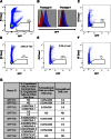
Transplanted GFP+ MSCs are capable of persistent engraftment in the marrow/endosteal region of Sca-1−/− for 6 months. (A–F): Flow cytometric analysis. (A): GFP signal of total bone marrow/compact bone cells from donor GFP-LUC mice. (B): GFP signal of donor MSCs at passage 0 and 1. (C): GFP signal of enriched cells from donor GFP-LUC mice. (D, E): Two examples of mice that displayed GFP+ cells. (F): GFP signal was absent from naïve Sca-1−/− mice. (G): Table displaying quantitative polymerase chain reaction detection of Y-chromosome engraftment; percent donor engraftment and engrafted cells per million of enriched nonhematopoietic/nonendothelial fraction of bone marrow/compact bone cells, percent donor engraftment, and cells per million in total nucleated bone marrow cells; percent donor engraftment and cells per million of total lung cells; and percent donor engraftment in total liver cells. Abbreviations: ctrl, control; GFP, green fluorescent protein; LUC, luciferase; MSCs, mesenchymal stromal cells; ND, not determined; PI, propidium iodide.
Figure 4.
Figure 4.
Sca-1−/− mice that received a single MSC transplant display increased rate of bone formation, osteoclast activity, and bone turnover 6 months after transplant. (A): Representative fluorescent images of calcein staining in WT, naïve Sca-1−/−, and MSC-transplanted Sca-1−/− mice. (B): Percent mineralizing surface normalized to bone surface. (C): Bone formation rate normalized to bone surface. (D): Bone formation rate normalized to bone volume. (E): Tartrate-resistant acid phosphatase (TRAP) analysis of osteoclast surface normalized to bone surface. (F): TRAP analysis of osteoclast number normalized to bone surface. (G): BSE imaging quantification of FWHMH, a measure of bone turnover. ∗, p ≤ .05; †0.1 ≥ p ≥ .05 [WT (n = 5 dynamic histo, n = 4 TRAP, BSE) Sca-1−/− (n = 6 dynamic histo, TRAP, n = 5 BSE), MSC Tx (n = 8, all)]. Abbreviations: BFR/BS, bone formation rate normalized to bone surface; BFR/BV, bone formation rate normalized to bone volume; BSE, backscattered electron; FWHMH, full width at half-maximal height; MS/BS, percent mineralizing surface normalized to bone surface; MSC, mesenchymal stromal cell; MSC Tx, MSC-transplanted; OcN/BS, osteoclast number normalized to bone surface; OcS/BS, osteoclast surface normalized to bone surface; WT, wild type.
Figure 5.
Figure 5.
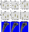
Sca-1−/− mice that received a single MSC transplant display improved trabecular microarchitecture 6 months after transplant. MicroCT analysis of proximal tibias of WT, naïve Sca-1−/−, and MSC-transplanted Sca-1−/− mice (MSC Tx). Parameters investigated were as follows. (A): Connectivity density. (B): Degree of anisotropy. (C): Bone volume/total volume. (D): Trabecular number. (E): Trabecular spacing. (F): Structural Model Index. (G): Representative microCT images of WT, Sca-1−/−, and MSC Tx mice. Scale bar = 1.0 mm. ∗, p ≤ .05; †, 0.1 ≥ p ≥ .05 [WT (n = 7), Sca-1−/− (n = 9), and MSC Tx (n = 8)]. Abbreviations: BV/TV, bone volume/total volume; Conn.D, connectivity density; DA, degree of anisotropy; microCT, microcomputed tomography; MSC, mesenchymal stromal cell; MSC Tx, MSC-transplanted; SMI, Structural Model Index; Tb.N, trabecular number; Tb.Sp, trabecular spacing; WT, wild type.

References

    1. Thompson D. On Growth and Form. New York, NY: Macmillan Co.; 1945. p. 1116.
    1. Wolff J. Das gesetz der transformation der knochen. Deutsche Medizinische Wochenschrift. 1892;19:1222–1224.
    1. Hodgskinson R, Currey J. The effect of variation in structure on the Young’s modulus of cancellous bone: A comparison of human and non-human material. Proc Inst Mech Eng H. 1990;204:115–121. - PubMed
    1. Goulet RW, Goldstein SA, Ciarelli MJ, et al. The relationship between the structural and orthogonal compressive properties of trabecular bone. J Biomech. 1994;27:375–389. - PubMed
    1. Manolagas SC, Parfitt AM. What old means to bone. Trends Endocrinol Metab. 2010;21:369–374. - PMC - PubMed

Publication types

MeSH terms