

Normative NeuroFlexor data for detection of spasticity after stroke: a cross-sectional study
- PMID: 26987557
- PMCID: PMC4797345
- DOI: 10.1186/s12984-016-0133-x
Normative NeuroFlexor data for detection of spasticity after stroke: a cross-sectional study
Abstract
Background and objective: The NeuroFlexor is a novel instrument for quantification of neural, viscous and elastic components of passive movement resistance. The aim of this study was to provide normative data and cut-off values from healthy subjects and to use these to explore signs of spasticity at the wrist and fingers in patients recovering from stroke.
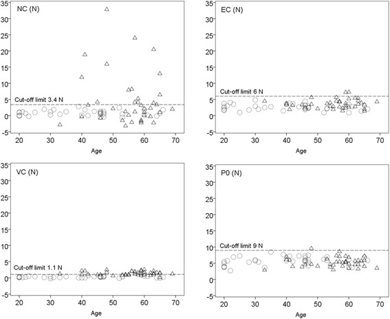
Methods: 107 healthy subjects (age range 28-68 years; 51 % females) and 39 stroke patients (age range 33-69 years; 33 % females), 2-4 weeks after stroke, were assessed with the NeuroFlexor. Cut-off values based on mean + 3SD of the reference data were calculated. In patients, the modified Ashworth scale (MAS) was also applied.
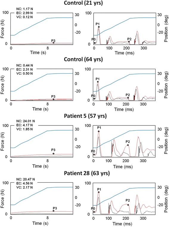
Results: In healthy subjects, neural component was 0.8 ± 0.9 N (mean ± SD), elastic component was 2.7 ± 1.1 N, viscous component was 0.3 ± 0.3 N and resting tension was 5.9 ± 1 N. Age only correlated with elastic component (r = -0.3, p = 0.01). Elasticity and resting tension were higher in males compared to females (p = 0.001) and both correlated positively with height (p = 0.01). Values above healthy population cut-off were observed in 16 patients (41 %) for neural component, in 2 (5 %) for elastic component and in 23 (59 %) for viscous component. Neural component above cut-off did not correspond well to MAS ratings. Ten patients with MAS = 0 had neural component values above cut-off and five patients with MAS ≥ 1 had neural component within normal range.
Conclusion: This study provides NeuroFlexor cut-off values that are useful for detection of spasticity in the early phase after stroke.
Keywords: Biomechanics; Muscle spasticity; Normative data; Stroke; Upper extremity.
Figures
Similar articles
-
Quantifying neural and non-neural components of wrist hyper-resistance after stroke: Comparing two instrumented assessment methods.Med Eng Phys. 2021 Dec;98:57-64. doi: 10.1016/j.medengphy.2021.10.009. Epub 2021 Oct 24. Med Eng Phys. 2021. PMID: 34848039
-
Validity, Intra-Rater Reliability and Normative Data of the Neuroflexor™ Device to Measure Spasticity of the Ankle Plantar Flexors after Stroke.J Rehabil Med. 2023 Mar 3;55:jrm00356. doi: 10.2340/jrm.v54.2067. J Rehabil Med. 2023. PMID: 36867093
-
Test-retest and inter-rater reliability of a method to measure wrist and finger spasticity.J Rehabil Med. 2013 Jul;45(7):630-6. doi: 10.2340/16501977-1160. J Rehabil Med. 2013. PMID: 23695917
-
Sensitivity of the NeuroFlexor method to measure change in spasticity after treatment with botulinum toxin A in wrist and finger muscles.J Rehabil Med. 2014 Jul;46(7):629-34. doi: 10.2340/16501977-1824. J Rehabil Med. 2014. PMID: 24850135
-
Measurement Properties of the NeuroFlexor Device for Quantifying Neural and Non-neural Components of Wrist Hyper-Resistance in Chronic Stroke.Front Neurol. 2019 Jul 3;10:730. doi: 10.3389/fneur.2019.00730. eCollection 2019. Front Neurol. 2019. PMID: 31379705 Free PMC article.
Cited by
-
Rigidity in Parkinson's disease: evidence from biomechanical and neurophysiological measures.Brain. 2023 Sep 1;146(9):3705-3718. doi: 10.1093/brain/awad114. Brain. 2023. PMID: 37018058 Free PMC article.
-
Robot-Aided Systems for Improving the Assessment of Upper Limb Spasticity: A Systematic Review.Sensors (Basel). 2020 Sep 14;20(18):5251. doi: 10.3390/s20185251. Sensors (Basel). 2020. PMID: 32937973 Free PMC article.
-
A domestic robotic rehabilitation device for assessment of wrist function for outpatients.J Rehabil Assist Technol Eng. 2020 Dec 4;7:2055668320961233. doi: 10.1177/2055668320961233. eCollection 2020 Jan-Dec. J Rehabil Assist Technol Eng. 2020. PMID: 33329903 Free PMC article.
-
Effects of 60 Min Electrostimulation With the EXOPULSE Mollii Suit on Objective Signs of Spasticity.Front Neurol. 2021 Oct 15;12:706610. doi: 10.3389/fneur.2021.706610. eCollection 2021. Front Neurol. 2021. PMID: 34721255 Free PMC article.
-
Changes in the Neural and Non-neural Related Properties of the Spastic Wrist Flexors After Treatment With Botulinum Toxin A in Post-stroke Subjects: An Optimization Study.Front Bioeng Biotechnol. 2018 Jun 15;6:73. doi: 10.3389/fbioe.2018.00073. eCollection 2018. Front Bioeng Biotechnol. 2018. PMID: 29963551 Free PMC article.
References
-
- Lance JW. Spasticity: disordered motor control. In: Feldman RG, Young RR, Koella WP, editors. Symposium Synopsis. 4. Chicago: Year Book Medical Publishers; 1980. pp. 485–94.
Publication types
MeSH terms
LinkOut - more resources
Full Text Sources
Other Literature Sources
Medical
