. 2016 May 20;291(21):11373-84.
doi: 10.1074/jbc.M115.699173. Epub 2016 Mar 17.

MP-4 Contributes to Snake Venom Neutralization by Mucuna pruriens Seeds through an Indirect Antibody-mediated Mechanism

Affiliations

MP-4 Contributes to Snake Venom Neutralization by Mucuna pruriens Seeds through an Indirect Antibody-mediated Mechanism

Ashish Kumar et al. J Biol Chem. .

Expression of concern in

Abstract

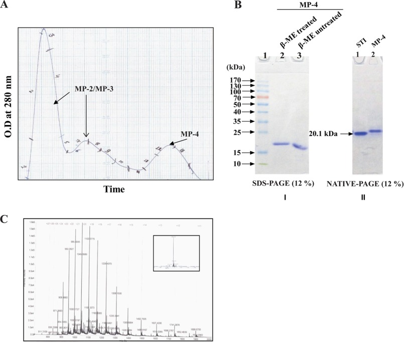
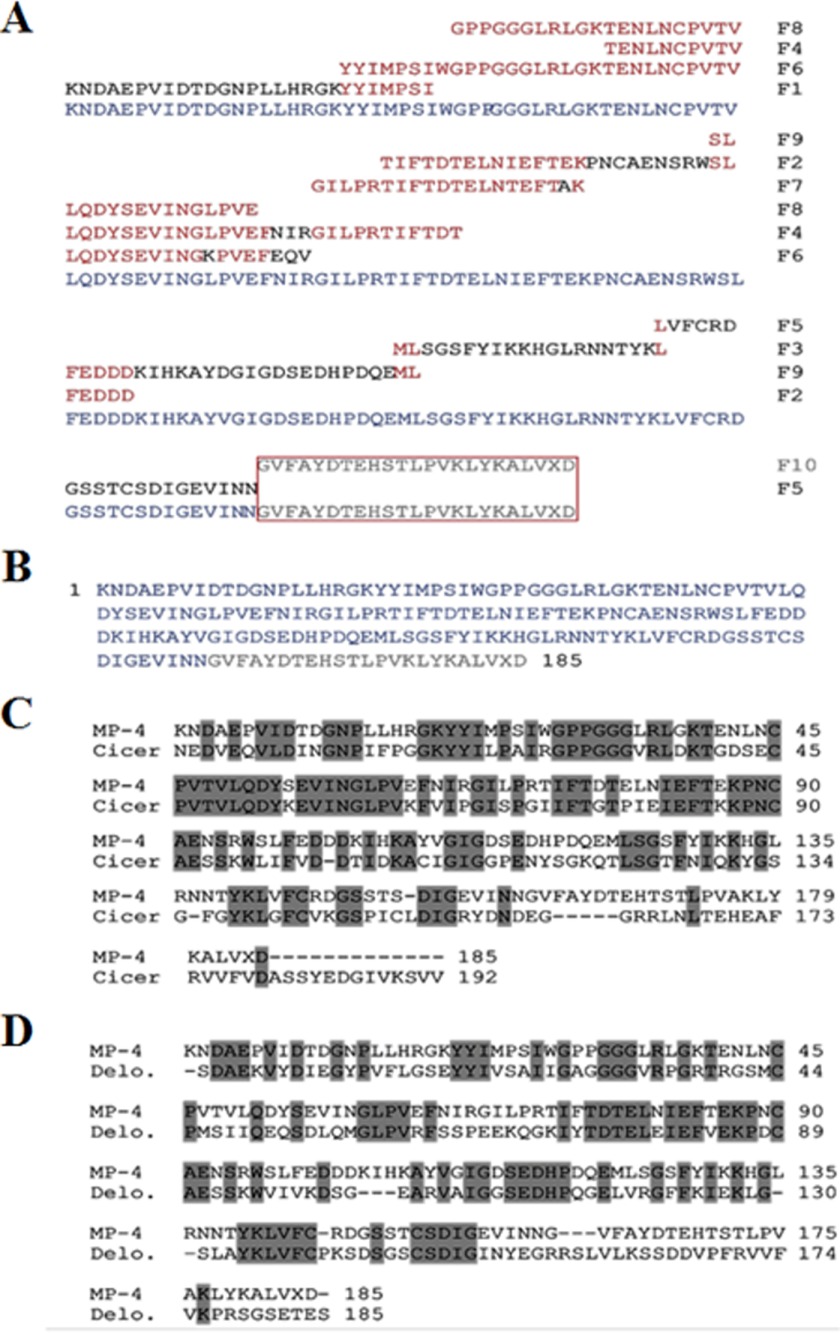
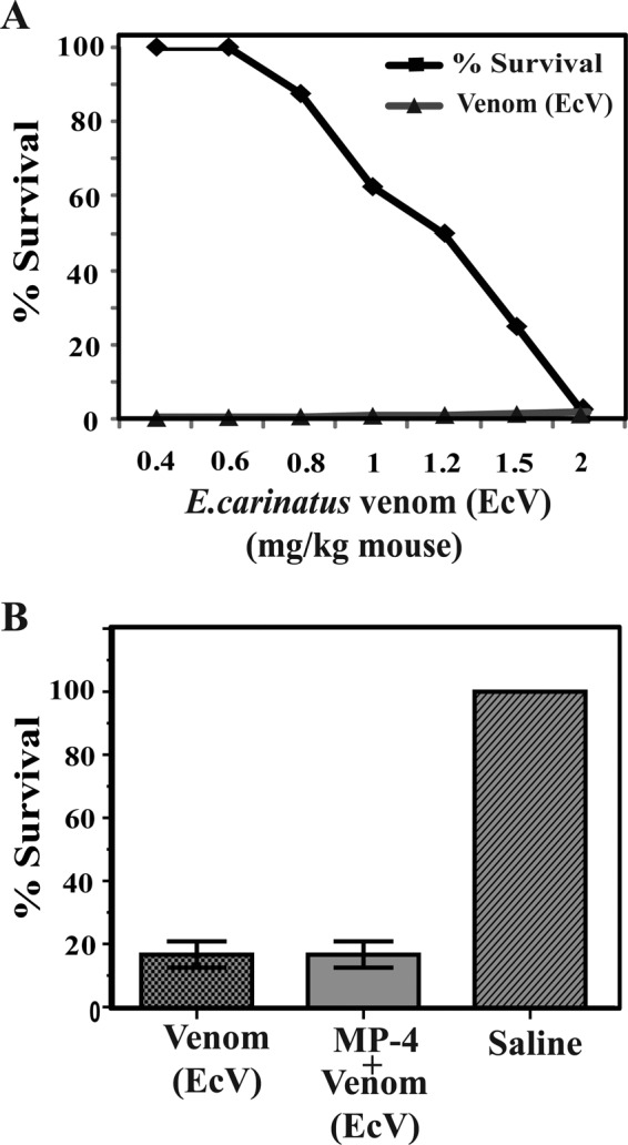
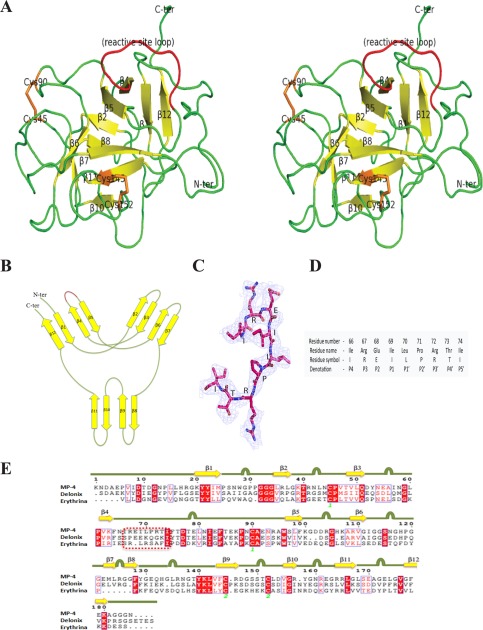
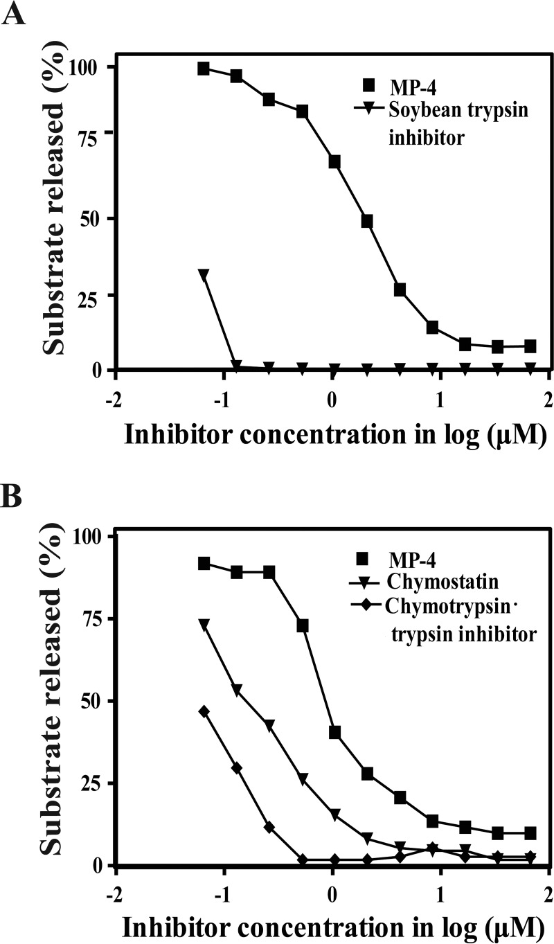
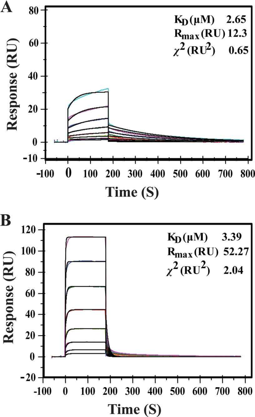
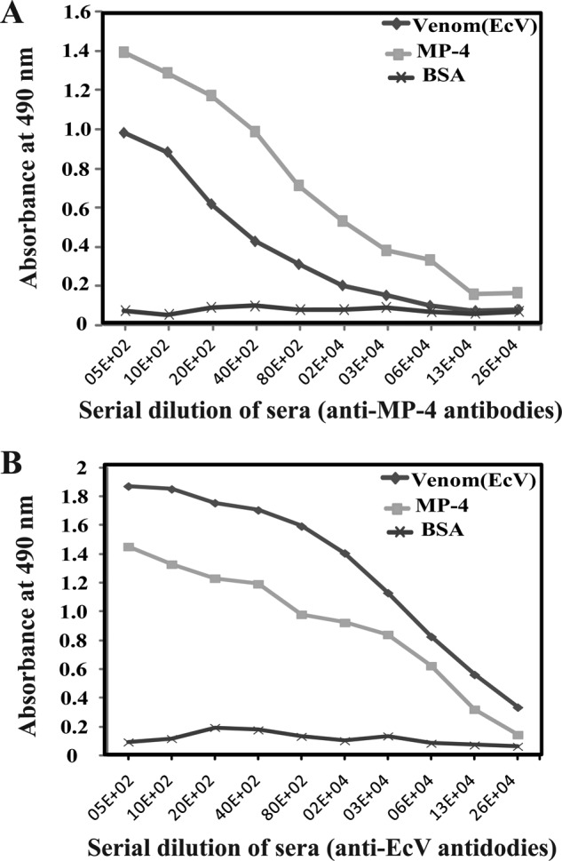
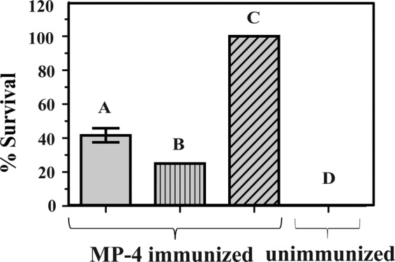
Mortality due to snakebite is a serious public health problem, and available therapeutics are known to induce debilitating side effects. Traditional medicine suggests that seeds of Mucuna pruriens can provide protection against the effects of snakebite. Our aim is to identify the protein(s) that may be important for snake venom neutralization and elucidate its mechanism of action. To this end, we have identified and purified a protein from M. pruriens, which we have named MP-4. The full-length polypeptide sequence of MP-4 was obtained through N-terminal sequencing of peptide fragments. Sequence analysis suggested that the protein may belong to the Kunitz-type protease inhibitor family and therefore may potentially neutralize the proteases present in snake venom. Using various structural and biochemical tools coupled with in vivo assays, we are able to show that MP-4 does not afford direct protection against snake venom because it is actually a poor inhibitor of serine proteases. Further experiments showed that antibodies generated against MP-4 cross-react with the whole venom and provide protection to mice against Echis carinatus snake venom. This study shows that the MP-4 contributes significantly to the snake venom neutralization activity of M. pruriens seeds through an indirect antibody-mediated mechanism.

Keywords: antibody; crystal structure; humoral response; protease inhibitor; snake venom.


Figures

FIGURE 1.
FIGURE 1.
Analysis of seed proteins of M. pruriens by 12% SDS-PAGE in reducing conditions after ammonium sulfate fractionation. A, lanes 2–7, 0–30%, 30–40%, 40–50%, 50–60%, 60–70%, and 70–80% fractions, respectively. In lane 1, low molecular weight protein markers (Sigma-Aldrich) were loaded. B, 40% fraction highlights two major protein bands, MP-1 and MP-6; the 60% fraction shows four major protein bands, MP-2, MP-3, MP-4, and MP-5. C, N-terminal sequences by Edman degradation method of all of the major proteins present in 40 and 60% ammonium sulfate fractions.
FIGURE 2.
FIGURE 2.
Purification and characterization of MP-4. A, size exclusion chromatography profile of 60% ammonium sulfate fraction depicting the first two peaks, corresponding to MP-2 and MP-3. The third peak corresponds to MP-4. B (left), SDS-PAGE (12%) analysis shows the presence of a single band for MP-4 in reducing (lane 2) and non-reducing (lane 3) conditions. Lane 1, prestained molecular weight markers (Thermo Scientific). B (right), native PAGE (12%) shows a single band of STI (Sigma-Aldrich) in lane 1, which is used as a molecular weight marker (20.1 kDa), and lane 2 shows the band of MP-4. C, mass spectrometry profile of MP-4 with a deconvoluted single sharp peak of 20.884 kDa shown in the inset.
FIGURE 3.
FIGURE 3.
Derivation of full-length sequence of MP-4 and sequence analysis. A, internal peptide fragments obtained from enzymatic and chemical digestion and their contig sequence alignment is displayed. The peptide fragment (F10) in the box was not clear in N-terminal sequencing, which was built on the basis of a homologous sequence present in the protein database (C-terminal 25 ambiguous residues highlighted in light gray). B, full-length amino acid sequence of MP-4 generated by fragment alignment. C, MP-4 sequence alignment with its closest ortholog from C. arietinum. This protein shows 57% sequence identity with MP-4. D, the closest MP-4 ortholog for which a structure is available is a Kunitz-type protease inhibitor from D. regia (Protein Data Bank code 1R8N). MP-4 shows 44% sequence identity to this protein. The identical residues are highlighted in dark gray.
FIGURE 4.
FIGURE 4.
Optimization and Evaluation of anti-snake venom activity of MP-4. A, optimization of EcV LD50, which was found to be 2 mg/kg. B, three groups of mice were administrated with venom EcV, preincubated mixture EcV + MP-4, and saline, separately. The percentage survival in the first and second groups of mice were identical.
FIGURE 5.
FIGURE 5.
Overall structure of MP-4. A, stereo ribbon diagram of MP-4 structure. Yellow and green, β-strands and loops, respectively. The reactive site loop is highlighted in red. Orange, cysteine amino acid. B, a schematic of the secondary structure elements in MP-4. In this wiring diagram, the red region indicates the reactive site loop location. C, reactive site loop with 2FoFc map at 1σ level. D, position, name, symbol, and denotation of the reactive site loop residues (P4–P5′) in a conventional representation. E, multiple sequence alignment of MP-4, Delonix, and Erythrina. Square dashed box indicates the reactive site loop in all of the three proteins. Yellow arrow for β-sheets and green line with bulge, loop regions in the MP-4 structure.
FIGURE 6.
FIGURE 6.
Protease inhibitory activity of MP-4. A, activity of MP-4 against trypsin. Lines marked with squares and triangles represent MP-4 and STI, respectively. B, activity of MP-4 against the chymotrypsin. Chymostatin and chymotrypsin·trypsin inhibitor are used as control. Lines marked with squares, triangles, and rhombi represent MP-4, chymostatin, and chymotrypsin·trypsin inhibitor, respectively.
FIGURE 7.
FIGURE 7.
Binding analysis of MP-4 with trypsin and chymotrypsin by SPR. A, SPR sensorgram for the binding of MP-4 (ligand) to trypsin (analyte) immobilized on the surface of a CM5 chip. The ligand was tested in the concentration range from 16 to 0.125 μm. B, binding of MP-4 (ligand) to chymotrypsin (analyte), where chymotrypsin was used in the range from 32 to 0.125 μm. Shown are the equilibrium rate constant, Rmax, and χ2 value for MP-4·trypsin and MP-4·chymotrypsin complex over the corresponding sensorgram. RU, response units.
FIGURE 8.
FIGURE 8.
Evaluation of cross-reactivity of antibodies generated against MP-4 and EcV by ELISA. A, antibodies generated against MP-4 cross-react with whole EcV. B, antibodies generated against EcV proteins cross-react with MP-4 protein. BSA was used as a control.
FIGURE 9.
FIGURE 9.
In vivo protection of MP-4 against EcV. Bar A, mice immunized with MP-4 and administered 1 mg/kg EcV; bar B, MP-4-immunized mice administered the MLD of EcV (2 mg/kg); bar C, MP-4-immunized mice administered saline; bar D, unimmunized mice that were subjected to the MLD of EcV (2 mg/kg). Each bar is a representation of the percentage survival of mice for experiments conducted three times with eight mice in each group. Error bar, S.D.

References

    1. Warrell D. A. (2010) Snake bite. Lancet 375, 77–88 - PubMed
    1. Kasturiratne A., Wickremasinghe A. R., de Silva N., Gunawardena N. K., Pathmeswaran A., Premaratna R., Savioli L., Lalloo D. G., and de Silva H. J. (2008) The global burden of snakebite: a literature analysis and modelling based on regional estimates of envenoming and deaths. PLoS Med. 5, e218. - PMC - PubMed
    1. Chippaux J. P. (1998) Snake-bites: appraisal of the global situation. Bull. World Health Organ. 76, 515–524 - PMC - PubMed
    1. Hsu J. (2015) Defanging snakebites. Sci. Am. 313, 14–16
    1. Koh D. C., Armugam A., and Jeyaseelan K. (2006) Snake venom components and their applications in biomedicine. Cell. Mol. Life Sci. 63, 3030–3041 - PMC - PubMed

Publication types

MeSH terms

LinkOut - more resources