

Usefulness of quantitative peritumoural perfusion and proton spectroscopic magnetic resonance imaging evaluation in differentiating brain gliomas from solitary brain metastases
- PMID: 26988081
- PMCID: PMC4977921
- DOI: 10.1177/1971400916638358
Usefulness of quantitative peritumoural perfusion and proton spectroscopic magnetic resonance imaging evaluation in differentiating brain gliomas from solitary brain metastases
Abstract
Objectives: The purpose of our study was to evaluate whether peritumoural perfusion weighted and proton spectroscopic magnetic resonance imaging can be used in differentiating between primary gliomas and solitary metastases.
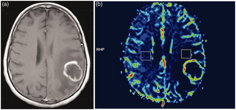
Methods: Ten low-grade gliomas, eight high-grade gliomas and 10 metastases were prospectively evaluated with magnetic resonance imaging, dynamic susceptibility contrast enhanced perfusion imaging and single-voxel proton magnetic resonance spectroscopy before surgical resection or stereotactic biopsy. Maximal relative cerebral blood volume values were calculated drawing three regions of interest of 2 cm(2) in the non-enhancing peritumoural areas. Maximal relative cerebral blood volume values were normalised to that of contralateral normal-appearing white matter. Maximal choline/creatine ratios were calculated from three voxels of 10 cm(3) placed in the peritumoural areas defined as non-enhancing peritumoural white matter surrounding the tumour. The tumour grade presumed with these values was compared to histopathological grading. Differences in the study parameters between groups were assessed using the Mann-Whitney test. A receiver operating characteristic analysis was performed to determine cut-off values.
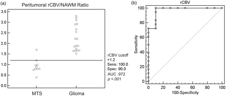
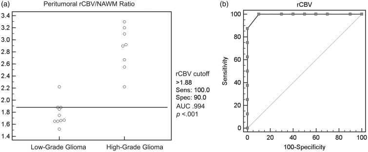
Results: A clear relative cerebral blood volume cut-off value of 1.88 was detected for differentiating low-grade gliomas from high-grade gliomas. A clear relative cerebral blood volume cut-off value of 1.20 was detected for differentiation of metastases from gliomas. The differences in the choline/creatine ratios in the peritumoural regions of high-grade gliomas and of solitary metastasis were statistically significant (P < 0.001) but a clear cut-off value was not found.
Conclusion: Our preliminary data support the hypothesis that peritumoural perfusion-weighted imaging can assist in preoperative differentiation between a glioma and a solitary metastasis.
Keywords: Brain glioma; brain metastases; magnetic resonance spectroscopy; perfusion weighted magnetic resonance imaging.
© The Author(s) 2016.
Figures
References
-
- Law M, Cha S, Knopp EA, et al. High-grade gliomas and solitary metastases: differentiation by using perfusion and proton spectroscopic MR imaging. Radiology 2002; 222: 715–721. - PubMed
-
- Aprile I, Torni C, Fiaschini P, et al. High-grade cerebral glioma characterization: usefulness of MR spectroscopy and perfusion imaging associated evaluation. Neuroradiol J 2012; 25: 57–66. - PubMed
MeSH terms
LinkOut - more resources
Full Text Sources
Other Literature Sources
Medical
