Comparative Study
. 2016 Mar 17:47:45.
doi: 10.1186/s13567-016-0331-3.

Comparative analysis of routes of immunization of a live porcine reproductive and respiratory syndrome virus (PRRSV) vaccine in a heterologous virus challenge study

Affiliations
Comparative Study

Comparative analysis of routes of immunization of a live porcine reproductive and respiratory syndrome virus (PRRSV) vaccine in a heterologous virus challenge study

Kang Ouyang et al. Vet Res. .

Abstract

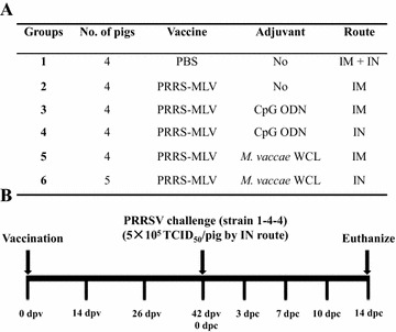
Porcine reproductive and respiratory syndrome (PRRS) is caused by PRRS virus (PRRSV), which infects primarily the respiratory tract of pigs. Thus intranasal (IN) delivery of a potent vaccine-adjuvant formulation is promising. In this study, PRRS-MLV (VR2332) was coadministered ± an adjuvant Mycobacterium vaccae whole cell lysate or CpG ODN through intramuscular (IM) or IN route as a mist, and challenged with a heterologous PRRSV 1-4-4 IN at 42 days post-vaccination (dpv). At 14 and 26 dpv, vaccine viral RNA copies were one log greater in the plasma of PRRS-MLV IM compared to IN vaccinated pigs, and the infectious replicating vaccine virus was detected only in the IM group. In PRRS-MLV ± adjuvant IM vaccinated pigs, reduced viral RNA load and absence of the replicating challenged virus was observed at 7, 10 and 14 days post-challenge (dpc). At 14 dpc, in BAL fluid ≥ 5 log viral RNA copies were detected in all the pig groups, but the replicating challenged virus was undetectable only in IM groups. Immunologically, virus neutralizing antibody titers in the plasma of IM (but not IN) vaccine groups was ≥ 8 against the vaccine and challenged viruses. At 26 dpv, PRRS-MLV IM (without adjuvant) received pigs had significantly increased population of CD4 and CD8 T cells in PBMC. At 14 dpc, relatively increased population of IFN-γ(+) total lymphocytes, NK, CD4, CD8 and γδ T cells were observed in the MLV-IM group. In conclusion, PRRS-MLV IM vaccination induced the virus specific T cell response in pigs, but still it is required to improve its cross-protective efficacy.


Figures

Figure 1
Figure 1
PRRS-MLV vaccination and challenge study work plan. A Conventional crossbred pigs were randomly divided into six Groups (4 or 5 per Group). PRRS-MLV was administered once without (saline) or with an adjuvant, M. vaccae WCL or CpG ODN, through intramuscular (IM) or intranasal (IN) route. B All the experimental pigs were challenged with a heterologous PRRSV strain 1-4-4 by IN route at 42 dpv. Blood samples were collected and rectal temperature and body weight were recorded at 0, 14, 26, 42 dpv and 3, 7, 10, 14 dpc and pigs were euthanized at 14 dpc.
Figure 2
Figure 2
Dynamics of body temperature and PRRSV load in the plasma and BAL fluid of pigs. (A) Rectal temperature of pigs was recorded at the indicated dpv and dpc. PRRSV RNA copies and replicating infectious virus load in the plasma (BD) and BAL fluid (E, F) were quantified by qRT-PCR and indirect immunofluorescence assay. The numbers above the bar in the graphs (E, F) represent the ratio of PRRSV positive pigs to the total in each group. Each data point or bar in the graph represents the average value from 4 or 5 pigs ± SEM. Asterisk marked in panel C indicates the virus titers in MLV-IM, MLV-CPG IM and MLV-IM Groups were significantly reduced compared to mock (PBS) challenged Group (P < 0.01) and MLV-CPG IN Group (P < 0.05) at 7 dpc. In panel D asterisks (*P < 0.05 and **P < 0.01) indicate statistical significant difference between the marked two pig Groups at 7 dpc.
Figure 3
Figure 3
PRRSV neutralizing antibody (NA) titers in the plasma and BAL fluid of pigs. Viral NA titers in the plasma (AC) and BAL fluid (DF) against challenged PRRSV strain 1-4-4 (A, D), PRRS-MLV parent strain VR2332 (B, E) and a genetically variant Type 2 PRRSV strain MN184 (C, F) were determined by indirect immunofluorescence assay. The NA titer of a sample was expressed as the reciprocal of the highest dilution that inhibited >90% of the virus induced immunofluorescence activity. Each data point or bar in the graph represents the average NA titer from 4 or 5 pigs ± SEM.
Figure 4
Figure 4
Population of PRRSV specific lymphocyte subsets in the blood of pigs. PBMC isolated from individual pigs were unstimulated (control) or stimulated with PRRSV (virus) or pooled five conserved peptides (peptides) and immunostained using indicated pig specific cell surface markers and analyzed by flow cytometry. Population of CD3ε+CD4αCD8α+, Th/memory cells (CD3ε+CD4α+CD8α+) and γδ T cells in the PBMC collected at 26 dpv (AC) and 14 dpc (DF) are shown. Each bar represents the average cell count of 50 000 events from 4 or 5 pigs ± SEM. Lowercase alphabets and asterisks indicate a statistically significant difference (a or *P < 0.05, b or **P < 0.01 and c P < 0.001) between control (PBS) and vaccine received pig groups under the same ex vivo stimulation condition, or between different stimulation conditions within the same treatment group.
Figure 5
Figure 5
Population of PRRSV specific IFN-γ + total lymphocyte response in the blood of pigs. PBMC isolated from individual pigs at 26 dpv (AC) and 14 dpc (DF) were unstimulated (control) or stimulated with PRRSV strain 1-4-4 (virus) or pooled five conserved peptides (peptides). IFN-γ secreting cells in total CD3ε+ lymphocytes and NK cells (CD3εCD4αCD8α+) are shown. Each bar represents the average cell counts of 50 000 events from 4 or 5 pigs ± SEM. Lowercase alphabet and asterisks indicate a statistically significant difference (a or *P < 0.05, **P < 0.01 and *** P < 0.001) between control (PBS) and vaccine received groups under the same ex vivo stimulation condition, or between different stimulation conditions within the same treatment group.
Figure 6
Figure 6
PRRSV specific IFN-γ + T lymphocyte subsets in the blood of pigs. PBMC isolated from individual pigs at 26 dpv (AC) and 14 dpc (DF) were unstimulated (control) or stimulated with PRRSV (virus) or pooled five conserved peptides (peptides). Cells were immunostained for T cell surface markers CD3ε, CD4α, CD8α, and δ chain (γδ T cells) and intracellular IFN-γ+ cells are shown. Each bar represents the average cell counts of 50 000 events from 4 or 5 pigs ± SEM. Lowercase alphabet and asterisks indicate a statistically significant difference (a or *P < 0.05, **P < 0.01 and c or ***P < 0.001) between control (PBS) and vaccine received groups under the same ex vivo stimulation condition, or between different stimulation conditions within the same treatment group.

References

    1. Neumann EJ, Kliebenstein JB, Johnson CD, Mabry JW, Bush EJ, Seitzinger AH, Green AL, Zimmerman JJ. Assessment of the economic impact of porcine reproductive and respiratory syndrome on swine production in the United States. J Am Vet Med Assoc. 2005;227:385–392. doi: 10.2460/javma.2005.227.385. - DOI - PubMed
    1. Holtkamp DJ, Kliebenstein JB, Neumann EJ, Zimmerman JJ, Rotto HF, Yoder TK, Wang C, Yeske PE, Mowrer CL, Haley CA. Assessment of the economic impact of porcine reproductive and respiratory syndrome virus on United States pork producers. J Swine Health Prod. 2013;21:72–84.
    1. Collins JE, Benfield DA, Christianson WT, Harris L, Hennings JC, Shaw DP, Goyal SM, McCullough S, Morrison RB, Joo HS, Gorcyca D, Chladek D. Isolation of swine infertility and respiratory syndrome virus (isolate ATCC VR-2332) in North America and experimental reproduction of the disease in gnotobiotic pigs. J Vet Diagn Invest. 1992;4:117–126. doi: 10.1177/104063879200400201. - DOI - PubMed
    1. Wensvoort G, Terpstra C, Pol JM, ter Laak EA, Bloemraad M, de Kluyver EP, Kragten C, van Buiten L, den Besten A, Wagenaar F, Broekhuijsen JM, Moonen PL, Zetstra T, de Boer EA, Tibben HJ, de Jong MF, van’t Veld P, Greenland GJ, van Gennep JA, Voets MT, Verheijden JH, Braamskamp J. Mystery swine disease in The Netherlands: the isolation of lelystad virus. Vet Q. 1991;13:121–130. doi: 10.1080/01652176.1991.9694296. - DOI - PubMed
    1. Conzelmann KK, Visser N, Van Woensel P, Thiel HJ. Molecular characterization of porcine reproductive and respiratory syndrome virus, a member of the arterivirus group. Virology. 1993;193:329–339. doi: 10.1006/viro.1993.1129. - DOI - PMC - PubMed

Publication types

MeSH terms

LinkOut - more resources