. 2016 Mar 18:6:23138.
doi: 10.1038/srep23138.

Influenza A viruses escape from MxA restriction at the expense of efficient nuclear vRNP import

Affiliations

Influenza A viruses escape from MxA restriction at the expense of efficient nuclear vRNP import

Veronika Götz et al. Sci Rep. .

Erratum in

Abstract

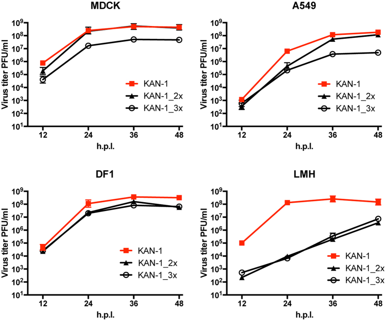
To establish a new lineage in the human population, avian influenza A viruses (AIV) must overcome the intracellular restriction factor MxA. Partial escape from MxA restriction can be achieved when the viral nucleoprotein (NP) acquires the critical human-adaptive amino acid residues 100I/V, 283P, and 313Y. Here, we show that introduction of these three residues into the NP of an avian H5N1 virus renders it genetically unstable, resulting in viruses harboring additional single mutations, including G16D. These substitutions restored genetic stability yet again yielded viruses with varying degrees of attenuation in mammalian and avian cells. Additionally, most of the mutant viruses lost the capacity to escape MxA restriction, with the exception of the G16D virus. We show that MxA escape is linked to attenuation by demonstrating that the three substitutions promoting MxA escape disturbed intracellular trafficking of incoming viral ribonucleoprotein complexes (vRNPs), thereby resulting in impaired nuclear import, and that the additional acquired mutations only partially compensate for this import block. We conclude that for adaptation to the human host, AIV must not only overcome MxA restriction but also an associated block in nuclear vRNP import. This inherent difficulty may partially explain the frequent failure of AIV to become pandemic.


Figures

Figure 1
Figure 1. Recombinant KAN-1 viruses carrying MxA escape mutations show a degree of attenuation in different cell culture systems.
Cells were infected at an MOI of 0.001 with wild-type KAN-1 (KAN-1) or the indicated mutant viruses. At the indicated hours post infection (h.p.i.), virus titers were determined by plaque assay. Error bars indicate the standard deviation of the mean of at least three independent experiments.
Figure 2
Figure 2. Additional adaptive mutations in NP affect viral polymerase activity in the presence of MxA.
(A) Structural model of an H5N1 virus nucleoprotein. Major amino acids allowing MxA escape are highlighted in blue. Additional mutations acquired during propagation of KAN-1_3x in tissue culture are marked in magenta. The program PyMOL was used to assign the indicated positions based on the structural model of A/HK/483/97(H5N1) NP (PDBcode: 2Q06). (B) Polymerase activity in the presence of MxA. HEK293T cells were transiently transfected with expression plasmids coding for PB2, PB1, PA of KAN-1, the indicated NP proteins, a minigenome encoding the firefly luciferase and a renilla luciferase expression plasmid to normalize for variations in expression efficiency. Polymerase activity in the presence of antivirally inactive MxA_T103A was used to normalize the data obtained with MxA (relative activity). Western blot analysis was performed to determine the expression levels of NP and MxA. Error bars indicate the standard deviation of the mean of at least 3 independent experiments. Student’s T test was performed to determine the P value **P < 0.01, ***P < 0.001, ****P < 0.0001, not significant (ns).
Figure 3
Figure 3. The additional mutations in NP of KAN-1_3x only partially restore viral fitness.
Cells were infected at an MOI of 0.001 of wild-type KAN-1 (KAN-1) or the indicated mutant viruses. At the indicated hours post infection (h.p.i.), virus titers were determined by plaque assay. Error bars indicate the standard deviation of the mean of at least three independent experiments.
Figure 4
Figure 4. MxA escape of KAN-1_3x is influenced by the additional mutations in NP.
(A) A549-MxA cells were treated with siRNAs targeting MxA (siMxA) or a non-targeting siRNA (siCtr). Three days post transfection, cells were infected with wild-type KAN-1 (KAN-1) or the indicated mutant viruses at a MOI of 0.001 and virus titers were determined by plaque assay 48 hours post infection. Error bars indicate the standard deviation from the mean of 3 independent experiments. Fold differences in virus titers are indicated. (BD) A459 cells expressing (MxA) or lacking MxA (shMxA) were infected at an MOI of 0.001 of wild-type KAN-1 (KAN-1) or the indicated mutant viruses. At the indicated hours post infection (h.p.i.), virus titers were determined by plaque assay. Error bars indicate the standard deviation of the mean of 3 independent experiments. Fold differences in virus titers are indicated.
Figure 5
Figure 5. Attenuation due to MxA escape is caused by inefficient nuclear import of incoming vRNPs.
(A) DF1 or LMH cells were infected with wild-type KAN-1 (WT) or KAN-1_2x (2x) at an MOI of 50 in the presence of 100 μg/ml cycloheximide and primary viral transcript levels were determined 3 hours later. Viral RNA species were detected by primer extension analysis using specific primers for the NA segment. (B) Detection of incoming vRNPs in LMH cells infected with wild-type KAN-1 (KAN-1) or KAN-1_2x at an MOI of 50 in the presence of 100 μg/ml cycloheximide. Infection was carried out on ice for 40 minutes to synchronize virus entry before incubation at 37 °C for the times indicated. Incoming vRNPs were visualized by NP staining and cell nuclei by TOPRO3 staining. Histograms indicate the NP fluorescence intensity along the yellow line in selected cells. Red dashed lines indicate the borders of the cell nuclei identified by TOPRO3 staining. (C) Equal amounts of virus like particles (VLP) (see supplementary Figure S7A), reconstituted with either wild-type (WT) or mutant NP proteins (2x, 3x) and a minigenome coding for a firefly luciferase, were used to infect the indicated cell lines. Luciferase activity was determined 10 h post infection. HA was omitted as a negative control (ctrl). Error bars indicate the standard deviation from the mean of at least 4 independent experiments. Student’s T test was performed to determine the P value *P < 0.05, **P < 0.01, ****P < 0.0001, not significant (ns).
Figure 6
Figure 6. Nuclear import of vRNPs carrying MxA escape mutations depends on importin-α/β.
(A) Equal amounts of KAN-1 VLP reconstituted with either wild-type (WT) or the indicated KAN-1 mutant NP proteins (2x; 3x; 3x + 16) and a minigenome coding for a firefly luciferase were used to infect LMH cells in the presence of no compound, 15 mM inactive N-(4-methoxyphenyl) (4-MPR) as a negative control, 15 μM N-(4-hydroxyphenyl) retinamide (4-HPR) or 10 μM ivermectin. Luciferase activity was determined 10 h post infection. HA was omitted as a negative control (ctrl). Error bars indicate the standard deviation from the mean of 2–4 independent experiments. Student’s T test was performed to determine the P value *P < 0.05, **P < 0.01, ***P < 0.001, ****P < 0.0001, not significant (ns). (B) Nuclear vRNP import in the presence of import inhibitors. LHM cells were pretreated with cycloheximide and infected with wild-type SC35M or mutant SC35M_2x at an MOI of 50 in the presence of 15 μM N-(4-hydroxyphenyl) retinamide (4-HPR) or 15 μM inactive N-(4-methoxyphenyl) (4-MPR) as a negative control. Infection was carried out on ice for 40 minutes to synchronize virus entry and further incubated at 37 °C. After 90 minutes, cells were fixed and incoming vRNPs were visualized using a NP-specific antibody. Cell nuclei were visualized by DAPI staining. Images were acquired using a Zeiss confocal laser microscope.
Figure 7
Figure 7. Model depicting the evolutionary bottleneck for avian influenza viruses encountering the host restriction factor MxA.
To establish a new lineage in the human population, avian influenza A viruses have to acquire several adaptive mutations in almost all viral proteins, including MxA escape mutations in NP. However, the acquisition of MxA escape amino acids in NP is associated with severely reduced viral fitness, due to impaired nuclear import of vRNPs. Stabilizing mutations in NP (e.g. 16D) are required to overcome this fitness restriction, but are not sufficient to restore viral growth properties. As a consequence further additional mutations in NP and probably other viral gene products are required.

References

    1. Klenk H. D. Influenza viruses en route from birds to man. Cell Host Microbe 15, 653–654 (2014). - PubMed
    1. Miller M. S. & Palese P. Peering into the crystal ball: influenza pandemics and vaccine efficacy. Cell 157, 294–299 (2014). - PubMed
    1. Hatta M. & Kawaoka Y. The continued pandemic threat posed by avian influenza viruses in Hong Kong. Trends Microbiol. 10, 340–344 (2002). - PubMed
    1. WHO. Influenza at the Human-Animal Interface (HAI) http://www.who.int/influenza/human_animal_interface/en/ (accessed 16 October 2015).
    1. Neumann G. & Kawaoka Y. Transmission of influenza A viruses. Virology 479–480, 234–246 (2015). - PMC - PubMed

MeSH terms