. 2016 Mar 17:14:21.
doi: 10.1186/s12915-016-0243-8.

The transcription factor scleraxis is a critical regulator of cardiac fibroblast phenotype

Affiliations

The transcription factor scleraxis is a critical regulator of cardiac fibroblast phenotype

Rushita A Bagchi et al. BMC Biol. .

Abstract

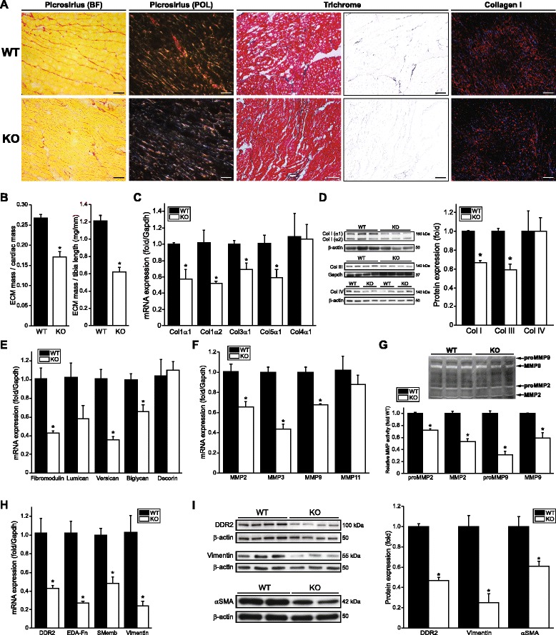
Background: Resident fibroblasts synthesize the cardiac extracellular matrix, and can undergo phenotype conversion to myofibroblasts to augment matrix production, impairing function and contributing to organ failure. A significant gap in our understanding of the transcriptional regulation of these processes exists. Given the key role of this phenotype conversion in fibrotic disease, the identification of such novel transcriptional regulators may yield new targets for therapies for fibrosis.

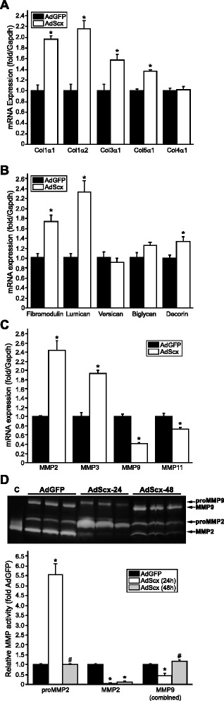
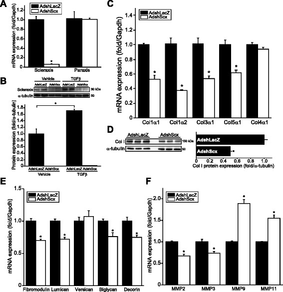
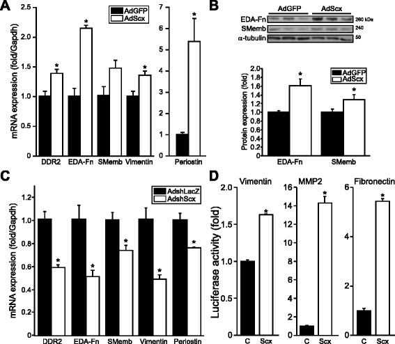
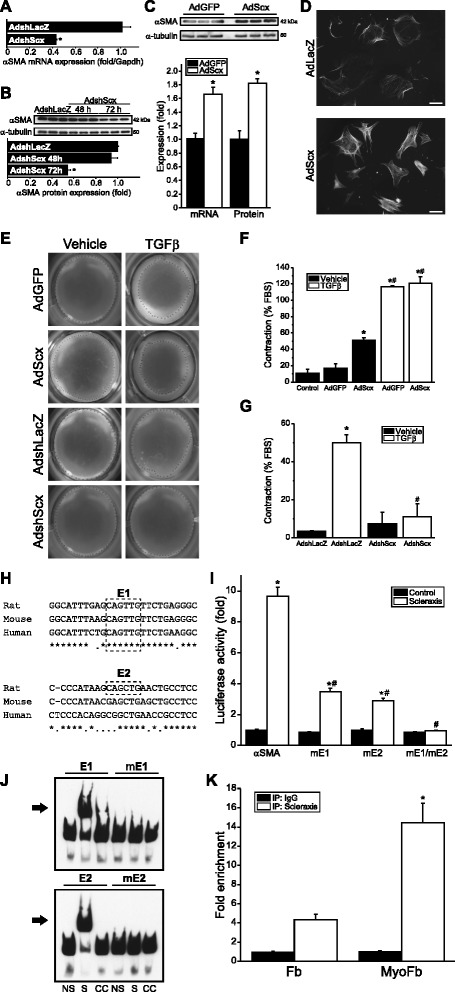
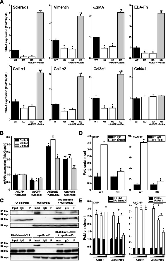
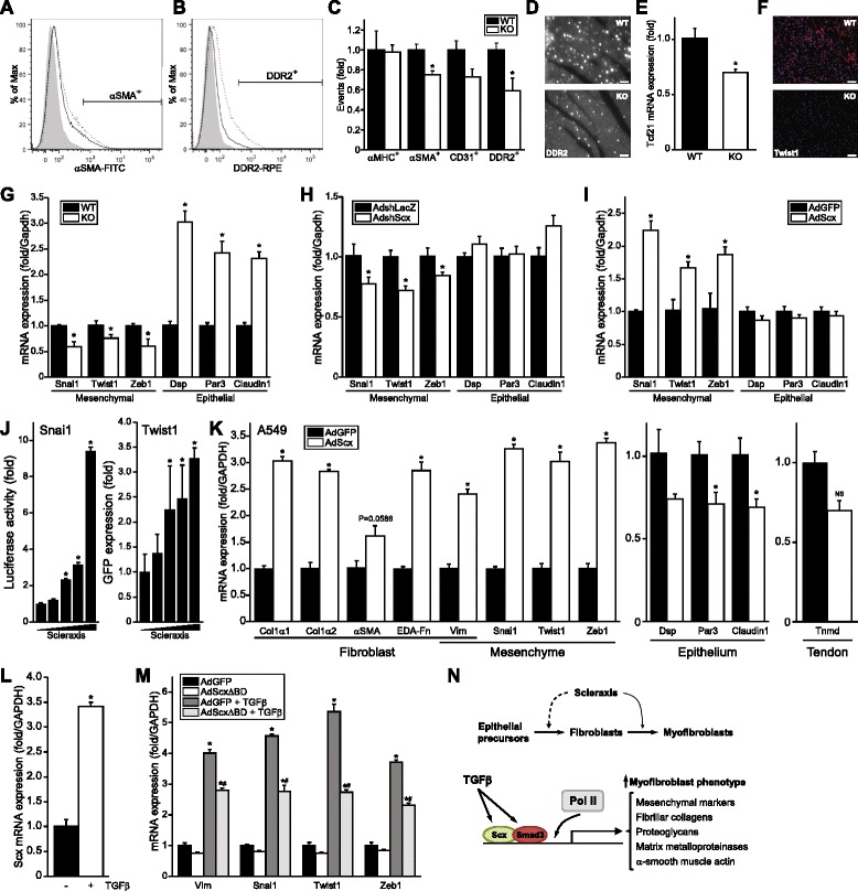
Results: Using explanted primary cardiac fibroblasts in gain- and loss-of-function studies, we found that scleraxis critically controls cardiac fibroblast/myofibroblast phenotype by direct transcriptional regulation of myriad genes that effectively define these cells, including extracellular matrix components and α-smooth muscle actin. Scleraxis furthermore potentiated the TGFβ/Smad3 signaling pathway, a key regulator of myofibroblast conversion, by facilitating transcription complex formation. While scleraxis promoted fibroblast to myofibroblast conversion, loss of scleraxis attenuated myofibroblast function and gene expression. These results were confirmed in scleraxis knockout mice, which were cardiac matrix-deficient and lost ~50% of their complement of cardiac fibroblasts, with evidence of impaired epithelial-to-mesenchymal transition (EMT). Scleraxis directly transactivated several EMT marker genes, and was sufficient to induce mesenchymal/fibroblast phenotype conversion of A549 epithelial cells. Conversely, loss of scleraxis attenuated TGFβ-induced EMT marker expression.

Conclusions: Our results demonstrate that scleraxis is a novel and potent regulator of cellular progression along the continuum culminating in the cardiac myofibroblast phenotype. Scleraxis was both sufficient to drive conversion, and required for full conversion to occur. Scleraxis fulfills this role by direct transcriptional regulation of key target genes, and by facilitating TGFβ/Smad signaling. Given the key role of fibroblast to myofibroblast conversion in fibrotic diseases in the heart and other tissue types, scleraxis may be an important target for therapeutic development.

Keywords: EMT; Extracellular matrix; Fibroblast; Gene expression; Myofibroblast; Phenoconversion; Transcription.


Figures

Fig. 1
Fig. 1
Scleraxis up-regulates matrix target genes. ac Assay of collagen (a), proteoglycan (b) and matrix metalloproteinase (c) mRNA expression by qPCR following over-expression of Scx (AdScx) in primary rat cardiac proto-myofibroblasts compared to controls (AdGFP) reveals numerous matrix genes are induced; n = 3. d Protein lysates from AdGFP- or AdScx-infected primary rat cardiac proto-myofibroblasts were assayed for MMP activity by gel zymography. Zymograms were obtained 24 or 48 h following infection and revealed transient induction of proMMP2 and loss of MMP9 following Scx over-expression; C, control recombinant human MMP2 protein; n = 3. *P < 0.05 vs AdGFP, #P < 0.05 vs corresponding 24 h time point
Fig. 2
Fig. 2
Cardiac matrix gene expression is attenuated by scleraxis knockdown. a Primary cardiac proto-myofibroblasts exhibited loss of Scx but not paraxis mRNA following infection with adenovirus encoding an shRNA targeting Scx (AdshScx) but not control shRNA (AdshLacZ) for 72 h (assayed by qPCR). Results were normalized to the respective AdshLacZ sample; n = 3. b Cells treated as in (a), with or without 10 ng/mL TGFβ or vehicle, were assayed for Scx protein expression 72 h after infection and results normalized to α-tubulin; n = 4. AdshScx attenuated Scx protein expression even in the presence of TGFβ. cd Fibrillar collagen mRNA (c) and nascent soluble 150 kDa collagen I protein (d) expression was down-regulated following Scx knockdown (assayed by qPCR or western blotting, respectively), with cell treatment and normalization as in (a); n = 3. ef The expression of mRNAs encoding several proteoglycans (e) and matrix metalloproteinases (f) (assessed by qPCR) was reduced following Scx knockdown; n = 3. *P < 0.05 vs corresponding AdshLacZ control
Fig. 3
Fig. 3
Scleraxis regulates cardiac fibroblast phenotype. ac Fibroblast/myofibroblast marker gene mRNA (a, c) and protein (b) expression was assayed in primary cardiac proto-myofibroblasts following Scx over-expression (AdScx) vs controls (AdGFP) (a, b), or following Scx knockdown (AdshScx) compared to controls (AdshLacZ) (c), assayed by qPCR or western blot; n = 3. Scx loss induced similar down-regulation of fibroblast and myofibroblast markers, while Scx over-expression induced marker expression. d Scx transactived the vimentin, MMP2 and fibronectin gene promoters compared to empty vector control (C) as determined by luciferase reporter assays in NIH-3T3 fibroblasts; n = 3 (vimentin, fibronectin) or n = 4 (MMP2). *P < 0.05 vs AdGFP (a, b), vs AdshLacZ (c) or vs control empty vector (d)
Fig. 4
Fig. 4
Scleraxis regulates cardiac myofibroblast α-smooth muscle actin expression and cell contraction. ab αSMA mRNA (a) and protein (b) expression were assayed by qPCR or western blot, respectively, following knockdown of Scx (AdshScx) in primary cardiac proto-myofibroblasts, showing attenuated αSMA expression compared to control (AdshLacZ); n = 3. c Scx over-expression (AdScx) up-regulated αSMA expression in primary rat cardiac proto-myofibroblasts as determined by mRNA or protein expression (qPCR or western blot, respectively); n = 3. d Induced αSMA was incorporated into stress fibers, as demonstrated by immunocytochemistry, indicating promotion of the myofibroblast phenotype by Scx; 20× objective, scale bar = 65 μm. e Primary cardiac proto-myofibroblasts seeded onto compressible collagen gel matrices were assayed for gel contraction following Scx over-expression or knockdown, with or without concomitant 10 ng/mL TGFβ1 to induce contraction. Crosses denote the detected margin of the collagen gel. fg Quantification of gel contraction images, reported as the percentage of the maximum contraction induced by 10 % FBS, demonstrates that Scx over-expression induces cell contraction (f), while Scx knockdown attenuates TGFβ1-induced contraction (g); n = 6 ((f) except AdGFP + TGFβ and AdScx + TGFβ; n = 3 each) or n = 3 (g). *P < 0.05 vs AdshLacZ (ab), vs AdGFP (c, f) or vs AdshLacZ + vehicle (g), #P <0.05 vs vehicle controls (f) or vs AdshLacZ + TGFβ (g). h Alignment of putative Scx-binding E-boxes (E1 and E2; dashed lines) in the proximal rat, mouse and human αSMA promoters. Asterisks and periods denote nucleotides conserved across three or two species, respectively. i Mutation of one or both E-boxes (mE1, mE2) attenuates transactivation of the rat αSMA proximal promoter by Scx as determined by luciferase reporter assay; n = 3. j Electrophoretic mobility shift assays demonstrate Scx-binding to E-boxes E1 and E2; this binding is abolished when the E-boxes are mutated (mE1 and mE2). NS, non-shifted lane; S, shifted lane; CC, 500× cold competition. The arrow denotes the shifted complex. k Scx-binding to the αSMA gene promoter is significantly enriched in cardiac myofibroblasts (MyoFb) compared to fibroblasts (Fb) as determined by chromatin immunoprecipitation assay with qPCR quantification of Scx-bound DNA; n = 3. *P < 0.05 vs control empty vector (i) or vs Fb (k), #P < 0.05 vs scleraxis + non-mutated promoter (i)
Fig. 5
Fig. 5
Scleraxis null mice exhibit reduced cardiac extracellular matrix and fibroblast marker gene expression. a Cross sections of hearts from wild type (WT) or Scx null (KO) hearts stained for fibrillar collagen using picrosirius red (BF, bright-field; POL, polarized light), Masson’s trichrome or via immunolabeling for total collagen I (red; plus DAPI nuclear staining in blue) revealed matrix loss in KO hearts; collagen I: 20× objective, scale bar = 35 μm; picrosirius/trichrome: 40× objective, scale bar = 66 μm. The blue color channel was extracted from the Masson’s trichrome sections for improved visualization. Samples are representative of at least three individual animals of each genotype. b Decellularized and dehydrated cardiac ventricular ECM from WT or KO mice was normalized to cardiac ventricular wet mass or tibia length and revealed ECM loss in KO hearts; n = 7 animals of each genotype. cd Fibrillar collagen gene expression by qPCR (c) or western blotting normalized to Gapdh or β-actin (d) was decreased in Scx KO hearts compared to WT; n = 3 ((c) or collagen I blot in (d)) or n = 4 (collagen III and IV blots in (d)). Collagen I protein expression was the sum of the α1 and α2 isoforms. ef The expression of mRNAs encoding several proteoglycans (e) and matrix metalloproteinases (f) (assessed by qPCR) was lost in Scx KO hearts; n = 3 animals of each genotype. g MMP2 and MMP9 activity (latent pro-form and mature, assayed by gel zymography) of protein lysates from WT or KO hearts was reduced in null animal hearts; n = 3 animals of each genotype. *P < 0.05 vs WT. hi Fibroblast/myofibroblast marker gene mRNA (h) and protein (i) expression was assayed in WT and Scx KO mice, assayed by qPCR or western blot; n = 3 ((h) and vimentin in (i)) or n = 4 (DDR2 and αSMA in (i)). Scx loss induced similar down-regulation of fibroblast and myofibroblast markers. *P < 0.05 vs WT
Fig. 6
Fig. 6
Requirement of scleraxis for Smad3-mediated gene expression. a Over-expression of Scx (AdScx) for 24 h rescues expression of vimentin, αSMA, ED-A fibronectin and fibrillar collagen mRNA (by qPCR) in primary cardiac proto-myofibroblasts obtained from Scx null mice compared to controls (AdGFP); n = 3 independent samples per genotype. Impaired collagen expression in Scx null cells is thus due to Scx loss and not a defect in the basal transcriptional machinery. b Up-regulation of fibrillar collagen mRNA expression by Smad3 (assessed by qPCR) is attenuated following Scx knockdown (AdshScx) compared to shRNA control in cardiac proto-myofibroblasts (AdshLacZ); n = 3. c Scx and Smad3 physically interact. Expression vectors for HA-tagged Scx and myc-tagged Smad3 were individually or jointly transfected into NIH-3T3 fibroblasts, then immunoprecipitated (IP) by anti-HA antibodies and subjected to western blotting with anti-myc antibodies (upper panels). Non-immunoprecipitated input and non-specific IgG antibodies were employed as positive and negative controls, respectively. Equal loading of transfected whole-cell lysates is shown by anti-HA and anti-myc western blots. Conversely, co-immunoprecipitation with Smad3 was not observed when a Scx mutant lacking its protein-interaction domain (HA-ScleraxisΔHLH) was employed (lower panels). d Smad3 and RNA polymerase II interaction with the Col1α2 gene promoter is significantly impaired in cardiac fibroblasts derived from Scx KO mouse hearts compared to WT, as assessed by chromatin immunoprecipitation (anti-Smad3 antibody vs non-specific IgG antibody) and re-ChIP (anti-RNA Pol II antibody vs non-specific IgG antibody) followed by qPCR, indicating that Scx loss impairs Smad3 signaling and Smad3-mediated recruitment of the transcription complex to the Col1α2 gene promoter; n = 3 independent samples per genotype. e Adenovirus-mediated over-expression of a DNA-binding Scx mutant (AdScxΔBD; 10, 50 or 100 MOI) in human ventricular myofibroblasts attenuates Smad3 and RNA polymerase II recruitment to the Smad-binding element of the Col1α2 gene promoter compared to control (AdGFP) performed as in (d); n = 3. *P < 0.05 vs WT (a) or vs AdGFP + AdshLacZ (b) or vs IgG (de), #P < 0.05 vs KO + AdGFP (a) or vs AdSmad3 + AdshLacZ (b) or vs WT (d) or as indicated (e)
Fig. 7
Fig. 7
Loss of cardiac fibroblasts following scleraxis deletion in vivo. ab Representative flow cytometry histograms of WT and Scx KO heart single-cell suspensions show cardiac cells stained for αSMA (a) or DDR2 (b) in WT (dotted line) and KO (solid line) mice. Unstained cell sample was used as control (shaded histogram). c Total αMHC+, αSMA+, CD31+ or DDR2+ stained events per 5 × 104 events in WT or KO mice normalized to WT counts demonstrating normal cardiomyocyte counts but reduced numbers of fibroblasts; n = 3 independent samples per genotype. d Representative cardiac sections from WT and KO mice immunolabeled for DDR2 confirm the loss of DDR2+ cells; 63× objective, scale bar = 20 μm. e qPCR assay of Tcf21 mRNA expression in WT and Scx KO mice; n = 3 independent samples per genotype. f Cardiac sections as in (d) were immunolabeled for Twist1 expression (red; DAPI nuclear staining in blue); 20× objective, scale bar = 64 μm. g Cardiac mRNA from WT and Scx KO mice was assayed for expression of mesenchymal and epithelial marker genes by qPCR, indicating reduced epithelial-to-mesenchymal transition in KO hearts; n = 5 independent samples per genotype. hi Cardiac proto-myofibroblasts were subjected to Scx knockdown (h) or over-expression (i) and EMT markers assessed by qPCR, indicating that Scx regulates mesenchymal marker gene expression; n = 3. j Scx dose-dependently transactivates the Snai1 and Twist1 proximal gene promoters as determined by luciferase assay or GFP western blot, respectively; n = 3. k Representative fibroblast/myofibroblast, mesenchymal, epithelial and tendon marker gene mRNA expression was assayed in A549 epithelial cells following Scx over-expression (AdScx) vs controls (AdGFP), assayed by qPCR; n = 3. l Scx mRNA expression was assayed by qPCR in A549 cells following treatment with 2.5 ng/mL TGFβ or vehicle; n = 4. m Mesenchymal marker gene mRNA expression was assayed in A549 cells following Scx dominant negative mutant over-expression (AdScxΔBD) vs controls (AdGFP), with or without treatment with 2.5 ng/mL TGFβ or vehicle; n = 3. *P < 0.05 vs WT (c, e, g), vs AdshLacZ (h), vs AdGFP (i, k, m), vs control transfected vector (j) or vs vehicle (l); #P <0.05 vs AdGFP + TGFβ (m). n Putative model of action of Scx. Top panel, Scx is required for cardiac fibroblast to myofibroblast phenotype conversion (solid arrow), and possibly for transition of epithelial precursors to the mesenchymal/fibroblast phenotype (dashed arrow). Bottom panel, Scx is sufficient to directly transactivate numerous genes that characterize the myofibroblast phenotype, and is required for TGFβ/Smad3-mediated gene expression by facilitating Smad3 and RNA polymerase II interaction at target gene promoters such as Col1α2

References

    1. Espira L, Czubryt MP. Emerging concepts in cardiac matrix biology. Can J Physiol Pharmacol. 2009;87(12):996–1008. doi: 10.1139/Y09-105. - DOI - PubMed
    1. Fan D, Takawale A, Lee J, Kassiri Z. Cardiac fibroblasts, fibrosis and extracellular matrix remodeling in heart disease. Fibrogenesis Tissue Repair. 2012;5(1):15. doi: 10.1186/1755-1536-5-15. - DOI - PMC - PubMed
    1. Santiago JJ, Dangerfield AL, Rattan SG, Bathe KL, Cunnington RH, Raizman JE, et al. Cardiac fibroblast to myofibroblast differentiation in vivo and in vitro: expression of focal adhesion components in neonatal and adult rat ventricular myofibroblasts. Dev Dyn. 2010;239(6):1573–84. doi: 10.1002/dvdy.22280. - DOI - PubMed
    1. Dobaczewski M, de Haan JJ, Frangogiannis NG. The extracellular matrix modulates fibroblast phenotype and function in the infarcted myocardium. J Cardiovasc Transl Res. 2012;5(6):837–47. doi: 10.1007/s12265-012-9406-3. - DOI - PMC - PubMed
    1. Wang J, Chen H, Seth A, McCulloch CA. Mechanical force regulation of myofibroblast differentiation in cardiac fibroblasts. Am J Physiol Heart Circ Physiol. 2003;285(5):H1871–81. doi: 10.1152/ajpheart.00387.2003. - DOI - PubMed

Publication types

MeSH terms

LinkOut - more resources