. 2016 Mar 18:6:23380.
doi: 10.1038/srep23380.

Pathobiological Characterization of a Novel Reassortant Highly Pathogenic H5N1 Virus Isolated in British Columbia, Canada, 2015

Affiliations

Pathobiological Characterization of a Novel Reassortant Highly Pathogenic H5N1 Virus Isolated in British Columbia, Canada, 2015

Yohannes Berhane et al. Sci Rep. .

Abstract

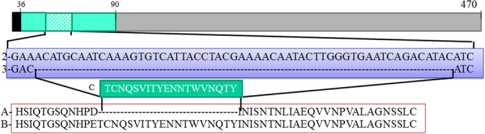
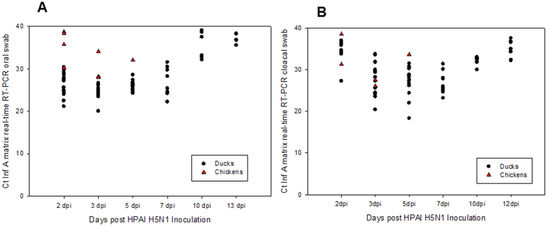
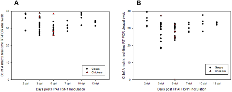
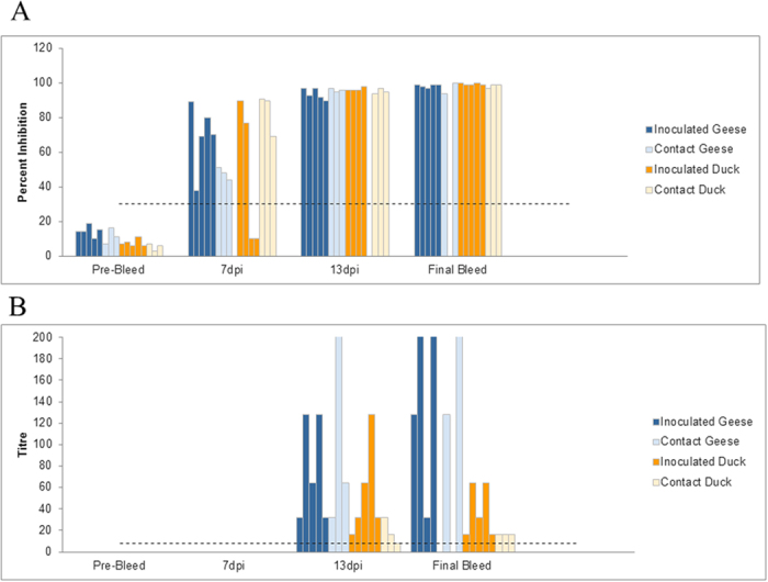
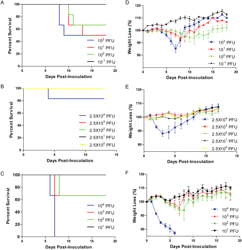
In the current study, we describe the pathobiologic characteristics of a novel reassortant virus - A/chicken/BC/FAV-002/2015 (H5N1) belonging to clade 2.3.4.4 that was isolated from backyard chickens in British Columbia, Canada. Sequence analyses demonstrate PB1, PA, NA and NS gene segments were of North American lineage while PB2, HA, NP and M were derived from a Eurasian lineage H5N8 virus. This novel virus had a 19 amino acid deletion in the neuraminidase stalk. We evaluated the pathogenic potential of this isolate in various animal models. The virus was highly pathogenic to mice with a LD50 of 10 plaque forming units (PFU), but had limited tissue tropism. It caused only subclinical infection in pigs which did result in seroconversion. This virus was highly pathogenic to chickens, turkeys, juvenile Muscovy ducks (Cairnia moschata foma domestica) and adult Chinese geese (Anser cynoides domesticus) causing a systemic infection in all species. The virus was also efficiently transmitted and resulted in mortality in naïve contact ducks, geese and chickens. Our findings indicate that this novel H5N1 virus has a wide host range and enhanced surveillance of migratory waterfowl may be necessary in order to determine its potential to establish itself in the wild bird reservoir.


Figures

Figure 1
Figure 1. Schematic description of full neuraminidase type 1 protein – 470 amino acids (1).
The NA domain corresponding to the stalk region is highlighted in green (36–90 AA) and the deletion of 19 AA in FAV-002 /H5N1 is highlighted in green/white squares. The globular head is highlighted in grey and the transmembrane domain in black. Alignment of the nucleotide sequence of the stalk region which is deleted in FAV-002/H5N1 (3) and AGT/H5N1 (2). Alignment of the protein sequence stalk region of FAV-002/H5N1 (A) and AGT/H5N1 (B). The deleted part of the NA stalk of FAV-002/H5N1 (C) is highlighted in green box.
Figure 2
Figure 2. The survival curves for SPF chickens infected with three different dilutions of FAV-002/H5N1.
The LD50 of FAV-002/H5N1 in chickens was determined to be 23 PFU.
Figure 3
Figure 3. The survival curves for domestic juvenile turkeys infected with 1000 PFU of FAV-005/H5N1 and naïve contact turkeys that were placed among the infected 24 hrs after infection.
The arrow indicates when naïve contact turkeys were placed among the infected turkeys at 24 hrs post-infection.
Figure 4
Figure 4. Survival curves of birds infected with 10,000 PFU of FAV-002/H5N1 and their contacts.
(A) shows the survival curves of Muscovy ducks that were infected with 10,000 PFU of FAV-002/H5N1. The arrow indicates when Muscovy ducks and chickens that were placed among the infected ducks at 24 hrs post-infection. (B) shows the survival curves of Chinese geese that were infected with 10,000 PFU of FAV-002/H5N1 and contact Chinese geese and chickens that were placed in the same animal cubicle at 24 hrs post-infection (arrow).
Figure 5
Figure 5. Virus shedding patterns of Muscovy ducks infected with 10,000 PFU of FAV-002/H5N1.
Total RNA was extracted from oral (A) and cloacal (B) specimens and was tested using the avian influenza matrix based real time RT-PCR assay.
Figure 6
Figure 6. Virus shedding patterns of Chinese geese infected with 10,000 PFU of FAV-002/H5N1.
Total RNA was extracted from oral (A) and cloacal (B) specimens and was tested using the avian influenza matrix based real time RT-PCR assay.
Figure 7
Figure 7. Serologic data for Chinese geese and Muscovy ducks that were infected with FAV-002/H5N1 virus and their corresponding contact groups.
(A) shows influenza A virus nucleoprotein antibodies levels at different intervals of time using cELISA. Values above 30% inhibition (dotted line) were considered positive. No significant differences were found among inoculated versus contact ducks and geese at each time point. Significant differences (P value < 0.001) were however found between final bleed versus pre-bleed, 13 dpi versus pre-bleed, 7 dpi versus pre-bleed, final bleed versus 7 dpi and 13 dpi versus 7 dpi groups based on two-way ANOVA followed by the Holm-Šídák multiple comparison test. (B) illustrates anti-H5 antibody levels based at different intervals of time. Antibody detection is based on the hemagglutination-inhibition test using homologous FAV-002/H5N1 antigen. HI titers above 8 (dotted line) were considered positive. A statistically significant (P = 0.023) interaction was found between animal groups (inoculated/contact geese and inoculated/contact ducks) and time post-inoculation. Statistically significant differences were observed in HI titers of animals between the following time points: final bleed versus pre-bleed (P < 0.001), final bleed versus 7 dpi (P < 0.001), 13 dpi versus pre-bleed (P = 0.027) and 13 dpi versus 7 dpi (P = 0.022). Inoculated geese showed significantly different HI titers at the following time points: final bleed versus pre-bleed (P < 0.001), final bleed versus 7 dpi (P < 0.001) and final bleed versus 13 dpi (P = 0.003). Finally significant differences in HI titers were observed in the contact geese group at the following time points: final bleed versus pre-bleed (P = 0.011) and final bleed versus 7 dpi (P = 0.013). Analysis was by two-way ANOVA followed by the Holm-Šídák multiple comparison test.
Figure 8
Figure 8. Microscopic lesions observed in geese, turkeys and ducks that were infected with FAV-002/H5N1.
(A) Extensive necrosis in the pancreas of a Chinese goose. (B) Presence of abundant viral antigen in the pancreas of a Chinese goose. (C) Presence of large areas hepatic necrosis with heterophil infiltration in a turkey. (D) Extensive area of hepatic necrosis associated with positive immuno-staining (arrow) in a goose. (E) Large multifocal areas of necrosis in the spleen of a turkey. (F) Positive immuno-staining in the multifocal areas of necrosis in the spleen of a Chinese goose. (G) Brain lesions in Muscovy ducks were subtle and variable and included necrosis with gliosis and meningitis, ependymitis and inflammation of the choroid plexus. (H) Extensive viral antigen (*) in the brain of the turkey.
Figure 9
Figure 9. Survival and weight loss of mice infected with H5N1 and H5N8 viruses.
Six- to 7-week-old female Balb/c mice were intranasally challenged with 10 fold serial dilutions of FAV-002/H5N1 (A,D), AGT/H5N1 (B,E) and AW-050/H5N8 (C,F) and observed for survival and weight loss. The percent survival was determined and mouse 50% lethal dose (MLD50) was calculated by the Miller and Tainter method (43). N = 6 mice per group.
Figure 10
Figure 10. Histopathology (HE) and immunohistochemistry (IHC) findings in lungs from H5N1 infected mice.
(A) Day 3: Mild to moderate bronchiolitis (arrow) and scattered foci of interstitial pneumonia (*). HE, bar = 100 μm. (B) Day 6: Large areas of necrotizing interstitial pneumonia (*) and bronchiolitis (arrow). HE, bar = 100 μm. (C) Day 3: Positive immunostaining for influenza A antigen is primarily observed in bronchiolar epithelial cells (arrow). IHC, bar = 50 μm. (D) Day 6: Viral antigen is detected in bronchiolar epithelial cells (arrows) as well as in macrophages (arrowheads). IHC, bar = 50 μm.
Figure 11
Figure 11. Histopathology (HE) and immunohistochemistry (IHC) findings in lungs from FAV-002/H5N1 infected pigs.
(A) Microscopic lesions in a lung characterized by interstitial infiltration of inflammatory cells (arrows), mild bronchiolar changes with degeneration and disorganization of epithelial cells (arrowhead) and increased numbers of macrophages in bronchiolar lumens and alveoli. HE, bar = 50 μm. (B) Positive immunostaining for influenza A viral antigen was detected in scattered bronchiolar epithelial cells. IHC, bar = 50 μm.
Figure 12
Figure 12. Neuraminidase enzyme kinetics and sensitivity of H5N1 viruses to oseltamivir.
(A) Neuraminidase kinetic curves for FAV-002/H5N1 and AGT/H5N1. Relative fluorescent unit (RFU) values for each virus (FAV-002/H5N1 and AGT/H5N1) were plotted against a 2-fold dilution series of MUNANA to determine the maximum velocity (Vmax) and the binding affinity (Km) to MUNANA for each virus. (B) Comparative sensitivity to oseltamivir for each virus is reflected by the percent inhibition of enzyme activity at each concentration of oseltamivir carboxylate, plotted as the ratio of RFU at each dose over the RFU value with no drug treatment. P value < 0.0001.

References

    1. Pasick J. et al. Reassortant Highly Pathogenic Influenza A H5N2 Virus Containing Gene Segments Related to Eurasian H5N8 in British Columbia, Canada, 2014. Sci. Rep. 5, 9484 (2015). - PMC - PubMed
    1. Prosser D. J. et al. Wild bird migration across the Qinghai-Tibetan plateau: a transmission route for highly pathogenic H5N1. PLoS One. 6, e17622 (2011). - PMC - PubMed
    1. Sims L. D. & Brown I. H. Multicontinental epidemic of H5N1 HPAI virus (1996–2007). In Avian Influenza (ed. Swayne D. E.) 251–286 (Blackwell Publishing, 2008).
    1. Keawcharoen J. et al. Wild ducks as long-distance vectors of highly pathogenic avian influenza virus (H5N1). Emerg. Infect. Dis. 14, 600–607 (2008). - PMC - PubMed
    1. Xu X. Y., Subbarao K., Cox N. J. & Guo Y. J. Genetic characterization of the pathogenic influenza A/Goose/Guangdong/1/96 (H5N1) virus: Similarity of its hemagglutinin gene to those of H5N1 viruses from the 1997 outbreaks in Hong Kong. Virology 261, 15–19 (1999). - PubMed

Publication types

MeSH terms