. 2016 May 1;76(9):2513-24.
doi: 10.1158/0008-5472.CAN-15-1325. Epub 2016 Mar 17.

Elucidation of the Roles of Tumor Integrin β1 in the Extravasation Stage of the Metastasis Cascade

Affiliations

Elucidation of the Roles of Tumor Integrin β1 in the Extravasation Stage of the Metastasis Cascade

Michelle B Chen et al. Cancer Res. .

Abstract

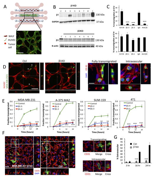
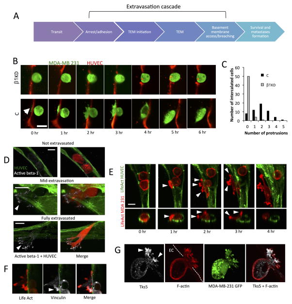
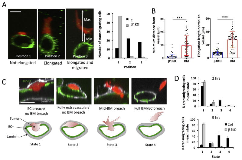
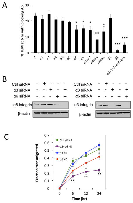
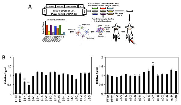
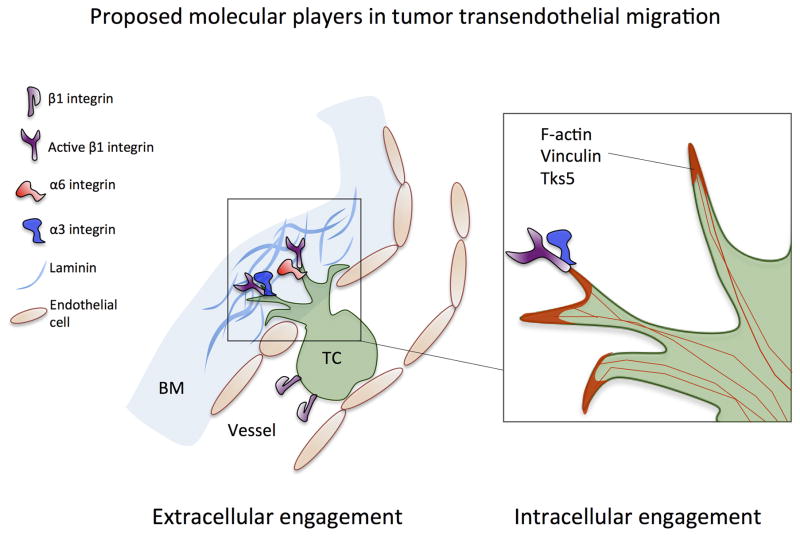
Tumor integrin β1 (ITGB1) contributes to primary tumor growth and metastasis, but its specific roles in extravasation have not yet been clearly elucidated. In this study, we engineered a three-dimensional microfluidic model of the human microvasculature to recapitulate the environment wherein extravasation takes place and assess the consequences of β1 depletion in cancer cells. Combined with confocal imaging, these tools allowed us to decipher the detailed morphology of transmigrating tumor cells and associated endothelial cells in vitro at high spatio-temporal resolution not easily achieved in conventional transmigration assays. Dynamic imaging revealed that β1-depleted cells lacked the ability to sustain protrusions into the subendothelial matrix in contrast with control cells. Specifically, adhesion via α3β1 and α6β1 to subendothelial laminin was a critical prerequisite for successful transmigration. β1 was required to invade past the endothelial basement membrane, whereas its attenuation in a syngeneic tumor model resulted in reduced metastatic colonization of the lung, an effect not observed upon depletion of other integrin alpha and beta subunits. Collectively, our findings in this novel model of the extravasation microenvironment revealed a critical requirement for β1 in several steps of extravasation, providing new insights into the mechanisms underlying metastasis. Cancer Res; 76(9); 2513-24. ©2016 AACR.


Conflict of interest statement

The authors disclose no potential conflicts of interest.

Figures

Fig 1
Fig 1. Knockdown of β1 integrin inhibits tumor-cell transendothelial migration in in vitro microvascular networks and in vivo extravasation assays
(A) Schematic (not to scale) of the microfluidic device for the formation of microvascular networks. A central gel region with suspended human umbilical cord vein cells (HUVECs) is flanked by two normal human lung fibroblast (NHLF) channels. Each gel region is flanked by two media channels (pink). Representative fluorescence image of a single field of view of the device at 20X (HUVEC LifeAct; green, MDA-MB 231; red, scale bar = 100 μm). (B) Western blot of β1 and β3 integrin expression after suppression via shRNA (C=control shRNA targeting firefly luciferase). (C) Effect of β1 and β3 knockdowns and integrin function-blocking antibodies on the TEM efficiency of MDA-MB 231 in in vitro microvascular networks at 6 hours (***p<0.001). (D) Representative field of views (20X) of live microvascular networks at 12 hr after seeding of control or β1 KD MDA-MB-231 cells (HUVEC; red, MDA-MB 231; green). Asterisks indicate fully transmigrated cells (scale bar = 100 μm). Immunostaining for CD31 show distinctions between transmigrated and non-transmigrated cells (scale bar = 20 μm). (E) Differences in kinetics of TEM between β1 KD and control shRNA cells were assessed for MDA-MB-231, A375 MA2, SUM-159 and 4T1 cells lines. Fraction transmigrated in human microvascular network devices is determined at the same field of view for time points of 0, 6, 12 and 24 hr (n=3, **p<0.01, ***p<0.001, bars represent mean +/− SEM). (F) Immunostaining for CD31 (red) in mouse lungs 24 hours after injection of 0.5 million MDA-MB 231 control or β1 KD cells (green). Asterisks indicate cells scored as transmigrated (scale bars = 20 μm). Cells in white dotted boxes are zoomed in to indicate examples of intravascular and extravascular cells. (G) Percentage of transmigrated control and β1 KD MDA-MB 231 cells in mice lungs 3hr, 16 hr and 24 hr after tail vein injection (n=8 mice per condition at 3 hr and 24 hr, 4 mice per condition at 16 hr, 100 randomly selected tumor cells analyzed per mouse) (**p<0.001, bars represent mean +/− SEM).
Fig 2
Fig 2. Activated β1 and actomyosin rich protrusion formation precede and are required for complete transmigration
(A) Depiction of the multiple steps involved in the extravasation cascade. Circulating tumor cells arrest due to tumor-endothelium adhesion or size restriction. This is followed by onset of TEM, which may involve initiation of protrusions breaching an originally intact endothelium. After complete TEM, tumor cells may invade past the vascular basement membrane. (B) Time-lapse confocal microscopy of representative transmigrating control and β1-5 KD cells (single plane) over a period of 6 hours (HUVEC cell tracker; red, MDA-MB 231; green, scale bar=15 μm). Arrow indicates the formation of initial protrusions into the subendothelial matrix in control cells. (C) Differences between control and β1-5 KD cells in the frequency of protrusion formation in single cells during TEM at 3 hours (55 intercalated cells over 3 devices per condition were chosen randomly and scored). Protrusion number (that was definable at 30X magnification) was quantified via 3D reconstructions of single transmigrating cells. (D) Immunostaining with an antibody against the activated conformation of β1 integrin (clone 12G10, white) in microvascular networks (green) (scale bar = 10 μm). Arrows indicate localization at protrusion tips. (E) Time-lapse imaging depicting spatial organization of tumor F-actin (red) during TEM in microvascular network devices. Arrows indicates areas in protrusions where F-actin appears as punctates (scale bar = 10 μm). (F–G) Immunostaining of vinculin and Tks-5 in tumor cells during mid-transmigration past the endothelium. Arrows indicate localization of indicated proteins at the protrusion tips (scale bars = 10 μm).
Fig 3
Fig 3. β1 integrins mediate invasion past the basement membrane
(A) Representative cross sections of lumens in microvascular devices (scale bars = 10 μm) depicting the 3 distinct states in which a transmigrated tumor cell can be found relative to the endothelium after 6 hours (Position 1: directly adjacent to abluminal surface, Position 2: adjacent and elongated normal to vessel wall, Position 3: migrated away from the endothelium) (scale bars = 10 μm). Quantification of the number of fully transmigrated β1KD or control cells found in each “state” (50 cells analyzed per condition). (B) Quantification of the migration distance of extravasated tumor cells away from the endothelium. Distance is defined as the shortest length between the endothelium (at the point where transmigration occurred) and the nearest point on the transmigrated tumor cell body. Elongation length of the same transmigrated tumor cells is defined as the maximum length of the cell normal (perpendicular) to the vessel wall at the point of transmigration (n=50 transmigrated cells per condition, ***p<0.001, bars represent mean +/− standard deviation). (C) Representative immunofluorescence staining depicting the possible position of transmigrated tumor cells relative to the sub-endothelial laminin layer (LN: white, HUVEC LifeAct: green, MDA-MB 231: red, scale bars=10 μm). State 1: breached ECs but not laminin, State 2: fully transmigrated but no breaching of laminin, State 3: simultaneously breaching EC and laminin layer, State 4: fully breached EC and laminin layers. (D) Percentage of total fully transmigrated control or total β1-5 KD cells that are found in states 1 to 4 relative to the laminin layer (n=2 experiments, 4 devices per condition).
Fig 4
Fig 4. Transendothelial migration is mediated by α3β1 and α6β1 integrins via interactions with laminin
(A) Percentage transendothelial migration in microvascular network devices at 6 hours when tumor cells are treated with function-blocking antibodies for various α and β subunits (n=3 experiments, 3 devices per condition, *p<0.05, **p<0.01, ***p<0.001, bars represent mean +/− SEM). (B) Western blot analysis of alpha 3 and alpha 6-integrin individual knockdowns and co-knockdowns in MDA-MB-231 via siRNA. (C) Kinetics of TEM in microvascular networks following the same regions at 0, 6, 12 and 24 hr after tumor cell seeding.
Fig 5
Fig 5. Knockdown of β1 integrin in 4T1 cells inhibits metastatic colonization
(A) Schematic of our Luminex-based approach for multiplexing tail vein metastasis assays (see Methods). 4T1 cells were stably transduced with uniquely barcoded (BC) vectors expressing miR30-based shRNAs targeting one integrin subunit per cell population, and knock down was confirmed by flow cytometry (see Table 1). Uniquely barcoded 4T1 integrin knockdown cell populations were then mixed in equal numbers and injected into the tail veins of syngeneic Balb/C mice. After 17 days, Genomic DNA was isolated from the metastasis-containing lungs, and the relative amount of each barcode (i.e. the relative number cells expressing each shRNA) was quantified using streptavidin-conjugated APC (Strep-APC) and the Luminex FlexMap 3D system. (B) Luminex-based quantification of the relative metastatic burden of each 4T1 integrin knockdown cell population. Graphs show the relative signal + S.E.M. from each shRNA relative to the signal for that shRNA in the starting mixed population (n=10 mice / mix analyzed in duplicate).
Fig 6
Fig 6. Proposed molecular players involved in tumor transendothelial migration
Extracellular engagement: activation of β1 integrin facilitates protrusion maintenance past retracted endothelium, via engagement to subendothelial ECM. Specifically, integrins α3β1 and α6β1 are both required for adhesion to vascular basement membrane laminin. Intracellular engagement: β1integrin-rich extracellular adhesions maintain protrusions, allowing focal adhesion proteins (e.g. vinculin) and F-actin to be recruited to the tips of protrusions, resulting in transmigration via acto-myosin contraction at the protruding edge.

References

    1. Heyder C, et al. Realtime visualization of tumor cell/endothelial cell interactions during transmigration across the endothelial barrier. J Cancer Res Clin Oncol. 2002;128:533–8. - PubMed
    1. Haier J. An Intravital Model to Monitor Steps of Metastatic Tumor Cell Adhesion Within the Hepatic Microcirculation. J Gastrointest Surg. 2003;7:507–515. - PubMed
    1. Ganguly KK, Pal S, Moulik S, Chatterjee A. Integrins and metastasis. 2013:251–261. - PMC - PubMed
    1. Hynes RO. Integrins: Bidirectional , Allosteric Signaling Machines In their roles as major adhesion receptors , integrins. 2002;110:673–687. - PubMed
    1. Orini MM, et al. THE α3 β1 INTEGRIN IS ASSOCIATED WITH MAMMARY CARCINOMA CELL METASTASIS , INVASION , AND GELATINASE B ( MMP-9 ) ACTIVITY. 2000;342:336–342. - PubMed

Publication types

Substances