. 2016 Jun;89(6):630-44.
doi: 10.1124/mol.116.103267. Epub 2016 Mar 17.

Repurposing Treprostinil for Enhancing Hematopoietic Progenitor Cell Transplantation

Affiliations

Repurposing Treprostinil for Enhancing Hematopoietic Progenitor Cell Transplantation

Zahra Kazemi et al. Mol Pharmacol. 2016 Jun.

Erratum in

Abstract

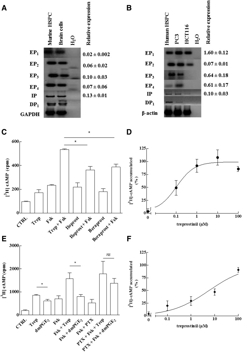
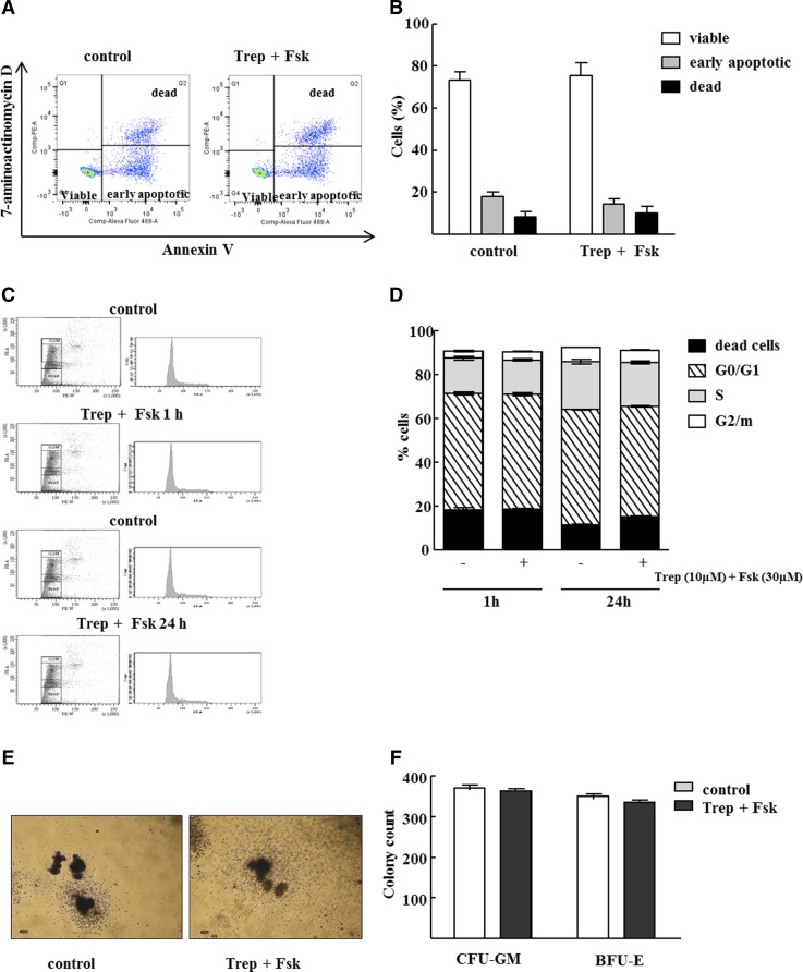
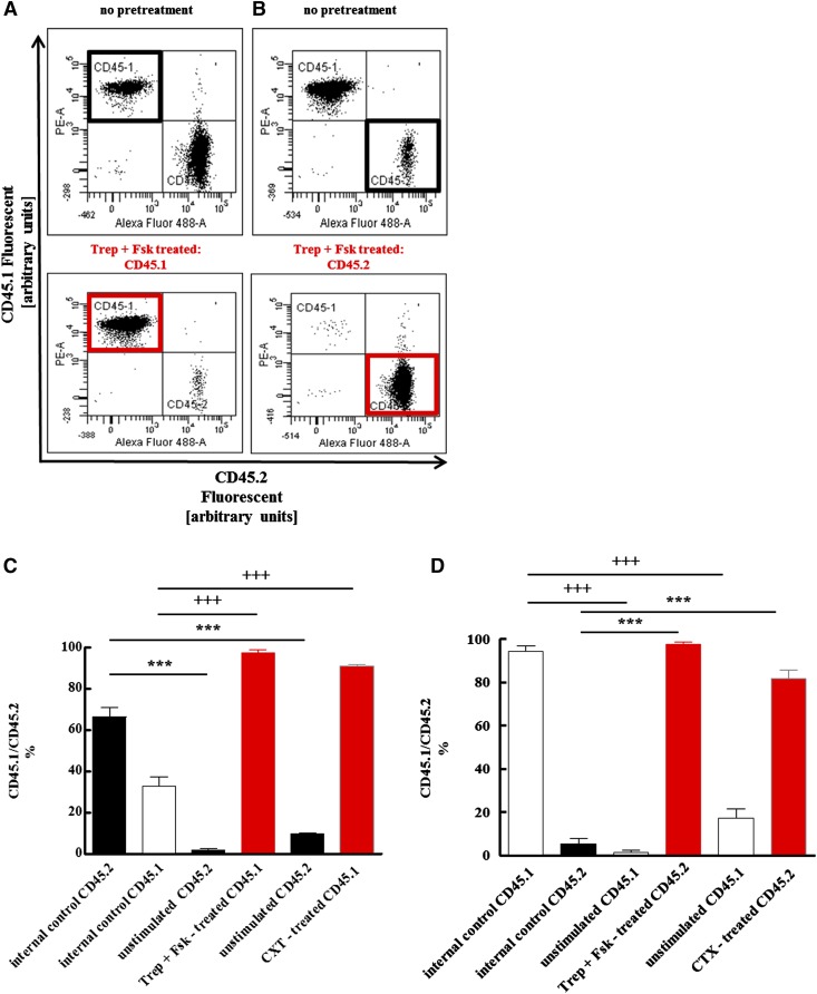
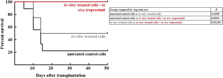
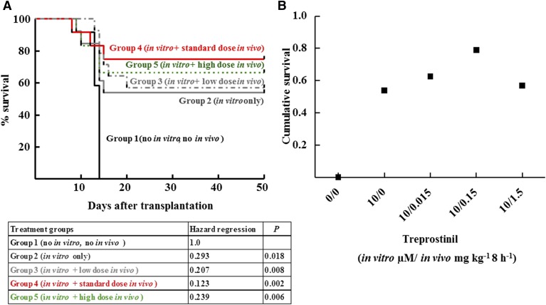
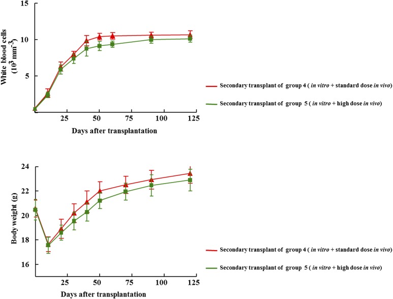
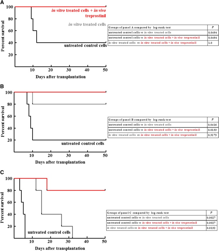
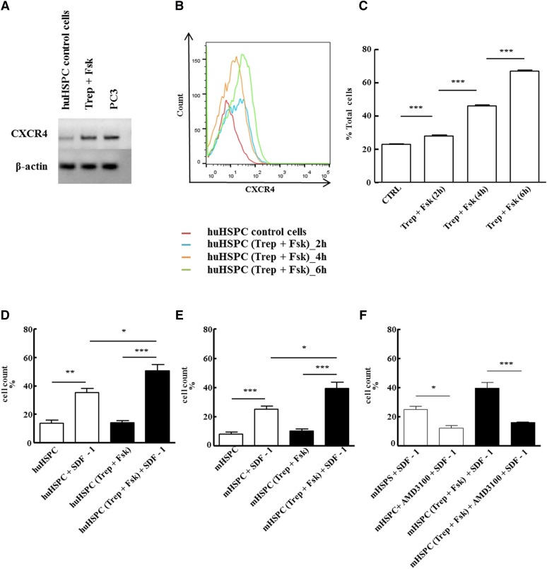
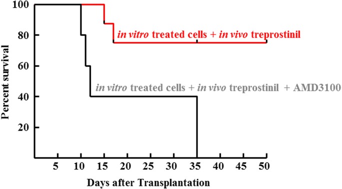
Activation of Gs-coupled receptors enhances engraftment of hematopoietic stem and progenitor cells (HSPCs). We tested the hypothesis that treprostinil, a prostacyclin analog approved for the treatment of pulmonary hypertension, can be repurposed to improve hematopoietic stem cell transplantation. Murine and human HSPCs were isolated from bone marrow and umbilical cord blood, respectively. Prostanoid receptor agonists and the combination thereof with forskolin were tested for their capacity to stimulate [(3)H]cAMP accumulation in HSPCs. Three independent approaches were employed to verify the ability of agonist-activated HSPCs to reconstitute the bone marrow in lethally irradiated recipient mice. The underlying mechanism was explored in cellular migration assays and by blocking C-X-C motif chemokine receptor 4 (CXCR4). Among several prostanoid agonists tested in combination with forskolin, treprostinil was most efficacious in raising intracellular cAMP levels in murine and human HPSCs. Injection of murine and human HSPCs, which had been pretreated with treprostinil and forskolin, enhanced survival of lethally irradiated recipient mice. Survival was further improved if recipient mice were subcutaneously administered treprostinil (0.15 mg kg(-1) 8 h(-1)) for 10 days. This regimen also reduced the number of HSPCs required to rescue lethally irradiated mice. Enhanced survival of recipient mice was causally related to treprostinil-enhanced CXCR4-dependent migration of HSPCs. Treprostinil stimulates the engraftment of human and murine hematopoietic stem cells without impairing their capacity for self-renewal. The investigated dose range corresponds to the dose approved for human use. Hence, these findings may be readily translated into a clinical application.


Figures

Fig. 1.
Fig. 1.
Prostanoid receptor expression (A, B) and stimulation of cAMP accumulation (C–F) in murine and human HSPCs. (A, B) RNA was isolated from murine (A) and human HSPCs (B) and reverse transcribed. RNA prepared from murine brain cells (mixed culture of neurons and glial cells) and the human prostate cancer cell lines PC3 and HCT116 served as positive controls. PCR-dependent amplification was done using primers listed in Table 1. Amplicons for all E prostanoid receptors (EP1–4, IP, and DP1) were electrophoretically resolved on an agarose gel and visualized by ethidium bromide staining. The lane labeled H2O denotes the control, where the amplification was done in the absence of prior reverse transcription. The mRNA encoding glyceraldehyde-3-phosphate dehydrogenase (GAPDH) was amplified as internal reference. Quantitative PCR was performed using Maxima SYBR Green/ROX qPCR Master Mix (2×) and equal amounts of cDNAs and primers. Human and murine HPRT1 was used as reference gene for normalization of qPCR experiments. Each reaction condition was performed in triplicate. Relative abundance was calculated using the 2–ΔCt method (gene-specific expression level relative to that of the endogenous reference gene HPRT1, which was set at 1). We failed to identify a primer pair capable of amplifying murine DP1 receptor transcripts with an efficiency close to 100%; accordingly, the quantification of DP1 receptor transcripts is not shown. (C–F) The cAMP response of murine (C, D) and CD34+ human HSPCs (E, F) was determined after metabolic labeling of the adenine nucleotide pool with [3H] adenine. In some instances (PTX), cells were concomitantly also preincubated with pertussis toxin (100 ng ml−1) for 16 hours. (C) Murine HSPCs were stimulated with treprostinil (10 μM; Trep), iloprost (30 μM), beraprost (30 μM), and forskolin (30 μM; Fsk) or the combination of indicated agonists and forskolin (30 μM). In the presence of 10 μM treprostinil, 30 μM forskolin was more efficacious than 30 μM iloprost and beraprost (p = 0.02, one-way ANOVA). (E) Human HSPCs were stimulated with treprostinil (10 μM), forskolin (30 μM), the combination thereof, or dmPGE2 (10 μM), or the combination thereof with forskolin (30 μM). Both, in the absence and presence of 30 μM forskolin, 10 μM treprostinil was more efficacious than 10 μM dmPGE2 (p = 0.03; Wilcoxon test). In contrast, in cells, which had been pretreated with pertussis toxin, stimulation with forskolin+treprostinil and forskolin+dmPGE2 did not result in any statistically significant difference in cAMP accumulation (ns). Panels D and F show the concentration-response curve for treprostinil-induced cAMP accumulation for murine (in the presence of 30 μM forskolin) and human HSPCs, respectively. The maximum levels of [3H]cAMP accumulation was 595 ± 60 cpm (D) and 1943 ± 262 cpm (F). Data are means ± S.D. (n = 3).
Fig. 2.
Fig. 2.
Pretreatment of murine and human HSPCs with treprostinil and forskolin does neither induce apoptosis nor alters cell cycle progression or differentiation potential. Human HSPCs were incubated with 10 μM treprostinil and 30 μM forskolin for 1h. Subsequently, (A, B) apoptosis induction and (C, D) cell cycle progression was assessed by flow cytometric analysis. Representative original pictures are depicted (A, C, left hand panel) and data obtained in three independent experiments was summarized (B, D, right hand panel). No difference in apoptotic cells or distribution of cells according to G0/1, S and G2 phase was detected between untreated and treated cells (one way ANOVA). (E, F) Murine HSPCs were isolated form bone marrow, pretreated and resuspended in a methylcellulose medium containing growth factors required for supporting the differentiation and growth of granulo-monocytic colony–forming units (CFU-GM) and of the erythrocyte lineage (BFU-E, burst-forming-unit erythroid). After 10 days the number of colonies was counted under a light microscope and shape and morphology of colonies was observed. Shown are representative photomicrographs and the quantification of three independent experiments. Data are means ± S.E.M. (n = 3).
Fig. 3.
Fig. 3.
Competitive advantage of HSPCs after pretreatment with the combination of treprostinil and forskolin or cholera toxin over unstimulated controls. Murine HSPCs were isolated from CD45.1+ and from CD45.2+ donor mice. Either CD45.1+ (A, C) or CD45.2+ (B, D) cells were incubated for 1 hour in medium in the absence of any additional stimulus, in the presence of the combination of 10 μM treprostinil and 30 μM forskolin (Trep + Fsk, pretreated) or of 10 μg ml−1 cholera toxin (as a positive control). First, cell suspensions were prepared that contained either equivalent amounts of both unstimulated CD45.1+ and CD45.2+ cells [top panel in (A); bars labeled “internal control” in (C)] or equivalent amounts of stimulated and untreated donor cells [bottom panel in (A)]. These suspensions containing a total of 106 cells were administered by tail vein injection to lethally irradiated recipients (A, C). Conversely, CD45.2+ cells, which were either not stimulated [top panel in (B); bars labeled “internal control” in (D)], or stimulated [bottom panel in (B)], and untreated CD45.1+ were mixed at a ratio of 1:10; a total of 5 × 10⁵ cells were injected into recipient mice (B, D). The proportion of bone marrow cells arising from the two sources was determined by flow cytometry 16 weeks after transplantation. Representative original dot plots are shown in (A) and (B); bar diagrams in (C) and (D) represent the means ± S.E.M. (n = 5 animals per group) from two independent experiments. The indicated statistical comparisons of CD45.2- (+) and CD45.1-positive cells (*) were done by analysis of variance (ANOVA) followed by Tukey’s multiple comparison (P = 0.0001).
Fig. 4.
Fig. 4.
Survival curves for lethally irradiated BALB/c mice after transplantation with murine HSPCs, which had been left untreated, treated with the combination of treprostinil and forskolin and further stimulated by in vivo administration of treprostinil. Murine HSPCs were incubated in the absence (no treatment; n = 9) or presence of the combination of 10 μM treprostinil and 30 μM forskolin (in vitro pretreatment); subsequently, the cell suspension (3 × 10⁵ cells per mouse) was administered by tail vein injection to lethally irradiated recipient mice. Some mice, which received pretreated cells, were also treated in vivo with treprostinil (in vitro and in vivo treatment; n = 10; 0.15 mg kg−1 s.c for 10 days), whereas others received no further treatment (in vitro pretreatment; n = 13). The differences between the survival curves are statistically significant: in vitro pretreatment versus in vitro/in vivo treatment, P = 0.01290; untreated control versus in vitro/in vivo treatment, P = 0.0003 (log-rank test).
Fig. 5.
Fig. 5.
In vivo administration of treprostinil enhances survival of recipient mice. Kaplan-Meier plots for survival of lethally irradiated recipient mice receiving 2 × 10⁵ untreated murine HSPCs (group 1, n = 12), HSPCs that had been solely pretreated in vitro with the combination of treprostinil and forskolin (group 2, n = 13), or mice that were transplanted with pretreated murine HSPC and administered—in addition—treprostinil by subcutaneous injection (group 3: 0.015 mg kg−1, n = 12; group 4: 0.15 mg kg−1, n = 14; group 5: 1.5 mg kg−1, n = 14). The experiment was performed as outlined in the legend to Fig. 4 and in Materials and Methods. In panel (B), survival is replotted as a function of the administered treprostinil dose to illustrate the bell-shaped nature of the concentration-response curve. The statistical comparison was done by a Cox proportional hazard regression analysis: There was a dose-dependent effect of treprostinil on survival; however, the relation was bell-shaped: at the highest dose (1.5 mg kg−1 8 h−1), survival was lower than at the intermediate dose (0.15 mg kg−1 8 h−1). The Cox proportional hazard regression analysis identified the effect of different treatment regimens on survival. Treatment groups were modeled as a categorical predictor variable. Regression was performed using IBM SPSS Statistics 20.0. Omnibus Test revealed that the inclusion of the covariate “treatment group” was of high statistical significance: The decrease in hazard ratio displayed treatment dependence (see tabulated values). Compared with the “no treatment” control (group 1), the risk of fatal events decreased progressively from group 2 (sole in vitro treatment) to group 4 (additional in vivo treatment with the standard treprostinil dose). This trend leveled off and declined in animals exposed to a high treprostinil dose (group 5) as expected from a bell shaped dose-response relationship.
Fig. 6.
Fig. 6.
Time-dependent change in peripheral white blood cell counts (upper graph) and body weight (lower graph) after transplantation of bone marrow cells obtained from primary recipients. One-hundred, sixty days after transplantation four mice originating from treatment group 4 and group 5 respectively, were sacrificed, their femurs were flushed with phosphate-buffered saline to recover bone marrow cells. Subsequently, 2 × 10⁶ bone marrow cells were transplanted into lethally irradiated secondary recipient mice. All mice that received this secondary transplant survived. At the indicated time points, blood was drawn, the white blood cell count was determined, and the weight of the mice recorded. Data are means ± S.E.M.
Fig. 7.
Fig. 7.
Survival curves for lethally irradiated NSG mice after transplantation with different amounts of human HSPCs. CD34+ human hematopoietic progenitor cells were isolated from umbilical cord blood and incubated in the absence (no treatment, group 1) or presence of the combination of treprostinil and 30 μM forskolin (in vitro pretreatment, groups 2 and 3); subsequently, the cell suspension (2.5 × 10⁵, 2 × 10⁵, and 1.5 × 10⁵ cells per mouse in the (A), (B), and (C) panels, respectively) was administered by tail vein injection to lethally irradiated recipient NSG mice. Some mice, which received pretreated cells, were also treated in vivo with treprostinil (in vitro and in vivo treatment, group 3; 0.15 mg kg−1 8 h−1, s.c. and for 10 days), whereas others received no further treatment (in vitro pretreatment – group 2). Each group was comprised of 5 animals. The tables summarize the comparisons of the individual Kaplan-Maier plots by log-rank test.
Fig. 8.
Fig. 8.
In vitro pretreatment with treprostinil and forskolin enhances the action of SDF-1 via CXCR4. (A) RNA was isolated from human HSPCs that had been incubated in the absence (control) or presence of the combination of 10 μM treprostinil and 30 μM forskolin (Trep + Fsk) for 1 hour. RNA prepared from the human prostate cancer cell line PC3 served as positive control. After reverse transcription, PCR-dependent amplification was done using primers listed in Table 1. Amplicons for CXCR4 were electrophoretically resolved on an agarose gel and visualized by ethidium bromide staining. The mRNA encoding β-actin was amplified as internal control. The data are representative of two additional experiments with similar results. (B, C) Human HSPCs were incubated in the absence (red trace = vehicle control) or in the presence of the combination of treprostinil (10 μM) and forskolin (30 μM) for 2 hours (blue trace), 4 hours (orange trace), and 6 hours (green trace) at 37°C and stained for CXCR4 with an allophycocyanin-labeled antibody. (B) FACS histograms from a representative experiment. (C) Bar diagram shows means ± S.E.M. from three independent experiments. Statistically significant differences were examined using repeated-measures ANOVA followed by Bonferroni’s multiple comparison. (D–F) Freshly isolated murine and human HPSC were pretreated in vitro with either vehicle (open bars) or 10 μM treprostinil and 30 μM forskolin (closed bars) for 1 hour at 37°C followed by washing steps. A suspension (2 × 10⁵ cells in 0.1 ml of medium containing growth factors) of human (D) or murine HSPCs (E, F) was added to the upper chamber of a Transwell and allowed to migrate toward SDF-1 (100 ng ml−1 in the lower chamber) for 4 hours. Cells that had migrated through the 5-μm filter were counted. HSPCs were also incubated in the absence and presence of 10 μM AMD3100 to confirm that the effects arose from a stimulation of CXCR4 (F). Data represent ± S.E.M. from three independent experiments carried out in triplicate. The statistical comparison was done by repeated-measures ANOVA followed by Tukey’s multiple comparison (*P < 0.05, **P < 0. 01, ***P < 0.001).
Fig. 9.
Fig. 9.
In vivo administration of CXCR4-antagonist (AMD3100) abrogates the beneficial effect of treprostinil on the survival of recipient mice. Murine HPSCs were pretreated in vitro with treprostinil and forskolin as outlined in the legend to Fig. 4 and injected (2 × 10⁵ per mouse) into lethally irradiated recipient mice. These were subsequently divided into two groups. Mice allocated to group 1 (n = 10) were further subjected to in vivo treatment with treprostinil (0.15 mg kg−1), and mice in group 2 (n = 10) received both treprostinil (0.15 mg kg−1 8 h−1) and AMD3100 (3.3 mg kg−1 8 h−1) by subcutaneous injection every 8 hours for 10 days. The difference between the two survival curves was statistically significant (P = 0.007, log-rank test).

References

    1. Adams GB, Alley IR, Chung UI, Chabner KT, Jeanson NT, Lo Celso C, Marsters ES, Chen M, Weinstein LS, Lin CP, et al. (2009) Haematopoietic stem cells depend on Galpha(s)-mediated signalling to engraft bone marrow. Nature 459:103–107. - PMC - PubMed
    1. Adams GB, Chabner KT, Alley IR, Olson DP, Szczepiorkowski ZM, Poznansky MC, Kos CH, Pollak MR, Brown EM, Scadden DT. (2006) Stem cell engraftment at the endosteal niche is specified by the calcium-sensing receptor. Nature 439:599–603. - PubMed
    1. Aksentijevich I, Flinn I. (2002) Chemotherapy and bone marrow reserve: lessons learned from autologous stem cell transplantation. Cancer Biother Radiopharm 17:399–403. - PubMed
    1. Alexander SP, Benson HE, Faccenda E, Pawson AJ, Sharman JL, Spedding M, Peters JA, Harmar AJ, CGTP Collaborators (2013) The Concise Guide to PHARMACOLOGY 2013/14: G protein-coupled receptors. Br J Pharmacol 170:1459–1581. - PMC - PubMed
    1. Aronoff DM, Peres CM, Serezani CH, Ballinger MN, Carstens JK, Coleman N, Moore BB, Peebles RS, Faccioli LH, Peters-Golden M. (2007) Synthetic prostacyclin analogs differentially regulate macrophage function via distinct analog-receptor binding specificities. J Immunol 178:1628–1634. - PubMed

MeSH terms