Case Reports
. 2016 Mar 17:2016:bcr2015213046.
doi: 10.1136/bcr-2015-213046.

Electrocardiographic clue for a mid-LAD lesion

Affiliations
Case Reports

Electrocardiographic clue for a mid-LAD lesion

Rohat Ak et al. BMJ Case Rep. .

Abstract

ECG is still the first diagnostic tool for coronary artery disease. It is possible to predict the localisation of affected vessel(s) through ST and T changes on ECG. Sometimes, reciprocal changes may be the only marker of acute myocardial ischaemia, as single T-wave inversion in lead aVL may represent a coronary artery lesion in the left anterior descending (LAD). A 49-year-old woman presented to the emergency department, with left-sided chest pain. Her initial ECG showed no ischaemic changes. On the third hour ECG there was T-wave inversion in leads aVL and V2, and troponin turned positive. Coronary angiography showed 90% mid-LAD occlusion. The importance of this case is that patients with ischaemic chest pain should be followed with serial ECG. Also, emergency physicians should be alert to identify new changes on ECG, as isolated T-wave inversion in lead aVL can be the only finding to take the patient into the catheterisation laboratory.


Figures

Figure 1
Figure 1
Twelve-lead ECG at presentation showing normal sinus rhythm and no ischaemic change.
Figure 2
Figure 2
Dynamic T-wave changes on ECG; T-wave inversions in leads aVL and V2.
Figure 3
Figure 3
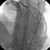
Coronary angiography showing mid-left anterior descending lesion (white arrow).
Figure 4
Figure 4
Coronary angiography showing ostial left anterior descending occlusion (white arrow).

References

    1. Glancy DL, Doghmi W. Use of indicative and reciprocal electrocardiographic changes to help localize the site of coronary occlusion. Proceedings (Bayl Univ Med Cent) 2001;14:104–5. - PMC - PubMed
    1. Kracoff OH, Adelman AG, Oettinger M et al. . Reciprocal changes as the presenting electrocardiographic manifestation of acute myocardial ischemia. Am J Cardiol 1993;71:1359–62. 10.1016/0002-9149(93)90556-R - DOI - PubMed
    1. Hassen GW, Costea A, Smith T et al. . The neglected lead on electrocardiogram: T wave inversion in lead aVL, nonspecific finding or a sign for left anterior descending artery lesion. J Emerg Med 2014;46:165–70. 10.1016/j.jemermed.2013.08.079 - DOI - PubMed
    1. Birnbaum Y, Wilson JM, Fiol M et al. . ECG diagnosis and classification of acute coronary syndromes. Ann Noninvasive Electrocardiol 2014;19:4–14. 10.1111/anec.12130 - DOI - PMC - PubMed
    1. Birnbaum Y, Sclarovsky S, Mager A et al. . ST segment depression in aVL: a sensitive marker for acute inferior myocardial infarction. Eur Heart J 1993;14:4–7. 10.1093/eurheartj/14.1.4 - DOI - PubMed

Publication types