. 2016 May 15;143(10):1674-87.
doi: 10.1242/dev.133728. Epub 2016 Mar 17.

Integrin signalling regulates YAP and TAZ to control skin homeostasis

Affiliations

Integrin signalling regulates YAP and TAZ to control skin homeostasis

Ahmed Elbediwy et al. Development. .

Abstract

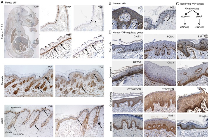
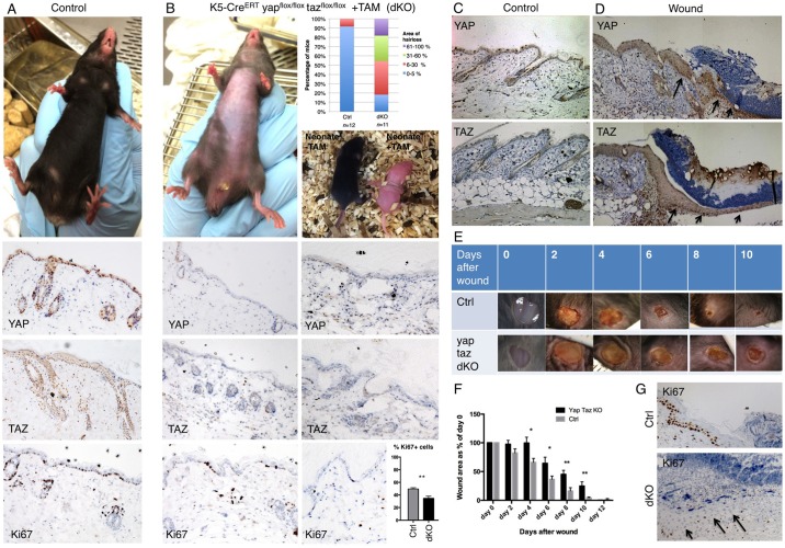
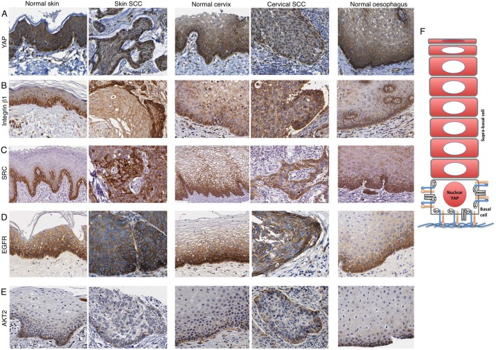
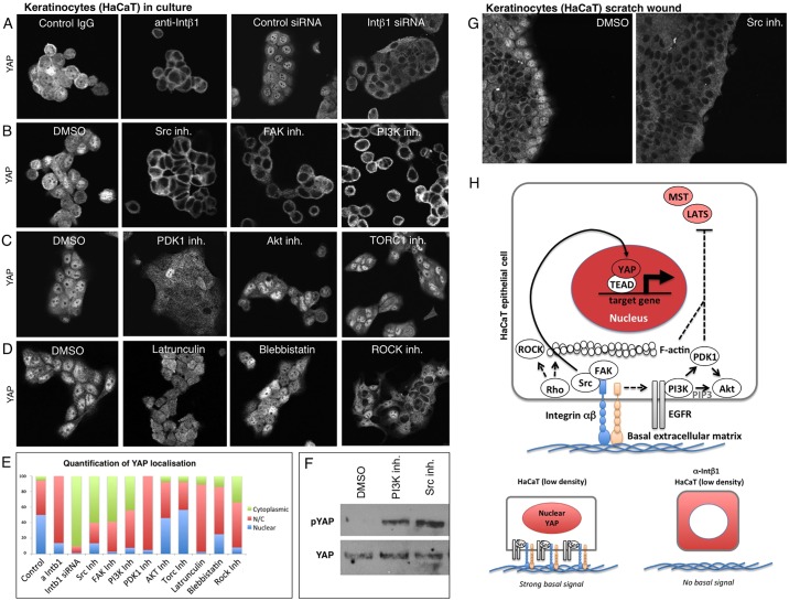
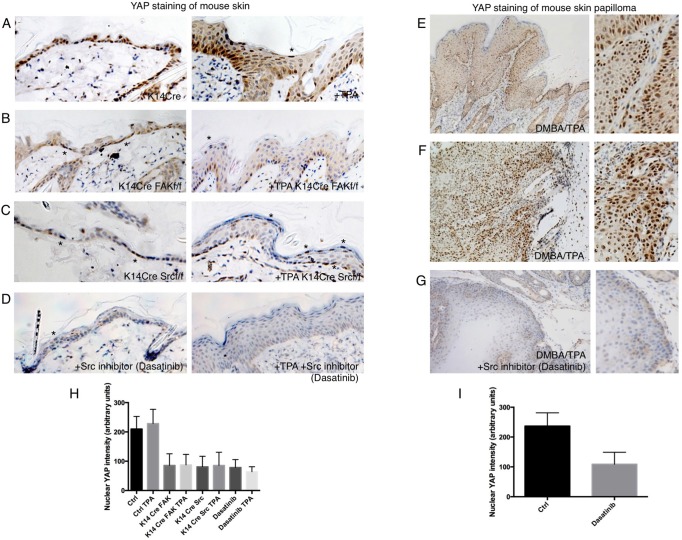
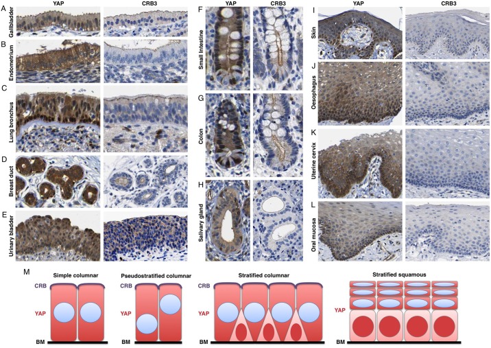
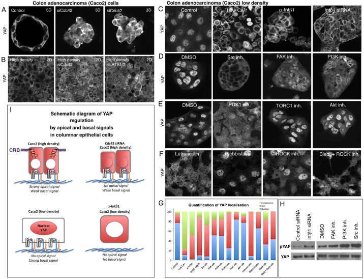
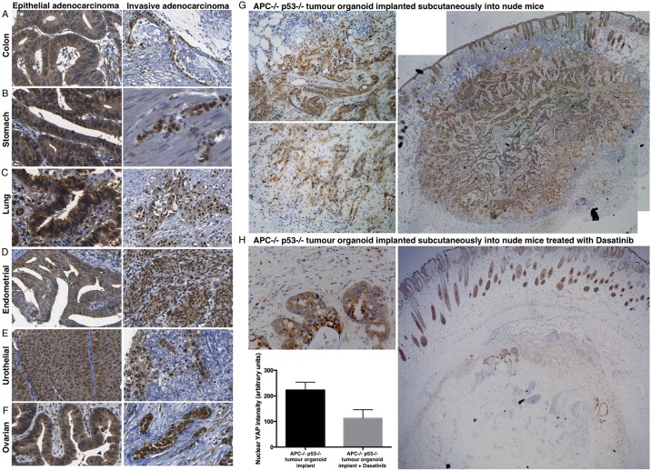
The skin is a squamous epithelium that is continuously renewed by a population of basal layer stem/progenitor cells and can heal wounds. Here, we show that the transcription regulators YAP and TAZ localise to the nucleus in the basal layer of skin and are elevated upon wound healing. Skin-specific deletion of both YAP and TAZ in adult mice slows proliferation of basal layer cells, leads to hair loss and impairs regeneration after wounding. Contact with the basal extracellular matrix and consequent integrin-Src signalling is a key determinant of the nuclear localisation of YAP/TAZ in basal layer cells and in skin tumours. Contact with the basement membrane is lost in differentiating daughter cells, where YAP and TAZ become mostly cytoplasmic. In other types of squamous epithelia and squamous cell carcinomas, a similar control mechanism is present. By contrast, columnar epithelia differentiate an apical domain that recruits CRB3, Merlin (also known as NF2), KIBRA (also known as WWC1) and SAV1 to induce Hippo signalling and retain YAP/TAZ in the cytoplasm despite contact with the basal layer extracellular matrix. When columnar epithelial tumours lose their apical domain and become invasive, YAP/TAZ becomes nuclear and tumour growth becomes sensitive to the Src inhibitor Dasatinib.

Keywords: Hippo pathway; Integrin; Stratified squamous epithelium; TAZ; Yes-associated protein.


Conflict of interest statement

Competing interests

The authors declare no competing or financial interests.

Figures

Fig. 1.
Fig. 1.
YAP and TAZ are expressed in both mouse and human skin and regulate gene expression in basal layer stem cells. (A) Mouse skin at three developmental stages, including embryonic (E17.5), neonate and adult. Tissue sections were stained for either YAP or TAZ to reveal their expression and subcellular localisation. (B) Human skin (adult) stained for either YAP or TAZ. Note the nuclear localisation in basal layer stem/progenitor cells as well as terminally differentiating flattened cells. Other differentiating cells have cytoplasmic YAP and TAZ localisation. Arrows indicate nuclear YAP/TAZ; asterisks indicate flattened suprabasal cells with nuclear YAP/TAZ. (C) Analysis of YAP-dependent gene expression by RNA-seq was performed by comparison of YAP gain and loss of function in keratinocytes (see Fig. S1). (D) YAP-regulated genes identified by RNA-seq analysed for their expression patterns in skin tissue by mining the Human Protein Atlas dataset (see Materials and Methods). Strong enrichment in basal layer stem/progenitor cells is evident for many target genes, indicating that YAP and TAZ are transcriptionally active in this population of cells.
Fig. 2.
Fig. 2.
Conditional inactivation of YAP and TAZ impairs skin homeostasis and wound repair in mice. (A) Control mice have a thick layer of hair (fur) covering their skin, which sections reveal is positive for YAP, TAZ and Ki67 (a marker of cell proliferation). (B) Double conditional knockout mice for YAP and TAZ treated with tamoxifen as adults or neonates exhibit dramatic hair loss. Adult skin sections are negative for YAP and TAZ, and have reduced levels of Ki67+ positive cells (quantified as a percentage of total interfollicular basal cells in each randomly selected 40× field of view. n=757 control cells; n=896 dKO cells). (C) Control mouse skin stained for YAP and TAZ. (D) Punch biopsy wound edge stained for YAP and TAZ. (E) Imaging of wound healing in control (n=8) and YAP/TAZ double conditional knockout mice (dKO; n=8). Note delayed healing in dKO. (F) Quantification of wound healing rates in control versus dKO animals. ImageJ was used to measure the wound area at each stage. (G) Proliferation of cells as marked by Ki67 staining is reduced in dKO wounds versus control animals. Values are means±s.e.m. *P<0.05, **P<0.01.
Fig. 3.
Fig. 3.
Integrin-Src and EGFR-PI3K localisation in human stratified squamous epithelia and squamous cell carcinomas (SSCs). The Human Protein Atlas dataset was mined to compare the expression and localisation of potential YAP regulators in human skin sections. YAP staining reveals basal layer nuclear localisation (A), ITGB1, SRC and EGFR staining reveals basal layer expression (B-D) and AKT2 staining reveals basal subcellular localisation (E) across squamous tissue types and cancers. (F) Model for YAP regulation in stratified squamous epithelia.
Fig. 4.
Fig. 4.
Basal integrin-Src signalling promotes nuclear localisation of YAP in human HaCaT keratinocyte epithelial cells. (A) YAP nuclear localisation is prevented by treatment of keratinocytes with anti-ITGB1 antibodies (PD52) or by ITGB1 siRNA treatment. (B) YAP nuclear localisation is prevented by treatment of keratinocytes with the Src inhibitor Dasatinib, by the FAK inhibitor PF573228 or by the PI3K inhibitor GDC0941, but not by treatment with DMSO solvent. (C) YAP nuclear localisation is reduced by treatment of keratinocytes with the PDK1 inhibitor BX795, but not by the AKT inhibitor MK2206, TORC1 inhibitor Everolimus or DMSO solvent. (D) YAP nuclear localisation is reduced by treatment of keratinocytes with the F-actin destabilising drug Latrunculin, the myosin II inhibitor Blebbistatin, or the Rho-kinase inhibitor Y27632. (E) Quantification of A-D. (F) Western blotting analysis of p-YAP levels in keratinocytes treated with either DMSO control, PI3K inhibitor or Src inhibitor. Total YAP levels are shown as a control. (G) Nuclear YAP localisation at the leading edge of a scratch wound in keratinocyte culture is abolished by treatment with the Src inhibitor Dasatinib. (H) Schematic diagram of YAP regulation in keratinocytes.
Fig. 5.
Fig. 5.
Basal integrin-Src signalling promotes YAP stability and nuclear localisation in mouse skin. (A) YAP staining in control and TPA-treated skin to induce hyperplasia. (B) YAP staining is reduced in FAK conditional KO skin before or after treatment with TPA. Note some residual nuclear YAP localisation in basal layer cells or highly flattened cells (asterisk). (C) YAP staining is reduced in Src conditional KO skin before or after treatment with TPA. Note that there is some residual nuclear YAP localisation in basal layer cells or highly flattened cells (asterisks). (D) YAP staining is reduced in Dasatinib-treated skin before or after treatment with TPA for 2 days. Note there is some residual nuclear YAP localisation in basal layer cells or highly flattened cells (asterisk). (E) YAP staining of mouse skin papilloma induced by DMBA-TPA treatment of mice expressing v-Ha-Ras (see Materials and Methods). Note stronger nuclear localisation in the basal layer. (F) YAP staining of mouse skin squamous cell carcinoma induced by DMBA-TPA treatment of v-Ha-Ras-expressing mice. (G) YAP staining is strongly reduced by treatment of DMBA-TPA induced papillomas with the Src inhibitor Dasatinib topically for 3 days. (H) Quantification of nuclear YAP intensity in A-D. (I) Quantification of nuclear YAP intensity in F,G. Values are means±s.e.m.
Fig. 6.
Fig. 6.
Apical-domain formation inhibits YAP nuclear localisation in human columnar epithelia. The Human Protein Atlas dataset was mined to compare the localisation of YAP with the presence of absence of the apical domain in different epithelia. YAP localises to the cytoplasm in columnar gallbladder epithelium (A) and columnar endometrial epithelium (B) which have a CRB3+ apical domain. (C) YAP localises to the nucleus of basal layer stem/progenitors, which lack CRB3 expression, and cytoplasm in columnar epithelial cells, which have a CRB3+ apical domain, in the bronchus. (D) YAP localises to the nucleus of basal layer stem/progenitors, which lack CRB3 expression, and cytoplasm in columnar epithelial cells, with a CRB3+ apical domain, in the breast. (E) YAP localises to the cytoplasm in pseudostratified columnar bladder epithelium, with a CRB3+ apical domain. (F) YAP localises to the nucleus of crypt base stem/progenitors, which lack a large CRB3+ apical domain, and cytoplasm in columnar epithelial cells, which feature a large CRB3+ apical domain, in the small intestine. (G) YAP localises to the nucleus of crypt base stem/progenitors, which have a small CRB3+ apical domain, and cytoplasm in columnar epithelial cells, which feature a large CRB3+ apical domain, in the colon. (H) YAP localises to the nucleus of basal layer stem/progenitors, which lack CRB3 expression, and cytoplasm in columnar epithelial cells, which have a CRB3+ apical domain, in the salivary gland. (I-L) YAP localises to the nucleus of basal layer stem/progenitors, and cytoplasm of differentiating squamous epithelial cells, even though the entire tissue lacks CRB3 expression. (M) Schematic diagram of YAP localisation in different epithelial tissue types.
Fig. 7.
Fig. 7.
Basal integrin-Src signalling promotes YAP nuclear localisation in human Caco2 epithelial cells when apical domain formation is blocked. (A) Caco2 colon adenocarcinoma cells form 3D cysts in cell culture that feature cytoplasmic YAP localisation. Silencing of CDC42 by siRNA knockdown disrupts apical-basal polarity and induces more nuclear YAP localisation. (B) Caco2 colon adenocarcinoma cells form 2D epithelial monolayers at high density. Silencing of CDC42 by siRNA knockdown disrupts apical-basal polarity and induces more nuclear YAP localisation, similar to silencing of LATS1/2. (C) YAP nuclear localisation is very strong when Caco2 cells are plated at low density to prevent apical domain formation. Nuclear localisation is prevented by treatment of Caco2 cells with low-calcium medium, anti-ITGB1 antibodies (PD52) or by ITGB1 siRNA treatment, but not in controls. (D) YAP nuclear localisation is prevented by treatment of Caco2 cells with the Src inhibitor Dasatinib, by the FAK inhibitor PF573228 or by the PI3K inhibitor GDC0941, but not by treatment with DMSO solvent. (E) YAP nuclear localisation is reduced by treatment of Caco2 cells with the PDK1 inhibitor BX795, but not by the AKT inhibitor MK2206, TORC1 inhibitor Everolimus or DMSO solvent. (F) YAP nuclear localisation is reduced by treatment of Caco2 cells with the F-actin destabilising drug Latrunculin, the myosin II inhibitor Blebbistatin or the Rho kinase inhibitor Y27632, or a combination of Blebbistatin and Y27532. (G) Quantification of C-F. (H) Western blotting analysis of p-YAP levels in Caco2 cells treated with control siRNAs or ITGB1 siRNAs, as well as DMSO control, FAK inhibitor, PI3K inhibitor or Src inhibitor. Total YAP levels are shown as a control. (I) Schematic diagram of YAP regulation in Caco2 cells.
Fig. 8.
Fig. 8.
YAP becomes nuclear in invasive adenocarcinomas, which become sensitive to Dasatinib. In the colon (A) and stomach (B) YAP localises to the cytoplasm of columnar epithelial cells in epithelial adenocarcinoma, and the nucleus of invasive adenocarcinoma cells, which have lost their columnar shape and lack a lumen. (C) In the bronchus, YAP localises to the nucleus of basal layer stem/progenitors and cytoplasm in columnar epithelial cells in epithelial adenocarcinoma, and the nucleus of invasive adenocarcinoma. (D) In the endometrial epithelium, YAP localises to the cytoplasm of columnar epithelial cells in epithelial adenocarcinoma, and the nucleus of invasive adenocarcinoma cells. (E) In urothelial epithelium, YAP localises to the cytoplasm in pseudostratified columnar cells in epithelial adenocarcinoma and to the nucleus of invasive adenocarcinoma. (F) YAP localises to the cytoplasm in ovarian adenocarcinoma, and to the nucleus of invasive ovarian adenocarcinoma. (G) YAP staining in Apc−/− p53−/− tumour organoids implanted subcutaneously into nude mice, which invade dramatically into the surrounding tissue. Note that cells at the invasive front feature nuclear YAP localisation, whereas columnar epithelial cells in the central regions of the tumour feature cytoplasmic YAP localisation. (H) YAP staining is strongly reduced by Dasatinib treatment of Apc−/− p53−/− tumour organoids implanted subcutaneously into nude mice. Invasive tumour cells are not visible. Quantification of YAP nuclear localisation was performed on n=200 tumour cells from G and H. Values are means±s.e.m.

References

    1. Akhtar N. and Streuli C. H. (2013). An integrin–ILK–microtubule network orients cell polarity and lumen formation in glandular epithelium. Nat. Cell Biol. 15, 17-27. 10.1038/ncb2646 - DOI - PMC - PubMed
    1. Anani S., Bhat S., Honma-Yamanaka N., Krawchuk D. and Yamanaka Y. (2014). Initiation of Hippo signaling is linked to polarity rather than to cell position in the pre-implantation mouse embryo. Development 141, 2813-2824. 10.1242/dev.107276 - DOI - PubMed
    1. Aragona M., Panciera T., Manfrin A., Giulitti S., Michielin F., Elvassore N., Dupont S. and Piccolo S. (2013). A mechanical checkpoint controls multicellular growth through YAP/TAZ regulation by actin-processing factors. Cell 154, 1047-1059. 10.1016/j.cell.2013.07.042 - DOI - PubMed
    1. Azzolin L., Panciera T., Soligo S., Enzo E., Bicciato S., Dupont S., Bresolin S., Frasson C., Basso G., Guzzardo V. et al. (2014). YAP/TAZ incorporation in the beta-catenin destruction complex orchestrates the Wnt response. Cell 158, 157-170. 10.1016/j.cell.2014.06.013 - DOI - PubMed
    1. Baumgartner R., Poernbacher I., Buser N., Hafen E. and Stocker H. (2010). The WW domain protein Kibra acts upstream of Hippo in Drosophila. Dev. Cell 18, 309-316. 10.1016/j.devcel.2009.12.013 - DOI - PubMed

MeSH terms