. 2015 Apr;10(2):154-164.
doi: 10.1007/s11515-015-1354-2. Epub 2015 Mar 30.

Emerging roles of autophagy in metabolism and metabolic disorders

Affiliations

Emerging roles of autophagy in metabolism and metabolic disorders

Altea Rocchi et al. Front Biol (Beijing). 2015 Apr.

Abstract

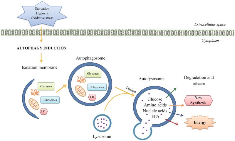
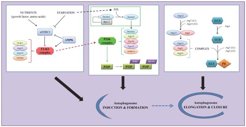
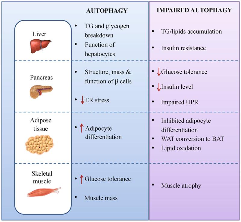
The global prevalence of metabolic disorders is an immediate threat to human health. Genetic features, environmental aspects and lifestyle changes are the major risk factors determining metabolic dysfunction in the body. Autophagy is a housekeeping stress-induced lysosomal degradation pathway, which recycles macromolecules and metabolites for new protein synthesis and energy production and regulates cellular homeostasis by clearance of damaged protein or organelles. Recently, a dramatically increasing number of literatures has shown that defects of the autophagic machinery is associated with dysfunction of multiple metabolic tissues including pancreatic β cells, liver, adipose tissue and muscle, and is implicated in metabolic disorders such as obesity and insulin resistance. Here in this review, we summarize the representative works on these topics and discuss the versatile roles of autophagy in the regulation of cellular metabolism and its possible implication in metabolic diseases.

Keywords: autophagy; diabetes; metabolic disease; metabolism; obesity; selective autophagy.


Figures

Figure 1
Figure 1
Autophagy in organelle turnover and nutrient recycling. Autophagy is initiated by the formation of an isolation membrane (or phagophore) under stress conditions, such as nutrient deprivation, hypoxia and oxidative stress. The isolation membrane enwraps bulk cytosol or specific cargos (misfolded proteins, mitochondria, glycogen, ribosomes, lipid droplets, etc.), and elongates and forms a double-membrane autophagosome. It then fuses with the lysosome into an autolysosome, where the resident hydrolytic enzymes digest cargos and various resulting metabolites and macromolecules (including amino acids, glucose, nucleic acids and free fatty acids) are released back to the cytosol as new building blocks or energy sources. LD, lipid droplets; FFA, free fatty acids.
Figure 2
Figure 2
Regulation of autophagosome formation. Under nutrient-rich conditions, active mTORC1 inhibits the ULK1 kinase complex; whereas starvation activates AMPK, suppresses mTORC1 and induces ULK1, which upregulates another kinase complex, the PtdIns3K complex. Recruitment of Beclin 1 to the PtdIns3K complex is essential for the kinase activity, and is negatively regulated by sequestration of Beclin 1 by Bcl-2 and positively regulated by release of Beclin 1 upon JNK-mediated phosphorylation of Bcl-2. The PtdIns3K complex generates PI3P, which recruits additional PI3P-binding proteins (including WIPI and DFCP1) to promote autophagosome formation. Elongation of autophagosome membrane requires two ubiquitin-like conjugation systems, Atg12-Atg5-Atg16 and LC3-PE; the former acts as an E3 ligase to assist the conjugation of LC3 to PE. All the above molecules and pathways cooperate on the induction and formation of autophagosomes under stress conditions.
Figure 3
Figure 3
Functions of autophagy in multiple metabolic tissues. The roles of autophagy in selective tissues are summarized, based on the results from both in vitro and in vivo studies. Impaired autophagy imposes opposite effects in specific organs, leading to diverse metabolic abnormalities and susceptibility to diseases. TG, triglycerides; UPR, unfolded protein response; WAT, white adipose tissue; BAT, brown adipose tissue.

References

    1. Axe EL, Walker SA, Manifava M, Chandra P, Roderick HL, Habermann A, Griffiths G, Ktistakis NT. Autophagosome formation from membrane compartments enriched in phosphatidy-linositol 3-phosphate and dynamically connected to the endoplasmic reticulum. J Cell Biol. 2008;182(4):685–701. - PMC - PubMed
    1. Baerga R, Zhang Y, Chen PH, Goldman S, Jin S. Targeted deletion of autophagy-related 5 (atg5) impairs adipogenesis in a cellular model and in mice. Autophagy. 2009;5(8):1118–1130. - PMC - PubMed
    1. Bjørkøy G, Lamark T, Brech A, Outzen H, Perander M, Øvervatn A, Stenmark H, Johansen T. p62/SQSTM1 forms protein aggregates degraded by autophagy and has a protective effect on huntingtin-induced cell death. J Cell Biol. 2005;171(4):603–614. - PMC - PubMed
    1. Boyle KB, Randow F. The role of ‘eat-me’ signals and autophagy cargo receptors in innate immunity. Curr Opin Microbiol. 2013;16(3):339–348. - PubMed
    1. Burman C, Ktistakis NT. Regulation of autophagy by phosphatidylinositol 3-phosphate. FEBS Lett. 2010;584(7):1302–1312. - PubMed

LinkOut - more resources